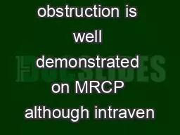
Holdorf OUTLINE PART 2 Pit OUTLINE PART 2 Laboratory values Gallbladder carcinoma Adenomyomatosis Biliary obstruction Common duct measurement Dilated Intrahepatic ducts Fatty meal Level of obstruction ID: 774916
Download Presentation The PPT/PDF document " Lecture 7 The Biliary Tract Part II" is the property of its rightful owner. Permission is granted to download and print the materials on this web site for personal, non-commercial use only, and to display it on your personal computer provided you do not modify the materials and that you retain all copyright notices contained in the materials. By downloading content from our website, you accept the terms of this agreement.
Slide1
Lecture 7The Biliary Tract Part II
Holdorf
Slide2OUTLINE PART 2
Pit
OUTLINE PART 2
Laboratory values
Gallbladder carcinoma
Adenomyomatosis
Biliary obstruction
Common duct measurement
Dilated Intrahepatic ducts
Fatty meal
Level of obstruction
Choledocholithiasis
Mirizzi syndrome
Bile duct carcinoma
Cholagnitis
Biliary atresia
Pneumobilia
Choledochal cyst
Caroli’s disease
Pancreatic adenocarcinoma
Slide3Laboratory Values
Bilirubin
Bilirubin is the end product of hemoglobin breakdown.
Total bilirubin = conjugated bilirubin + unconjugated bilirubin.
Conjugation
is the process of removing protein (albumin) from unconjugated bilirubin making it soluble. Conjugation is important for bilirubin disposal. This reaction not only renders the pigment excretable by the liver, but also detoxifies this waste product.
Slide4Laboratory Values
Increased INDIRECT or UNCONJUGATED bilirubin results from:
Increased bilirubin production (hemolysis) or
Decreased liver conjugation.
Increased DIRECT or CONJUGATED bilirubin (soluble) results from:
Decreased biliary excretion (bile duct obstruction).
Slide5Laboratory Values
Alkaline Phosphatase (ALP)
ALP is an enzyme found in bone, liver and placenta. ALP increases in diseases that impair bile formation, specifically BILE DUCT OBSTRUCTION. ALP also increases with bone growth (growing children) and pregnancy. ALP is also a sensitive indicator of liver and bone cancer.
ALP increases with:
Biliary obstruction
Liver disease (Hepatitis, Hepatocellular Ca)
Bone growth and diseases
Hyperparathyroidism
Pregnancy
Slide6Gallbladder Carcinoma
Presents with an:
Intraluminal mass
Asymmetric wall thickening or
A mass that fills the gallbladder lumen.
Associated findings include:
Liver metastases
Lymphadenopathy
Bile duct dilatation (Intra-hepatic)
Cholelithiasis
Slide7Gallbladder Carcinoma
Rare: 2-4 % of all gastrointestinal malignancies
Usually unresectable
5 year survival rate of 1%
Slide8Gallbladder Carcinoma
Slide9Metastatic GB Ca
Slide10Adenomyomatosis
Hyperplastic changes involving the gallbladder wall causing overgrowth of the mucosa, thickening of the wall, and formation of diverticula.
Diverticula within the gallbladder wall (Rokitansky-Aschoff sinuses or RAS) accumulate stones or sludge within them.
Slide11Adenomyomatosis
Sonograpically seen as hyperechoic foci within a thickened gallbladder wall.
Associated with a comet-tail (reverberation) artifact.
Differentiating between adenomyomatosis and cholesterolosis may be difficult.
Slide12Adenomyomatosis with comet tail artifacts
Slide13Adenomyomatosis
Slide14Biliary Obstriction
In the majority of patients, biliary obstruction is due to pathology of the distal CBD:
Gallstones or Carcinoma of the Head of the Pancreas.
These are the two most common lesions. Biliary obstruction is considered clinically when the patient presents with jaundice. There may or may not be associated RUQ pain and or a palpable RUQ mass.
Slide15Biliary Obstruction
Serum alkaline phosphatase and conjugated bilirubin levels are typically elevated.
Obstruction of the distal CBD results in progressive dilatation of the extrahepatic and intrahepatic biliary tree (distal to proximal).
Causes of biliary obstruction:
Choledocholithiasis (Biliary duct stone)
Pancreatic carcinoma
Cholangiocarcinoma (Carcinoma of the biliary ducts)
Cholangitis (Inflammation of the bile ducts
Mirizzi Syndrome
Choledochal cyst (cystic dilatation of the bile ducts)
Gallbladder carcinoma
Slide16Biliary Obstruction Distal CBD
Slide17Biliary Obstruction- Enlarged CBD and Pancreatic Duct
Slide18Common Duct Measurement
The common hepatic duct is routinely measured (lumen only) at the point where the right hepatic artery courses between the portal vein and the biliary duct.
The size of the extrahepatic bile duct is the most sensitive means of distinguishing medical vs. surgical jaundice. (Medical-due to acute or chronic hepatitis or cirrhosis: Surgical-due to stones, tumors or strictures).
Slide19Common Duct Measurement
The diameter of the common duct increases with age with 10 mm being the upper normal value in elderly patients.
Measurement criteria:
1. One millimeter per decade rule-the common duct diameter increased with age. One millimeter per decade is a acceptable rate of size increase. Thus, at age 60, duct size may normally be 6 mm and at age 80, duct size may normally be 8 mm.
Slide20Common Duct Measurement
2. Typically the normal common duct diameters are:
Normal
<
5 mm
Equivocal 6 to 7 mm
Dilated
>
8 mm
Post cholecystectomy, the common duct acts as a reservoir for bile storage and may increase up to 10 mm or 1 cm
Slide21Common duct dilated S/P cholecystectomy
Slide22Common duct – Shotgun or parallel channel sign
Slide23Dilated intrahepatic ducts
Criteria to differentiate intrahepatic bile ducts from portal veins:
Parallel channel sign or Shotgun sign:
refer to the dilated hepatic duct adjacent to the portal vein.
Irregular and tortuous bile ducts:
biliary ducts are more tortuous than the accompanying portal vein.
Stellate confluence:
bile ducts branch in a star-shaped configuration.
Acoustic enhancement-
bile structures attenuate sound less than blood producing posterior acoustic enhancement.
Slide24Irregular dilated intrahepatic bile ducts
Slide25Fatty Meal
Biliary dynamics, gallbladder contractility, or obstruction, can be assessed by administering a fatty meal.
Cholecystokinin- a hormone that is released into the blood by the ingestion of fatty foods, causes gallbladder contraction.
Equivocal bile duct dilation or abnormal lab values (conjugated bilirubin, alkaline phosphatase) would be reasons to administer a fatty meal.
Negative result-unchanged or decreased size.
Positive result- duct increase in size
Slide26Level of Obstruction
The part of the biliary tree that dilates as a result of obstruction depends on the level of obstruction.
Slide27Distal Common Bile Duct Obstruction- entire system distends including the gallbladder
Slide28Common Hepatic Obstruction.Only proximal ducts will distend. The gallbladder will be contracted.
Slide29Obstruction at the junction of the right and let hepatic ducts – Intrahepatic ducts dilate- GB contracted.
Slide30Slide31Biliary radicals
Slide32Choledocholithiasis
Formation or presence of calculi in the bile ducts. Most common cause of extrahepatic obstructive jaundice.
Symptoms include:
Biliary colic (RUQ pain)
Jaundice
Laboratory values that increase: (all three will increase)
Conjugated serum bilirubin
Alkaline phosphatase
GGT
Slide33Definitions
Conjugated serum bilirubin
Refer back to the laboratory section.
GGT (Gamma-glutamyltransferease)
GGT is predominantly used as a diagnostic marker for liver diseased. Elevated levels suggests diseases of the liver, biliary system, and pancrease
Slide34Choledocholithiasis cont.
Complications of choledocholithiasis include:
Biliary cirrhosis
Cholangitis
Pancreatitis
Diagnostic Tests include:
ERCP (endoscopic retrograde cholangiopancreatography)
PTC Percutaneous transhepatic cholangiography
CT
Ultrasound
Slide35Homework:
Just what is an ERCP and PTC?
Slide36Mirizzi Syndrome
Extrahepatic biliary obstruction due to an impacted stone in the cystic duct causing extrinsic mechanical compression of the common duct.
Associated findings include:
Intrahepatic duct dilation
Cystic duct stone
Curved segmental stenosis of CHD
Cholescystocholedocal fistula
Slide37ERCP showing Mirizzi Syndrome
Slide38Homework
Just what is a Cholescystocholedocal fistula?
Slide39Bile Duct CarcinomaCholangiocarcinoma
Bile duct adenocarcinomas usually originate within extrahepatic bile ducts (CHD or CBD).
A
klatskin Tumor
is a cholangiocarcinoma located at the hepatic hilum (junction of right and left hepatic duct) resulting in intrahepatic but not extrahepatic biliary dilation.
Predisposing conditions include:
Ulcerative colitis
Sclerosing cholangitis
Carol’s disease
Choledochal cyst
Parasitic infection
Chemical toxins.
Slide40Homework: Definitions
Ulcerative colitis
Sclerosing cholangitis
Carol’s disease (soon to come)
Choledochal cyst
Slide41Bile Duct carcinoma cont.
Diagnostic test include:
Ultrasound
CT
Choleangiography-
Choleangiography is the imaging of the
bile ducts (also known as the biliary tree) by
x-rays. There are at least two kinds of cholangiography:
percutaneous transhepatic cholangiography (PTC): Examination of
liver and bile ducts by x-rays. This is accomplished by the insertion of a thin needle into the liver carrying a contrast medium to help to see blockage in liver and bile ducts.
Endoscopic retrograde cholangiopancreatography (ERCP). Although this is a form of imaging, it is both diagnostic and therapeutic, and is often classified with surgeries rather than with imaging.
Slide42Cholangitis
Infection and inflammation of the biliary ducts resulting in wall thickening that compromises the lumen of the bile duct.
Associated with
Choledocholithiasis
Biliary tract interventions
Ulcerative colitis
Symptoms
Fever
Jaundice
RUQ pain
Slide43Cholangitis cont.
Results in increased:
Serum conjugated bilirubin
Serum alkaline phosphatase
Serum amylase and lipase
Leukocytosis
Slide44Cholangitis
Slide45Biliary Atresia
Jaundice persisting beyond 4 weeks is most commonly due to biliary atresia or neonatal hepatitis.
Absence of the extrahepatic biliary tree.
Associated with polysplenia syndrome with abdominal heterotaxia.
Sonographically seen as two-vessel portal triad (portal vein and proper hepatic artery seen only)
Slide46Biliary atresia is suspected in infants with jaundice.
Surgical drainage with the Kasai Portoenterostomy is the most successful in the treatment of biliary atresia if perfumed before 90 days of life.
Slide47Definitions
Polysplenia syndrome
A congenital disease manifested by multiple small accessory spleens.
Heterotaxia
A rare congenital defect in which the major visceral organs are distributed abnormally within the chest and abdomen.
Kasai Portoenterostomy
A surgical treatment performed on infants with
biliary atresia to allow for
bile drainage. In these infants, the
bile is not able to drain normally from the small
bile ducts within the
liver into the larger bile ducts that connect to the
gall bladder and
small intestine.
Slide48Pneumobilia
Pneumobilia is air in the biliary system. It is commonly associated with an ERCP (endoscopic retrograde cholangiopancreatogram).
Ultrasound findings include variable length echogenic foci in the intrahepatic bile ducts.
Comet-tail or reverberation artifacts are typically associated with air in the biliary tree.
Arterial calcification may mimic pneumobilia. Plain film radiography can differentiate air from calcifications.
Slide49Pneumobilia
Slide50Choledochal cyst
Congenital anomalies of the bile ducts consisting of cystic dilatation of the intra and extrahepatic bile ducts.
Sonographic findings include:
Two cystic structures in the RUQ
(gallbladder and dilated extrahepatic ducts)
Intrahepatic bile duct dilatation
Slide51Choledochal cyst cont.
Choledochal cysts are prevalent in Asia. More than 33% of reported cases are from Japan. Symptoms usually occur before age 10.
Pancreatitis
Cholangitis
Hepatic abscesses
Cirrhosis
Portal hypertension
cholangiocarcinoma
Slide52Choledochal cyst
Slide53Caroli’s Disease
Congenital abnormality of the biliary tract characterized by multifocal segmental dilatation of the intrahepatic bile ducts. This is a specific type of choledochal cyst.
Common in childhood and second to third decades of life.
Associated with
Congenital hepatic fibrosis
Portal hypertension
Medullary sponge kidney
Infantile polycystic kidney disease
Renal tubular ectasia
Choledochal cyst
Slide54Caroli’s disease cont.
Ultrasound findings
Multiple cystic structures that converge toward the porta hepatis communicating with the bile ducts. Sludge and calculi may accumulate in these ectatic (dilated) ducts resulting in posterior acoustic shadowing.
Complications include:
Recurrent cholangitis
Biliary calculi and obstruction
Hepatic abscess
Cholangiocarcinoma
Slide55Caroli’s disease
Slide56Caroli’s Disease
Slide57Pancreatic Adenocarcinoma
Most common cause of malignant neoplasm obstructing the biliary tree.
Pancreatic adenocarcinoma at the head of the pancreas typically causes
“Courvoisier gallbladder”.
This is an enlarged, non-diseased gallbladder due to a mechanical obstruction of the common bile duct.
Slide58Courvoisier gallbladder
Slide59Homework for Part 2
What are Rokitansky-Aschoff Sinuses?
What pathology is associated with Rokitansky-Aschoff Sinuses?
What is the sonographic presentation of gallbladder Carcinoma?
What three other findings should be investigated to confirm the diagnosis of gallbladder carcinoma?
What are the two most common causes of biliary tract obstruction?
What lab values are most likely to be elevated due to biliary tract obstruction?
What are the two signs that indicate intrahepatic bile duct dilatation?
Name three extra hepatic biliary ducts.
What pathology causes intrahepatic dilatation without extra hepatic biliary dilatation?
Describe the sonographic appearance of pneumobilia (air in the biliary tree.)
What is the most common reason for pneumobilia?
Choledochal cyst usually occurs in Asian women. Symptoms of pain, jaundice, and an abdominal mass may be present. What are the sonographic features associated with a choledochal cyst?
What is the sonographic appearance of Caroli’s disease?
What other genetically acquired conditions are associated with Caroli’s disease?
What is a Courvoisier gallbladder?
Describe the sonographic appearance of milk of calcium bile.
Bile duct carcinoma (cholangiocarcinoma) is an uncommon cancer. What predisposing conditions are associated with this cancer?
What is the name of the cholangiocarcinoma located at the hepatic hilum?