/
Movie Movie

Movie - PowerPoint Presentation

pamella-moone
pamella-moone . @pamella-moone
Follow
397 views
Uploaded On 2016-03-07

Movie - PPT Presentation

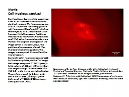
CellNucleusstackavi Confocal Laser Scanning Microscpy image stack of a Chironomus tentans salivary gland cell nucleus The fluorescence was due to the protein hrp36 conjugated to redfluorescent AlexaFluor647 which was microinjected into the cytoplasm After transport into the nucl ID: 245822

confocal pinhole cell psf pinhole confocal psf cell chemistry size nucleus image data spread movie shown kubitscheck siebrasse panel

Share:

Link:

Embed:

Download Presentation from below link

Download Presentation The PPT/PDF document "Movie" is the property of its rightful owner. Permission is granted to download and print the materials on this web site for personal, non-commercial use only, and to display it on your personal computer provided you do not modify the materials and that you retain all copyright notices contained in the materials. By downloading content from our website, you accept the terms of this agreement.


Presentation Transcript

Slide1

MovieCell-Nucleus_stack.avi

Confocal Laser Scanning

Microscpy image stack of a Chironomus tentans salivary gland cell nucleus. The fluorescence was due to the protein hrp36 conjugated to red-fluorescent AlexaFluor647, which was microinjected into the cytoplasm. After transport into the nucleus, hpr36 was incorporated into nascent RNA-particles, such that active transcription sites were marked. The brighter oval region in the image center is the cell nucleus. The polytene chromosomes showed their characteristic banding pattern. The images were aquired 15 min after after microinjection.Complete cell in fluorescence color-coded by the look-up-table „red hot“ of ImageJ Each image comprises 772x512 pixels, a total volume of 185 µm x 29 µm x 46 µm was imaged using a C-Apochromat 40X (NA 1.2 , water immersion) objective lens. The pinhole was set to 1.2 Airy units. Excitation 633nm (5% output); main dichromat UV/488/543/633; emission band pass filter LP650.

Data courtesy of Dr. Jan Peter

Siebrasse

and Dr. Ulrich Kubitscheck, Institute of Physical and Theoretical Chemistry,

Rheinische

Friedrich-

Wilhelms

-

Universtität

Bonn, 2011 (For further information on the biological contents, please refer to:

Siebrasse

. J.P.,

T.Kaminski

and

U.Kubitscheck

(2012) Nuclear export of single native mRNA molecules observed by light sheet fluorescence microscopy.

Proc

Natl

Acad

Sci

USA 109: 9426-31)

 Slide2

MovieCell_Nucleus_rotation.avi

The above image stack was processed and optimized for a 3D rotation presentation. All data processing was done using

ImageJ by Wayne Rasband, NIH (see http://rsb.info.nih.gov/ij/).Data courtesy of Dr. Jan Peter Siebrasse and Dr. Ulrich Kubitscheck, Institute of Physical and Theoretical Chemistry, Rheinische Friedrich-Wilhelms-Universtität Bonn, 2011 (For further information on the biological contents, please refer to:Siebrasse. J.P., T.Kaminski and U.Kubitscheck. 2012. Nuclear export of single native mRNA molecules observed by light sheet fluorescence microscopy. Proc Natl Acad Sci USA 109: 9426-31)Slide3

MovieConfocal_PSF.mp4

Confocal Point-Spread-Function

Calculated isointensity surfaces of the three-dimensional confocal point-spread-function are shown for a 0.7 NA objective with a 1 AU pinhole. The integrated intensity of the confocal psf is projected onto the individual axes and colored according to the color bar on the right. The isointensity surface shows all values of the psf having the same intensity relative to the maximum of the psf and is plotted using the same color table. We gratefully acknowledge Nicolai Hartmann, Department of Chemistry and Center for Nanoscience (CeNS), Ludwig-Maximilians-University Munich, for producing this movie.Slide4

MoviePSF_Pinhole.mp4

Confocal Point Spread Function as a function of Pinhole size

This video shows how the confocal point spread functions (psf) changes as the size of the pinhole is varied. The excitation psf without pinhole is shown in the upper left panel and the total psf is shown in the right upper panel. The fraction of light that is transmitted through the pinhole is shown on the graph in the lower panel with the green cursor marking the current pinhole size for the calculated psf. The smaller the pinhole size yields a higher overall resolution but at a significant loss in detection yield. The lower panel shows that a pinhole with the size of approximately 0.8 – 1 AU is the optimal compromise between resolution and detection yield. We thank Achim Hartschuh, Physical Chemistry, Department of Chemistry and Center for Nanoscience (CeNS), Ludwig-Maximilians-University of Munich, Germany, for producing this movie.Slide5

MovieConfocal_GFP_Cell.mov

Confocal Image of a Huh7 cell with

eGFP-a-tubulin. This movie shows a three-dimensional reconstruction of a z-stack of confocal images from a Huh7 cell, where a-tubulin has been tagged with eGFP. The microtubules are clearly visible, demonstrating the capability of confocal microscopy for three-dimensional imaging. The diameter of the cell is approximately 60 𝜇m. Dr. Ralf Bausinger, Department of Chemistry, University of Konstanz, Germany, is gratefully acknowledged for collecting and processing the data and for making this video available to us.Slide6

MovieSingle_Molecules

Single

DiI molecules diffusing in a fluid glass-supported lipid bilayer. The bilayer was prepared by vesicle-fusion from the lipid dioleyl-phosphatidylcholine (DOPC) with low amounts of the lipohilic dye DiI (molar fraction of ~10-8). The detail has a size of 30 x 30 µm2, recording rate was 20 frames per second, the movie plays at real-time. Data Courtesy by E. Klotzsch and G.J.Schütz, Institute of Applied Physics, Vienna University of Technology, Austria