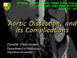
/
Aortic Lumen Detection Brad Wendorff, ECE 539 Aortic Lumen Detection Brad Wendorff, ECE 539

Aortic Lumen Detection Brad Wendorff, ECE 539 - PowerPoint Presentation

pasty-toler
pasty-toler . @pasty-toler
Follow
371 views
Uploaded On 2018-02-14

Aortic Lumen Detection Brad Wendorff, ECE 539 - PPT Presentation

Background Extremely important diagnostic tool eliminates need for exploratory surgery XRay Computed Tomography CT 3 Steps Injection of radioopaque dye iodine Acquisition and 3D reconstruction of 2D images ID: 631450

cluster lumen aortic reconstruction lumen cluster reconstruction aortic processing pre thrombus computed iodine sections terarecon attenuation results tomography regions

Share:

Link:

Embed:

Download Presentation from below link

Download Presentation The PPT/PDF document "Aortic Lumen Detection Brad Wendorff, EC..." is the property of its rightful owner. Permission is granted to download and print the materials on this web site for personal, non-commercial use only, and to display it on your personal computer provided you do not modify the materials and that you retain all copyright notices contained in the materials. By downloading content from our website, you accept the terms of this agreement.


Presentation Transcript

Slide1

Aortic Lumen Detection

Brad Wendorff, ECE 539Slide2

Background

Extremely important diagnostic tool – eliminates need for “exploratory surgery”

X-Ray Computed Tomography (CT)

3 Steps

Injection of radio-opaque dye (iodine)

Acquisition and 3D reconstruction of 2D images

Creation of angiograms via 3D reconstruction or

reprojection

of 2D sectionsSlide3

Motivation

Physicians are often interested in specific regions

Pre-processing may be required to remove impeding or irrelevant structures

Current pre-processing methods require manual tracing of regions of interest

TIME INTENSIVE – CT scans contain hundreds of 2D images

Manual pre-processing is difficult to reproduceIncrease accuracy and efficiency by automatingSlide4

Design Considerations

Attenuation within blood vessels may vary thus affecting Hounsfield Unit values

Measured attenuation may be corrupted by CT artifacts

Calcium

Thrombus

Iodine enhances only vascular lumen – It does not perfuse into areas of thrombus uniformlySemiautomatedSlide5

3D Reconstruction

Aortic LumenSlide6

Method of DetectionSlide7

K-means Clustering

Assign data points (voxels) to the cluster with the closest center

Continues to aggregate data points into each cluster until no changes occur

Implement this strategy on a series of axial slices

Extract cluster representing the aortic lumenSlide8

Analysis of Results

Quality of results is based on a comparison with segmentation produced by Industry Standard program

TeraRecon

iNtuition

Cluster diameters will be compared to manually edited segmentation in TeraReconSlide9

Questions?Slide10

References

S.

Shiffman

, G. D. Rubin, and S.

Napel

, Semiautomated editing of computed tomography sections for visualization of vasculature, vol. 2707, SPIE, 1996. http://www.siue.edu/~sumbaug/RetinalProjectPapers/Review%20of%20Blood%20Vessel%20Extraction%20Techniques%20and%20Algorithms.pdf