/
Connective and Supportive tissues:- Connective and Supportive tissues:-

Connective and Supportive tissues:- - PowerPoint Presentation

pasty-toler
pasty-toler . @pasty-toler
Follow
353 views
Uploaded On 2020-01-11

Connective and Supportive tissues:- - PPT Presentation

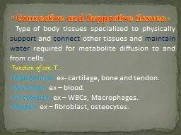
Connective and Supportive tissues Type of body tissues specialized to physically support and connect other tissues and maintain water required for metabolite diffusion to and from cells ID: 772537

cells fibers tissue connective fibers cells connective tissue con collagen dense reticular fiber function cell tissues ground blood type

Share:

Link:

Embed:

Download Presentation from below link

Download Presentation The PPT/PDF document "Connective and Supportive tissues:-" is the property of its rightful owner. Permission is granted to download and print the materials on this web site for personal, non-commercial use only, and to display it on your personal computer provided you do not modify the materials and that you retain all copyright notices contained in the materials. By downloading content from our website, you accept the terms of this agreement.


Presentation Transcript

Connective and Supportive tissues:- Type of body tissues specialized to physically support and connect other tissues and maintain water required for metabolite diffusion to and from cells. Function of con. T. : Mechanical: ex- cartilage, bone and tendon. Nutrition: ex – blood. Protection: ex – WBCs, Macrophages. Repair: ex – fibroblast, osteocytes.

General Con. T. composed of: First : Fibers of con. T.: Fiber : - Elongated structures formed from proteins. Types of fiber are:- Collagen (white) fiber : Fibers sheets and networks extremely strong and resistance to normal tearing forces. Elastic (yellow) fibers : thin single fibers form a network with ability of elasticity. Reticular fibers : small fibers appeared black in color .

Second : cells of con. T. : Mesenchyme cells ------- common cells, produce fibers and interstitial material. Adiposities ----- store fat. Plasma cells -----antibody producing cells. Macrophages ------- phagocytosis. Mast cells ---------- release histamine and cytokines. White blood cells (WBC). Pigment cells ---- in skin.

Macrophage Fibroblast Lymphocyte Fat cell Mast cell Neutrophil Capillary Cell types Extracellular matrix Fibers • Collagen fiber • Elastic fiber • Reticular fiber Ground substance

Third : ground substance. Watery unstained extracellular material contains water, polysaccharide mucouprotein and protein such as albumin, glycoprotein, and globulin. Classification of con. T. : First type of con. T. : General con . T. : Loose ( areolar ) con. T .:- many cells and little collagen, randomly distributed, thick layer beneath the epithelial in the digestive system.

Figure 4.8a Connective tissues. ( a) Connective tissue proper: loose connective tissue, areolar Description: Gel-like matrix with all three fiber types; cells: fibroblasts, macrophages, mast cells, and some white blood cells. Function: Wraps and cushions organs; its macrophages phagocytize bacteria; plays important role in inflammation; holds and conveys tissue fluid. Location: Widely distributed under epithelia of body, e.g., forms lamina propria of mucous membranes; packages organs; surrounds capillaries. Photomicrograph: Areolar connective tissue, a soft packaging tissue of the body (300x). Epithelium Lamina propria Fibroblast nuclei Elastic fibers Collagen fibers

Reticular tissue : specialized con. T. consist of collagen fiber produced by reticular cells .this tissue form a delicate network supports various type of cells and lymphocytes in most lymphoid organs (spleen) . ● Dense con. T . : less flexible.more resistance than loose con. T. , adopted to offer stress resistance and protection ..

Figure 4.8c Connective tissues. ( c) Connective tissue proper: loose connective tissue, reticular Description: Network of reticular fibers in a typical loose ground substance; reticular cells lie on the network. Function: Fibers form a soft internal skeleton (stroma) that supports other cell types including white blood cells, mast cells, and macrophages. Location: Lymphoid organs (lymph nodes, bone marrow, and spleen). Photomicrograph: Dark-staining network of reticular connective tissue fibers forming the internal skeleton of the spleen (350x). Spleen White blood cell (lymphocyte) Reticular fibers

Dense regular con. T :- consists of bundles of collagen fibers and fibroblasts forms tendons, ligaments and aponeuroses ( flat sheet of dense fibrous). Function : provide strong attachment between various structures Classified according to fiber arrangement to

Figure 4.8d Connective tissues. (d) Connective tissue proper: dense connective tissue, dense regular Description: Primarily parallel collagen fibers; a few elastic fibers; major cell type is the fibroblast. Function: Attaches muscles to bones or to muscles; attaches bones to bones; withstands great tensile stress when pulling force is applied in one direction. Location: Tendons, most ligaments, aponeuroses. Photomicrograph: Dense regular connective tissue from a tendon (500x). Shoulder joint Ligament Tendon Collagen fibers Nuclei of fibroblasts

Dense irregular con. T . :- F ew cells much collagen fibers randomly arranged. P roviding resistance to stress from all direction. ex – Dermis of the skin and sub mucosa of digestive system.

Figure 4.8e Connective tissues. (e) Connective tissue proper: dense connective tissue, dense irregular Description: Primarily irregularly arranged collagen fibers; some elastic fibers; major cell type is the fibroblast. Function: Able to withstand tension exerted in many directions; provides structural strength. Location: Fibrous capsules of organs and of joints; dermis of the skin; submucosa of digestive tract. Photomicrograph: Dense irregular connective tissue from the dermis of the skin (400x). Collagen fibers Nuclei of fibroblasts Fibrous joint capsule

Second type of con. T. :- Embryonic con. T. : Mesenchyme :- Con. Tissue developing mainly from mesoderm in embryo. Composition : Ground substance. Sparse collagen fibers,  reticular fibers.Mesenchyme cells Function: secretion of ground substance,  fibersproliferation and differentiation into different connective tissue cell types, smooth muscle cells, blood cells.

Mucoid (mucous) con. T. Location : umbilical cord Composition :Ground substance  rich in hialuronic acid Collagen fibersMucocytes (fibroblasts) Function: secretion of ground substance, fibers.