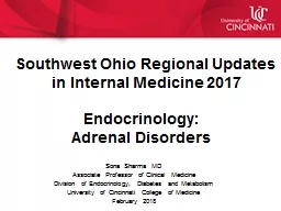
Sona Sharma MD Associate Professor of Clinical Medicine Division of Endocrinology Diabetes and Metabolism University of Cincinnati College of Medicine February 2018 Southwest Ohio Regional Updates ID: 751671
Download Presentation The PPT/PDF document "Endocrinology: Adrenal Disorders" is the property of its rightful owner. Permission is granted to download and print the materials on this web site for personal, non-commercial use only, and to display it on your personal computer provided you do not modify the materials and that you retain all copyright notices contained in the materials. By downloading content from our website, you accept the terms of this agreement.
Slide1
Endocrinology:Adrenal Disorders
Sona Sharma MDAssociate Professor of Clinical MedicineDivision of Endocrinology, Diabetes and MetabolismUniversity of Cincinnati College of MedicineFebruary 2018
Southwest Ohio Regional Updates
in
Internal Medicine 2017Slide2
Mineralocorticoids like aldosterone
Renin-Angiotensin
System
Renin
Catecholamines
(Norepinephrine, epinephrine)
Zona
Reticularis
Zona Glomerulosa
Medulla
Zona
Fasciculata
Glucocorticoids
like cortisol
CRH (Hypothalamus)
ACTH (Pituitary)
Hypothalamus
Pituitary
Adrenal Glands: Normal Anatomy and Physiology
Cortex Slide3
Adrenal
Hormones: Pathophysiology
Hormones
Tumors/ Syndromes associated with overproduction
Tumors/ Syndromes associated with under production
Cortex
Mineralocorticoids (Aldosterone)
Primary
Aldosteronism
Primary Adrenal insufficiency
Hypoaldosteronism
Glucocorticoids (Cortisol)
Cushing syndrome
Primary Adrenal insufficiency
Adrenal androgens (DHEAS, DHEA)
Virilizing
or feminizing tumors
Medulla
Catecholamines
(epinephrine, norepinephrine)
PheochromocytomaSlide4
Mineralocorticoids like aldosterone
Renin-Angiotensin
System
Renin
Catecholamines
(Norepinephrine, epinephrine)
Zona
Reticularis
Zona Glomerulosa
Medulla
Zona
Fasciculata
Glucocorticoids
like cortisol
CRH (Hypothalamus)
ACTH (Pituitary)
Hypothalamus
Pituitary
Applying
n
ormal
p
hysiology to work up of adrenal diseases
Cortex Slide5
Tests of overproduction and underproductionApplying concepts from physiology:-
Using diurnal rhythm of Cortisol productionCortisol production peaks in morning and nadir at nightSuspecting Cortisol deficiency –> check level when it is expected to be highest (8 am morning cortisol) Suspecting excess –> check level when it is expected to be lowest (midnight cortisol)
Using feedback regulation of adrenal hormone production
Suspecting
deficiency –>
stimulation tests
Suspecting excess –> suppression
tests Slide6
Cushing Syndrome/disease
Williams’s Textbook of endocrinology
History/Symptoms
Weight gain/centripetal obesity
Menstrual irregularity
Osteoporosis/osteopenia
Hypertension
Prediabetes or diabetes
Easy bruising
Acne/Hirsuitism
Examination findings
Purple
striaeDorsocervical,Supraclavicular fat padsMoon facies
Proximal myopathy
Definition:- large group of signs & symptoms that reflect prolonged and inappropriately high exposure of tissue to
glucocorticoids (exogenous and endogenous)Slide7
Diagnostic work up forCushing syndrome/disease
Step 1: Exclude exogenous steroids intake (most common cause of Cushing syndrome) Slide8
Cortisol (with
diurnal rhythm)ACTH (under CRH from
hypothalamus
)
-
Low
dose
Dexamethasone
Suppression Test (DST)= Failure
to suppress Endogenous Cortisol by 1 mg Dexamethasone 24 hour Urinary Free
Cortisol = Excessive cortisol production=> 24 UFC X2 with Creatinine Midnight salivary cortisol (MSC X2) =
Loss of normal diurnal rhythm
Step 2: Biochemical w/u to confirm endogenous Cushing
Pituitary
AdrenalsSlide9
Step 3: Establish level/cause/source of Cushing syndrome
Pituitary
Adrenals
ACTH
Ectopic
Source
of ACTH
production
Cortisol
ACTH
ACTH:- First step
ACTH
High Dose Dexamethasone suppression tests
MRI
sella
CT/MRI of chest/abdomen
Inferior Petrosal Sinus Sampling Slide10
Adrenal InsufficiencyDeficiency of glucocorticoid hormones (cortisol) with or without deficiency of mineralocorticoids (aldosterone)
Clinical Presentation:- Anorexia, nausea/vomiting, involuntary weight loss, generalized weakness, volume depletion, hypotension, abdominal painShock: Adrenal crisis: a medical emergencyHyponatremia, hypoglycemia
H
yperkalemia
,
hyperpigmentation (primary)Slide11
Causes of Adrenal Insufficiency
Primary (adrenal)
Central (Hypothalamus/Pituitary)
Autoimmune
adrenalitis
(Addison's)
m
ost common cause
Infections (e.g. TB, HIV, fungal)Infiltrations Adrenal hemorrhage (trauma, coagulopathy, sepsis)Drugs (e.g. mitotane
, ketoconazole)MetastasisBilateral adrenalectomyCongenital Adrenal Hyperplasia Adrenoleukodystrophy in males
Supraphysiologic doses of exogenous steroids is most common cause Megace, IpilimumabPituitary disease (e.g. tumor, trauma)
After surgery for Cushing diseaseSlide12
Diagnostic work up of Adrenal Insufficiency
Step 1: Confirm diagnosis Am cortisol < 5 ug/dL suggests adrenal insufficiency Standard 250 mcg Cosyntropin Stimulation test: Peak cortisol levels <18 μg/dL at 30 or 60 minutes confirms adrenal insufficiency
Step 2: Identify level of
defect
=> ACTH is the main test
Level of defect
Cortisol
ACTH
Aldosterone Renin
Primary AdrenalsLow
High
Low High
Central
Secondary (pituitary)
Tertiary (hypothalamus) Low Low or Normal
NormalNormal
Step 3: Work up of underlying causeSlide13
Treatment of Adrenal InsufficiencySevere
adrenal insufficiency/adrenal crisis is medical emergencyStart glucocorticoids (hydrocortisone) urgently without waiting for results of diagnostic tests Start intravenous fluids Chronic treatment:- Primary adrenal insufficiency needs both: Glucocorticoids (hydrocortisone, dexamethasone, prednisone) Mineralocorticoids (fludrocortisone)
Secondary/tertiary adrenal insufficiency need only glucocorticoids
Stress dosing of steroids during illness or
surgery
Medic alert/Intramuscular kit for prn use at home Slide14
Primary Aldosteronism (PA)
Definition of PA:- excessive, inappropriate, autonomous production of aldosterone hormone Too much aldosterone => Plasma Aldosterone Concentration (PAC) Suppressed renin => Plasma renin activity (PRA)
Clinical Presentation:-Most
common cause of
secondary HTN. Suspect
PA if:-
Severe or resistant HTN
+/- hypokalemia (even on diuretic) Early onset of HTNHTN + adrenal tumorFamily h/o early onset HTN, strokes <40 yrs, PA Slide15
Diagnostic work up of Primary Aldosteronism (PA)
Step 1: Biochemical Confirmation of Primary Aldosteronism (PA)Case detection/Screening tests:- PAC (ng/dL) => Aldosterone to renin ratio (ARR)PRA (ng/mL/min) ARR > 30 suggestive of PA (some authors also include PAC >10-15)ARR >30, positive screening tests, need confirmatory tests in most cases
Most antihypertensive meds can usually be continued except MRB, K sparing diuretics
(stop
for >/= 4 weeks prior to test)
Confirmatory
tests:- Measure aldosterone in 24 hour urine after oral salt loading or serum after IV saline infusionSlide16
Diagnostic work up of Primary Aldosteronism (PA)
Step 2: Subtype Classification of Primary Aldosteronism (PA)Imaging:- Abdomen CT scan is initial imaging unless contraindicatedAdrenal Vein Sampling (AVS):- to differentiate between unilateral vs bilateral aldosterone
overproduction (done only in surgical candidates)Slide17
Treatment of Primary Aldosteronism (PA)
Treatment: Medical vs surgicalUnilateral overproduction of aldosterone: Adrenalectomy (only in surgical candidates)Bilateral overproduction of aldosterone or patient not surgical candidate or declines surgerymedical treatment with mineralocorticoid receptor blockers (MRB: Spironolactone or Eplerenone) Slide18
Pheochromocytoma
Definition: Pheochromocytoma: tumor arising from adrenal medulla that commonly produces catecholamines“Extra adrenal pheochromocytomas”: ParagangliomaClinical presentation: HTN (can be labile, resistant, severe with hypertensive crises), sweating, palpitations, headaches, orthostatic hypotensionCan occur in familial conditions (MEN2, NF1, VHL, SDH mutations)
Can
be malignantSlide19
Diagnostic work of Pheochromocytoma
Lab tests: 24 hour urinary fractionated metanephrines with creatinine or Plasma free metanephrines (normetanephrine, metanephrine) Stress, meds (TCA, sympathomimetic, cocaine): false positive testsImaging: CT scan- Initial choice of imaging Usually large, heterogeneous, vascular, high HU, marked contrast enhancement
Other modalities:-
MRI
MIBG
scan (I-
Metaidodobenzylguanidine
) PET ScanSlide20
Pheochromocytoma
Treatment:-Surgery is mainstay but is high risk: Hypertensive crises/tachyarrhythmias Requires careful preoperative management:Control of HTN: Alphablockers, followed by betablockers Volume expansion: High sodium and fluid intake
Careful hemodynamic monitoring
Lifelong
annual biochemical and clinical monitoring requiredSlide21
Adrenal Incidentaloma
Definition:- adrenal mass discovered incidentally on imaging done for reasons other than adrenal diseaseClinical significance:- can be malignant or hormonally overproducing tumors. Most are benign and nonfunctionalRadiological exam is most helpful diagnostic testImaging features suggestive of benignity on CT scanSize:- < 4 cmImaging characteristics:- <10 HU on noncontrast CT scan, homogeneous, smooth bordered
Absence of these features: suspicious or
indeterminate
Other
d
iagnostic modalities:- MRI/PET Scan
Biopsy is only RARELY indicatedSlide22
Adrenal Incidentaloma
Recommend hormonal evaluation on initial presentation in:-All patients for pheochromocytoma (24 hour urinary fractionated metanephrines or plasma free metanephrines)
All patients for Cushing by 1 mg (low dose) DST
Only patients with HTN for
aldosteronism
(PRA, PAC)
Only patients with symptoms of
virilization in women or feminization in men for androgen secreting tumors (DHEAS, estradiol)Slide23
Adrenal Incidentaloma
Indications for surgery:-Most tumors > 4 cm in size (except adrenomyelolipoma)All pheochromocytoma Cushing and aldosteronoma in some people
Tumors that enlarge on follow up imaging
Need clinical, biochemical or imaging monitoringSlide24
Question 1
46 year old lady History:- resistant hypertension X 3 years + hypokalemia X 2 years No palpitations, headaches or sweating Periods are regular. No hirsuitism. No h/o CAD or PVD Meds: Lisinopril 40 mg/day, Metoprolol 25 mg X 2/day, Amlodipine 10 mg/day, HCTZ 25 mg/day, KCL 40 meq X 2/dayExam: BMI 25 kg/m2, BP 160/100 mm Hg, pulse 80/minute. No centripetal obesity or purple striae
Labs:- Na 143
meq
/L, K 3.3
meq
/L. No worsening of Cr after starting LisinoprilSlide25
Question 1
What would be the next step to diagnose her condition?Plasma metanephrines CT scan to look at adrenal glands Duplex ultrasound for renal arteries Plasma renin activity (PRA) and aldosteroneDexamethasone suppression test (DST)Slide26
She probably has primary aldosteronism (high BP+ hypokalemia)A-Plasma
metanephrines: IncorrectNo signs or symptoms of pheochromocytomaB-CT scan to look at adrenal glands: IncorrectBiochemical diagnosis should precede imaging C-Duplex ultrasound for renal arteries: IncorrectDuplex ultrasound is done to r/o renal artery stenosis (RAS) but she has no suggestive features of RAS
D-Plasma renin activity (PRA) and
aldosterone:
Correct
Check
PRA & aldosterone (for primary hyperaldosteronism
)E-Dexamethasone suppression test (DST): IncorrectDST is done to r/o Cushing but she has no signs or symptoms of CushingAnswersSlide27
Question 265 year old man
History:- referred after his colonoscopy got cancelled because BP rose to > 200/110 mm Hg prior to getting procedure Also c/o episodes of sudden rise in BP, sweating, palpitations, headaches in past few months Current meds: Losartan 50 mg, HCTZ 25 mgExam: BP 156/96 mm Hg, pulse 104/min, BMI 27 kg/m2Labs: normal serum K, PRA, PAC, elevated plasma metanephrines and normetanephrinesCAT scan abdo: 4.5 cm right adrenal mass with marked contrast enhancementSlide28
Question 2
What is the best next step in his management?Monitor him since BP is already getting betterRefer to surgeon for urgent adrenalectomy Start alphablocker like phenoxybenzamineStart betablockers like metoprololIncrease hydrochlorothiazide to 50 mg daily Slide29
He has pheochromocytoma: clinical symptoms + elevated plasma metanephrines
+ adrenal mass on CT ScanA- Monitor him since BP is already getting better: Incorrect Pheochromocytoma should be removed since it can be fatal. He is also symptomatic.B- Refer to surgeon for urgent adrenalectomy: IncorrectAdrenalectomy is a high risk surgery and requires preoperative and perioperative medical preparationC- Start
alphablocker
like
phenoxybenzamine
:
CorrectS
tart alphablocker before sending him for surgeryD- Start betablockers like metoprolol: IncorrectStarting betablockers before alpha blockers can precipitate hypertensive crisis because of unopposed alpha adrenergic actionE- Increase hydrochlorothiazide to 50 mg daily: Incorrect: HCTZ not enough to control HTN in him. May cause orthostasis
AnswersSlide30
Question 3
42 year old woman History: Unexplained weight gain of 20 lbs Worsening of DM over past 6 monthsIrregular periods, facial hair Exam: BMI 35 kg/m2, BP 158/86 mm HgCentripetal obesity, dorsocervical fat pads, purple striaeLabs: blood glucose 230 mg/dl, K 3.9 meq/L, am cortisol 16.7 mcg/dL, ACTH 5 pg/mLSlide31
Question 3
What is the next best test to confirm the diagnosis?250 mcg cosyntropin test Low dose dexamethasone test (DST)High dose dexamethasone suppression test (DST)Abdominal CT scan to look at adrenal glandsSellar MRI to look at pituitary glandSlide32
She has clinical signs/symptoms suggestive of Cushing. Needs biochemical confirmation of diagnosisA- 250 mcg
cosyntropin test: IncorrectCosyntropin test is to diagnosis adrenal insufficiencyB- Low dose dexamethasone test (DST): CorrectLow dose DST is an appropriate test to confirm diagnosis of Cushing disease/syndromeC- High dose dexamethasone suppression test (DST): Incorrect
H
igh
dose DST is done
to
differentiate between pituitary vs ectopic source and is done after diagnosis of Cushing has been confirmed
D- Abdominal CT and E Sellar MRI: IncorrectImaging tests are done after biochemical confirmation of Cushing AnswersSlide33
Question 439 year old man seen for the first time
History: T1DM since age 12 years and hypothyroidism C/o increased, unexplained hypoglycemia for past few monthsC/o some n/v, dizziness, weight loss of 6 lbs in last 3 monthsExam: BP 90/56 mm Hg, pulse104 beats/min. Increased pigmentation in his hands Current meds: glargine and aspart insulin, levothyroxineLabs: Na 132
meq
/L, K 5.6
meq
/L,
Tsh 5.5 U/mL (ULN 5.2), 8 am cortisol 2.6 mcg/dL with ACTH 670
pg/mLSlide34
Question 4
In addition to decreasing his insulin dose, what is the next best step in his management?Order an urgent brain MRI to rule out pituitary tumor Increase dose of levothyroxine to normalize tshStart hydrocortisone as soon as possibleCheck for celiac disease Order insulin induced hypoglycemia test to confirm adrenal insufficiencySlide35
A Order an urgent brain MRI to rule out pituitary tumor: wrongEven though ACTH is high, since his corresponding cortisol is low, he has adrenal insufficiency and not an ACTH producing
tumorB Increase dose of levothyroxine to normalize tsh: IncorrectBorderline high tsh can not explain his clinical picture/lab abnormalities. Glucocorticoids should be replaced before replacing/increasing LevothyroxineC Start hydrocortisone as soon as possible: CorrectHigh likelihood of primary adrenal insufficiency based on his symptoms and labs. Glucocorticoids should be started as soon as possibleD Check for celiac disease: Incorrect
C
eliac
disease cannot explain low
cortisol
E
Do insulin induced hypoglycemia test to confirm adrenal insufficiency: IncorrectIn a stable patient, test of choice to confirm adrenal insufficiency is Cosyntropin testAnswersSlide36
Question 5
62 year old man History: presented to ER for abdominal painAbdominal CT scan revealed a round, 2.2 cm adrenal mass, HU 9 No h/o HTN or hypokalemiaNo sweating or palpitationsExam: BMI 32 kg/m2, BP 132/80 mmHg. No purple striae, dorsocervical fat padSlide37
Question 5
What should be included in the diagnosis work up of this adrenal tumor?Biopsy of adrenal tumor No further tests needed since tumor looks benign on CT scanPRA and PAC8 mg (high dose) DSTPlasma or 24 hour urine metanephrinesSlide38
A Biopsy of adrenal tumor: IncorrectBiopsy is rarely indicated and only in specific situations. Always r/o
pheochromocytoma before performing biopsy B No further tests needed since tumor looks benign on CT scan: IncorrectAll patients with adrenal incidentaloma should be tested for Cushing and pheochromocytoma C PRA and PAC: IncorrectIn setting of adrenal tumor only hypertensive patients need to be tested for PA
D 8 mg (high dose)
DST:
Incorrect
1
mg or low dose DST (not high dose DST), is
the test of choice for adrenal incidentaloma to r/o cushingE Plasma or 24 hour urine metanephrines: CorrectDo Plasma or 24 hour urine metanephrines to rule out pheochromocytomaAnswersSlide39
References
The Management of Primary Aldosteronism: Case detection, Diagnosis, and Treatment: An Endocrine Society Clinical Practice Guideline (2016)Pheochromocytoma and Paraganglioma: An Endocrine Society Clinical Practice Guidelines (2014)Diagnosis and Treatment of Primary Adrenal Insufficiency: An Endocrine Society Clinical Practice Guideline (2015)The Diagnosis of Cushing's Syndrome: An Endocrine Society Clinical Practice Guideline (2008)American Association of Clinical Endocrinologists and American Association of Endocrine Surgeons Medical Guidelines for the Management of Adrenal Incidentaloma (2009)Management of adrenal incidentalomas: European Society of Endocrinology Clinical Practice Guideline in collaboration with the European Network for the Study of Adrenal Tumors (2016)