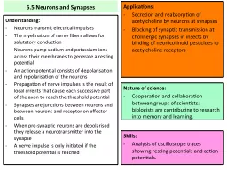
Understanding Neurons transmit electrical impulses The myelination of nerve fibers allows for salutatory conduction Neurons pump sodium and potassium ions across their membranes to generate a resting potential ID: 473701
Download Presentation The PPT/PDF document "6.5 Neurons and Synapses" is the property of its rightful owner. Permission is granted to download and print the materials on this web site for personal, non-commercial use only, and to display it on your personal computer provided you do not modify the materials and that you retain all copyright notices contained in the materials. By downloading content from our website, you accept the terms of this agreement.
Slide1
6.5 Neurons and Synapses
Understanding:Neurons transmit electrical impulsesThe myelination of nerve fibers allows for salutatory conductionNeurons pump sodium and potassium ions across their membranes to generate a resting potential An action potential consists of depolarization and repolarization of the neuronsPropagation of nerve impulses is the result of local currents that cause each successive part of the axon to reach the threshold potential Synapses are junctions between neurons and between neurons and receptor on effector cellsWhen pre-synaptic neurons are depolarized they release a neurotransmitter into the synapseA nerve impulse is only initiated if the threshold potential is reached
Applications:Secretion and reabsorption of acetylcholine by neurons at synapsesBlocking of synaptic transmission at cholinergic synapses in insects by binding of neonicotinoid pesticides to acetylcholine receptors
Skills:Analysis of oscilloscope traces showing resting potentials and action potentials.
Nature of science:
Cooperation and collaboration between groups of scientists: biologists are contributing to research into memory and learning.Slide2
Two systems:
Endocrine systemNervous systemRole of system Organs involved What does each organ do?Communication in the bodySlide3
Endocrine
NervousSlide4Slide5
Endocrine
Consists of glands, pancreas and reproductive organsReleases hormonesSlide6
Nervous
Consists of nerve cells called neuronsCentral and peripheral 85 billion neurons in humansSlide7
Neuron
Label the following:Cell bodyAxonDendriteAxon terminalNucleusMyelin sheath
Understanding:Neurons transmit electrical impulsesSlide8
NeuronSlide9
Non- Myelinated
No myelin sheathNerve impulse 1 metre/second
Understanding:The myelination of nerve fibers allows for salutatory conductionSlide10
Myelination
Many layers of phospholipids bilayersSchwann cells deposit myelin when they grows round the axonNode of Ranvier = gap between myelin deposited by adjacent Schwann cells.
Understanding:The myelination of nerve fibers allows for salutatory conductionSlide11
Myelination
Nerve impulse jumps from one node of Ranvier to the next.Saltatory conduction100 metres/second
Understanding:The myelination of nerve fibers allows for salutatory conductionSlide12
Non Myelinated
Myelinated Slide13
Non Myelinated
No myelin layersImpulse is 1m/s
MyelinatedSchwann Cells make myelin that wraps round axonImpulse jumps between Nodes of Ranvier 100m/s Slide14
Resting Potential (–70mV)
Overall there is a charge imbalanceNegative charge on the inside Positive charge on the outside
Understanding:Neurons pump sodium and potassium ions across their membranes to generate a resting potential Slide15
Resting Potential (–70mV)
No transmission = resting potentialThere IS a potential difference across the membraneDue to an imbalance of positive and negative charges across the membrane
Understanding:Neurons pump sodium and potassium ions across their membranes to generate a resting potential Slide16
Resting Potential (–70mV)
Sodium potassium pump pumps 3 sodium ions out for every 2 potassium ions inMembrane is also 50 times more permeable to potassium ions Potassium ions leak back across the membrane faster than sodium ionsBuild up of positive ions on outside of membrane creates difference in charge Sodium concentration gradient very steep
Understanding:Neurons pump sodium and potassium ions across their membranes to generate a resting potential Slide17
Action potential
Rapid change in membrane potential Depolarisation – negative to positiveRepolarisation – positive to negative(inside membrane of axon)
Understanding:An action potential consists of depolarization and repolarization of the neuronsSlide18
Depolarisation (+30mV)
Due to opening of sodium channelsAllows sodium ions to diffuse into neuron down concentration gradientReverses the charge imbalanceInside becomes positive
Understanding:An action potential consists of depolarization and repolarization of the neuronsSlide19
Repolarisation (–70mV)
Rapidly after depolarisationSodium channels closePotassium channels open Potassium ions diffuse out of ion down their concentration gradientInside of cell becomes negative again
Understanding:An action potential consists of depolarization and repolarization of the neuronsSlide20
Resting Potential (–70mV)
Returns to resting potential before another impulse can be sentSodium potassium pump moving sodium ions out, and potassium ions in
Understanding:An action potential consists of depolarization and repolarization of the neuronsSlide21
Label the diagrams
Summarise what happens during each stageInclude the following on the diagrams:Label pumps/proteinsWhat are pumps/proteins doing at each pointK+ and Na+ movementOverall charges inside and outsideThe potential inside the neuron (+30/-70mV)
Understanding:An action potential consists of depolarization and repolarization of the neuronsSlide22
Depolarisation
RepolarisationSodium channelsSodium ionsPotassium channels
Potassium ionsCharge change inside neuronOverall charge in neuron afterAction PotentialSlide23
Depolarisation
RepolarisationSodium channelsOpenCloseSodium ionsDiffuse into neuronStay inside neuronPotassium
channelsClosedOpenPotassium ionsStay inside neuronDiffuse out of neuronCharge change inside neuronNegative to positivePositive to negativeOverall charge in neuron afterPositive (+30mV)Negative (-70mV)Action PotentialSlide24
Nerve impulses
Start at one end of a neuron Propagated along axon to other end of neuronIon movements that depolarise one part of the neuron trigger depolarisation of the neighboring part
Understanding:Propagation of nerve impulses is the result of local currents that cause each successive part of the axon to reach the threshold potential
Only move in one direction as they are only initiated at one end of the neuronSlide25
Local Currents
The propagation of an action potential along the axon is due to sodium ion movementsSodium ion concentration increases inside the axon during depolarisationSome sodium ions then diffuse inside the axon to the area that has not yet been depolarisedThe same happens outside of the axon, however sodium ions move from the area that has not been depolarised to the area that has
Understanding:Propagation of nerve impulses is the result of local currents that cause each successive part of the axon to reach the threshold potential Slide26
Local Currents
This causes the membrane potential in the unpolarised area to rise from -70mV to -50mVThe Sodium channels are voltage-gated and open when a membrane potential of -50mV has been reachedThis is known as the threshold potential
Understanding:Propagation of nerve impulses is the result of local currents that cause each successive part of the axon to reach the threshold potential Slide27Slide28
Oscilloscope Traces
Membrane potentials can be measuredDisplayed in an oscilloscope Time on x-axisMembrane potential on y-axisResting potential = -70mVLocal currents = rise to -50mVAction potential = narrow spike to +30mVReturns to resting potential = -70mV
Skills:Analysis of oscilloscope traces showing resting potentials and action potentials.Slide29Slide30
Synapses
Junctions between cells in the nervous systemChemicals (neurotransmitters) send signals across synapses
Understanding:Synapses are junctions between neurons and between neurons and receptor on effector cellsSlide31Slide32
Synaptic Transmission
Nerve impulse propagated along pre-synaptic neuron until it reaches the end of the neuron and the pre-synaptic membrane
Understanding:Synapses are junctions between neurons and between neurons and receptor on effector cellsSlide33
Synaptic Transmission
2. Depolarisation of pre-synaptic membrane causes calcium ions (Ca2+) to diffuse through channels in the membrane into the neuron
Understanding:Synapses are junctions between neurons and between neurons and receptor on effector cellsSlide34
Synaptic Transmission
3. Influx of Ca2+ causes vesicles containing neurotransmitter to move to the pre-synaptic membrane and fuse with it
Understanding:Synapses are junctions between neurons and between neurons and receptor on effector cellsSlide35
Synaptic Transmission
4. Neurotransmitter is released into the synaptic cleft by exocytosis
Understanding:When pre-synaptic neurons are depolarized they release a neurotransmitter into the synapseSlide36
Synaptic Transmission
5. Neurotransmitter diffuses across the synaptic cleft and binds to receptors on the post synaptic membrane
Understanding:When pre-synaptic neurons are depolarized they release a neurotransmitter into the synapseSlide37
Synaptic Transmission
6. The binding of the neurotransmitter to the receptors causes adjacent sodium channels to open
Understanding:When pre-synaptic neurons are depolarized they release a neurotransmitter into the synapseSlide38
Synaptic Transmission
7. Sodium ions diffuse down their concentration gradient into the post-synaptic neuron, causing it to reach its threshold potential
Understanding:A nerve impulse is only initiated if the threshold potential is reachedSlide39
Synaptic Transmission
8. An action potential is triggered in the post-synaptic membrane and is propagated along the neuron
Understanding:Synapses are junctions between neurons and between neurons and receptor on effector cellsSlide40
Synaptic Transmission
9. The neurotransmitter is rapidly broken down and removed from the synaptic cleft
Understanding:Synapses are junctions between neurons and between neurons and receptor on effector cellsSlide41Slide42
Acetylcholine
Used as a neurotransmitter at many synapsesProduced from choline (diet) and acetyl (aerobic respiration)Loaded into vesicles and released into synaptic cleftBinding sites are specific
Applications:Secretion and reabsorption of acetylcholine by neurons at synapsesSlide43
Acetylcholinesterase
Breaks acetylcholine into chlorine and acetateReabsorbed back into pre-synaptic neuron where it is converted back into a neurotransmitter
Applications:Secretion and reabsorption of acetylcholine by neurons at synapsesSlide44
Neonicotinoids
What are they?What do they do?Advantages?Arguments against?
Applications:Blocking of synaptic transmission at cholinergic synapses in insects by binding of neonicotinoid pesticides to acetylcholine receptors