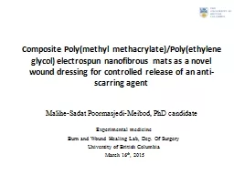
electrospun nanofibrous mats as a novel wound dressing for controlled release of an antiscarring agent Malihe Sadat PoormasjediMeibod PhD candidate Experimental medicine Burn and Wound Healing Lab Dep Of Surgery ID: 792591
Download The PPT/PDF document "Composite Poly(methyl methacrylate)/Poly..." is the property of its rightful owner. Permission is granted to download and print the materials on this web site for personal, non-commercial use only, and to display it on your personal computer provided you do not modify the materials and that you retain all copyright notices contained in the materials. By downloading content from our website, you accept the terms of this agreement.
Slide1
Composite Poly(methyl methacrylate)/Poly(ethylene glycol) electrospun nanofibrous mats as a novel wound dressing for controlled release of an anti-scarring agentMalihe-Sadat Poormasjedi-Meibod, PhD candidate
Experimental medicine
Burn and Wound Healing Lab, Dep. Of Surgery
University of British Columbia
March 16
th
, 2015
Slide2Background-Wound healing process
Slide3Background-Wound healing spectrum
Wound healing spectrum
Normal wound healing
Non-healing wounds
Diabetic foot ulcer
Pressure ulcer
Post-burn
h
ypertrophic
scars
3
Skin fibrosis
Slide4Background-HSC current treatments
4
4
Slide5Background-KynA as an anti-fobrogeic agent
A
C 6.25 12.5 25 50 100 150 (µg/ml)
β
-actin
Collagen-I
KynA
MMP1
Normal
Control
Vehicle
10x
2x
KynA
B
C
Poormasjedi-Meibod
et al.,
PLOSone
, 2014
**
**
Slide6HypothesisKynA can be incorporated into nanofibers to develop anti-fibrogenic wound dressings which slowly release the drug and improve the wound healing outcome.
Slide7Electrospinning process and parametersPolymerPEG 1KD(W/W%)
Voltage
(kV)
Syringe pump
(mm/min)
KynA
(W/W%)
Solvent
PMMA
(350 KD)024
0.16DMF
PMMA (350 KD)
1
24
0.1
6
DMF
PMMA
(350 KD)
2.5
24
0.07
6
DMF
PMMA
(350 KD)
5
24
0.07
6
DMF
PMMA
(350 KD)
10
24
0.05
6
DMF
PMMA
(350 KD)
20
24
0.05
6
DMF
7
Electrospinning
. Current
approaches to
electrospun
nanofibers
for tissue engineering
Nae
Gyune
Rim et al 2013 Biomed. Mater. 8 014102 doi:10.1088/1748-6041/8/1/
014102
Slide8SEM images of PMMA-PEG nanofibers
10%
2
0%
1%
2.5%
5%
PEG
1K
5
K
10K
PMMA
Slide9Dressing’s hydrophobicity and wetting
9
PMMA PMMA+10%PEG
A
B
Slide10Release study in PBS, total immersion settingA
10
Slide11Release study is PBS, 2 chamber well settingB
11
A
PBS
Hydrogel
NF+KynA
B
**
**
**
**
**
Assessment of medicated mat’s
cytocompatibility
12
Control
NF
NF+ KynA
Fibroblasts
C
Ethidium homodimer
Calcein
KynA
Dermal fibroblast
NF+KynA
DMEM media
A
Slide13Dressing’s biological activity assessment
*
*
*
*
**
A
Col-I
GAPDH
Con
NF
NF+KynA
KynA
MMP1
13
B
C
Slide1410x
2x
A
Normal
Control
NF
NF+
KynA
KynA cream
14
KynA
-loaded dressings reduces skin fibrosis
**
**
A
NF+KynA
KynA
cream
**
**
B
Nor Con NF
NF+KynA
KynA
cream
Slide15ConclusionPMMA+10% PEG nanofibers can be used as an effective slow releasing drug delivery system for KynA.Nanofiber-released KynA effectively modulates the expression of ECM components in vitro and in vivo.Application of KynA-loaded nanofibers can improve the wound healing outcome in patients prone to develop skin fibrosis.
Slide16Dr. Aziz Ghahary
Sanam Salimi
Dr. Layla Nabai
Ryan Hartwell
Dr. Reza Jalili
Dr.
Yunyuan Li
Dr. Ruhi Kilani
Acknowledgements
16
Collaborators:
Dr. Hellen Burt
John Jackson
Dr. Frank Ko
Victor Leung
Dr. Emma Guns
Dr. Azadeh Taba
Dr. Yun Zang
Ali Farokhi
Dr. Mohsen
Khosravi
Dr. Saman
Pakyari
Slide17Thank you for your attention!