/
Lecture three   ( Streptococcus) Lecture three   ( Streptococcus)

Lecture three   ( Streptococcus) - PowerPoint Presentation

piper
piper . @piper
Follow
66 views
Uploaded On 2023-08-25

Lecture three   ( Streptococcus) - PPT Presentation

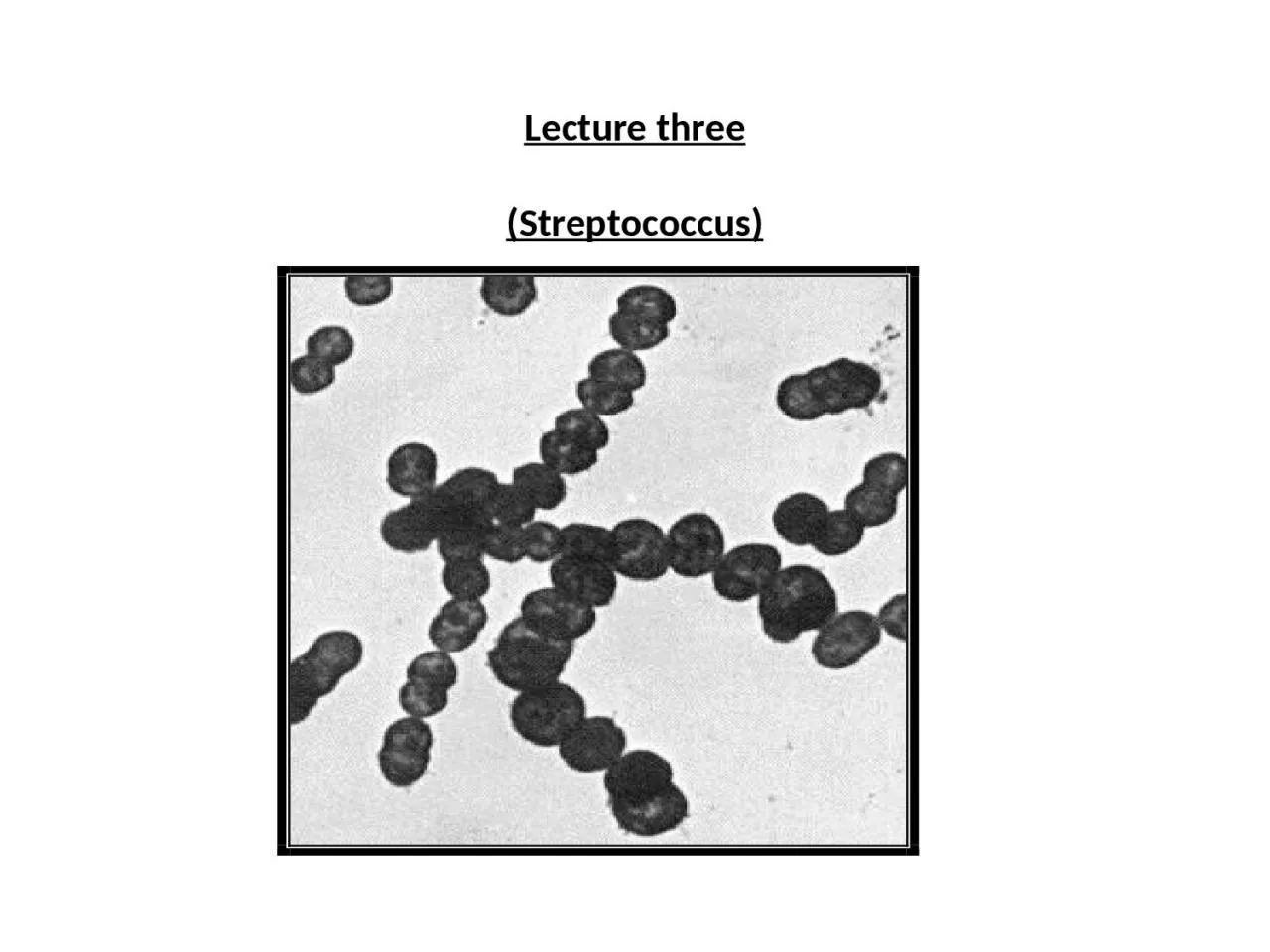
Greek streptus flexible coccus sphere spherical or ovoid cells occurring in short or long chins pairs not in groups Capsules are not regularly formid but develop in some species ID: 1014310

group streptococcus streptococci blood streptococcus group blood streptococci protein agar specific haemolysis antigens streptococcal serum test broth culture medium

Share:

Link:

Embed:

Download Presentation from below link

Download Presentation The PPT/PDF document "Lecture three   ( Streptococcus)" is the property of its rightful owner. Permission is granted to download and print the materials on this web site for personal, non-commercial use only, and to display it on your personal computer provided you do not modify the materials and that you retain all copyright notices contained in the materials. By downloading content from our website, you accept the terms of this agreement.


Presentation Transcript

1. Lecture three (Streptococcus)

2. Greek-streptus =flexible: coccus = sphere spherical or ovoid cells, occurring in short or long chins, pairs, not in groups. Capsules are not regularly formid but develop in some species. Gram positive, better growth on enriched media, non motile, non sporing catalase negative, responsible for a variety of diseases of man and animals. Some are commensals in the upper respiratory and intestinal tracts. Many are aerobes or facultative anaerobes. Classification of streptococci: Streptococci are placed in the family streptococcaceae . Strptococcus pyogenes (Streptococcus haemolyticus) The streptococci are gram positive spherical cells, measuring 0.8 -1 μm in diameter, they divide in only one plane and the tendency of the cells to remain united results in the development of the characteristic chains of sphere giving the organisms their name. chain formation results form the fact that a connecting link between the cocci probably composed of material link that forming the cell wall, is retained following the cell division. These inter cellular bridges are not easily broken.

3. Classification of streptococci  Aerobic and facultative anaerobes Obligate anaerobes   With soluble haemolysin with out soluble haemolysin Peptostreptococcus Beta-haemolysis Alpha haemolysis Gamma haemolysis (non-haemolytic) Serological grouping on the basis Strepto. viridans The enterococcus Of group specific polysaccharide-C group.Of Lancefield calassification. Classified into (5) Classified into (4) Species by physiological species by (19) Lancefield groups A,B,C,.H. and biochemical properties. Physiological K,L,M,……………U. and biochem. Properties. Group A- Streptococcus pyogenes Str. salivarus Str. faecalis Str. mutans Str. durans Str. sangius Str. faeciumOn the basis of Griffith serological Str. mitior Str. bovis Typing based on M-protein farther Str. milleri Classified into   Over (60) serotypes.

4.

5. Chains do not elongate indefinitely, because in some cases streptococci produce a dechaining enzyme. Non spore forming bacteria and are non motile the majority of strains in group-A are encapsulated and capsular material consists of hyaluronic acid. Cultural characters:The majority are aerobes and facultative anaerobes . the optimum temperature for growth is 37Ċ. Fastidious bacteria grow on medium containing blood or serum on serum broth or glucose broth: after 24hs granular growth is obtained and the medium remains clear with granules or powdery deposit.

6. .  On blood agar :This is the medium of choice, and after 24hr the colonies are circular, translucent, low convex, showing beta haemolysis (complete haemolysis) on fresh blood agar medium. The bacteria require about 15 amino acids and almost all the known members of the vitamin B complex are needed. Human blood is inferior to both horse and sheep blood in media for the identification of streptococci. The beta-haemolysis produced by pyogenic streptococcus on blood agar plates incubated aerobically is usually due to the action of an oxygen stable S-haemolysin improvement in haemolysis by anaerobic incubation is due to additional action of the oxygen labile haemolysis-O. streptococci may produce a haemolysin but may not cause beta-haemolysis. Streptococcus pneumoniae which causes greening broth aerobically and anaerobically, forms an oxygen sensitive haemolysin. ر

7. The mucoid character of some strains is associated with the production of hyaluronic acid capsules. (( group-A streptococci are significantly more sensitive to bacitracin than strains of all other groups )) 0.1unit of bacitracin per disc is employed on blood agar as a convenient method of differentiating Str. Pyogenes from other haemolytic streptococci. Biochemical reactions: These reactions are replaced by serological procedure to identify and classify these organisms. Streptococci are catalase negative with the possible exception of Peptostreptococcus and a few strains of group-D. Antigenic structure:Antigenic structure of group-A streptococcus comprise of: 1)Group-specific polysaccharide-C. 2)Type-specific protein antigens –M. 3)Other surface protein antigens. 1)Group-specific polysaccharide-C:It is present as structural components of the cell wall, and is a major component of streptococcus, on the basis of which Lancefield classified the streptococcus into serological groups.

8. 2)M-Protein:These are most important of the surface proteins and are antigens which determine the type specificity of group-A streptococcus. Also it is an important factor in virulence, as this antigen inhibits phagocytosis. On the basis of M-protein, group A streptococcus has been classified into approximately 60 different antigenic types. Type specific M-proteins are firmly attached to the cell surface. 3)Other surface protein antigens:These antigens occur at or near the surface and have been designated T-antigen and R-antigen. T-antigens are present in most of the strains of group-A streptococci. These antigens are given this name due to the fact that they were originally considered to be type specific antigens, but T-antigens may frequently occur along with M-antigens. These antigens have no relationship to virulence or protection. R-antigens are third class surface proteins which participate in agglutination reactions. Toxins and enzymes:More than 20 extra cellular products which are antigenic are elaborated by group A-streptococci, the important toxins and enzymes formed by Streptococcus pyogenes are:

9. 1-Streptolysin-S2-Streptolysin-O3-Erythrogenic toxin4-Streptokinase5-Diphoshopyridine nucleotidase (DPN-ase)6-Hyaluronidase7-Deoxyribonuclease8-Streptococcal proteinase9-Amylase10-Streptococcal opacity factor 1-Streptolycin-S:Is responsible for the zones of haemolysis which surrounded colonies of Streptococcus pyogenes on the surface of blood agar plates. The designation "S" refers to serum soluble due to the fact their haemolysin appears to be extracted from living streptococci which they are shaken with serum. It is oxygen stable and sensitive to heat and acid. Streptolysin-S is cell bound and its release from the cell depends on its association with some carrier molecules such as serum albumin or ribonucleic acid. It is not antigenic and no antibodies are formed.

10. . 2-Streptolysin-O:Oxygen labile and produced in serum free broth. Under aerobic conditions it fails to produce haemolytic zone around colonies of streptococcus, but under reducing conditions such as deep colonies in pour plate technique or anaerobic culture haemolysin is produced. All strains of group-A streptococci produce streptolysin-O. Streptolysin-O is protein in nature and is strongly antigenic. Antibody to it is found in the sera of many patients following streptococcal infection. Antibody is known antistreptolysin. Antistreptolysin-O (ASO) titer of 200 or more is generally considered to be indicative of recent infection. Measurement of serum antistreptolysin-O has become widely used as a test for establishing the occurrence of recent streptococcal infection. Streptolysin-O is toxin for leucocytes and has cardiotoxicity. If the organism produce only SLO (Streptolycin O),hemolysis may not detected ,when the culture incubation aerobically. 3-Erythrogenic toxin:It is a protein , antigenic, relatively heat stable but destroyed by boiling for one hour. This toxin is responsible for characteristic skin rash with pharyngitis and tonsillitis in scarlet fever and its mode of action is not known. It is also known as Dick toxin after its discoverer.

11. . Dick test:The skin test depending on the intradermal injection of erythrogenic toxin in humans, is called Dick test a positive Dick test is an erythrematous and often oedematous area of more than 10mm diameter on intradermal injection of 0.2ml of 1/1000 dilution of a filtrate of broth culture containing erythrogenic toxin. It appears with in 6-24 hours. The effect of the toxin is neutralized by antibody. A susceptible child shows positive Dick test. The same toxin, previously heated to destroy the Dick toxin is used as a control on the opposite arm. Inhibition of the effects of the toxin by antibody is further demonstrated by the fact that the intradermal injection of specific potent antitoxin into an area of scarlet fever rash, will result in blanching of the rash in 6-18hours during early stage of scalet fever and this is known as Schultz and Charlton reaction. 4-Streptokinase:It is known as streptococcal fibrinolysin. It transforming the plasminogen of human plasma into plasmin, an active proteolytic enzyme which digests fibrin and other proteins. 5-Diphosphopyridine nucleotidase:It is antigenic and associated with leucotoxicity and thus produces destruction of leucocytes. 6-Hyaluronidase: (spreading factor)It is an enzyme which splits hyaluronic acid, an important component of the ground substance of connective tissue. Thus hyaluronidase acid in spreading microorganisms (spreading factor). 7-Deoxyribonuclease: (streptodornase)It is an enzyme which cause depolymeriztion of DNA. 8-Streptococcal proteinase:It is not known whether streptococcal proteinase causes. Tissue damage in the course of streptococcal infection. Some believe that it is capable of breaking down the M-protein on which the virulence of the organisms depends and thus might contribute to the patients recovery.

12.  9-Amylase:This enzyme hydrolyses glycogen, amyloprotein and starch. 10-Streptococcal opacity factor: (OF) It is an alpha-lipoproteinase produced by certain serotypes which rise to opacity in serum broth. Pathogenesis:A)Acute infections: it may be 1)Tonsillitis and quinsy 2)Pharyngitis 3)otitis media 4)Paranasal sinusitis 5) Meningitis 6)Scarlet fever 7)Pneumonia 8) Erysipelas 9)Cellulitis 10)Impetigo 11)Puerperal sepsis 12)Septicaemia.B)Non suppurative complications: Acute glomerulonephritis. ASOT (Rheumatic fever)Laboratory diagnosis: The specimens to be obtained for the diagnosis depends on the nature of streptococcal infection. A)Acute pyogenic infection: by demonstrating the causative organism. The morbid material depends on the site of the lesion. It may be throat swab, pus swab, CSF, blood. The morbid material may be subjected to the following examination. 1- Smear examination2-Cultural examination:Blood agar is the medium of choice for the growth of haemolytic streptococcus, special medium for the isolation of streptococci is crystal violet blood agar, contains 1/106concentiation of crystal violet. Aerobic and anaerobic incubation may be done, depending on the site of the lesion. 3-Fluorescent antibody technique:Smear from serum broth culture of throat swab 2-3 hours old, may be stained with group A specific antibody conjugated with fluorescent dye, for the most rapid identification of group A streptococci in clinical disease.

13. . 4-Bacitracin sensitivity :A disc impregnated with 0.1 unit of bacitracin is placed on the surface of a heavily inoculated blood agar culture plate before incubation to ensure rapid recognition of group A strains which are sensitive to this antibiotic .Bacitracin may be false positive with some strains of groups B,C ,G and some alpha-hemolysis will susceptibly also.5-Serotyping of streptococci:Is of academic interest and a research tool which can be only carried out in laboratories well equipped with different antisera. B)In non suppurative complication: In rheumatic fever and acute glomerulonephritis, the diagnosis of streptococcal infection may be established by demonstrating high levels of antibody to streptococcal toxins. The most frequent and widely employed test is ''antistreptolysin-O-titer(A.S.O.T.). higher levels are generally found in rheumatic fever than in acute glomerulonephritis'' Chemotherapy:Penicillin is the treatment of choice in most cases of severe streptococcal infections. ***********  

14. Streptococcus viridans Many streptococci isolated from human source do not possess Lancefield polysaccharide, and they do not belong to any serological group. They produce alpha haemolysis on blood agar. Biochemical reactions:Based on biochemical reactions, Streptococcus viridans group have been classified into five species(Streptococcus salivarus ; Strep. mutans ;Strep. sangius ;Strep. mitior and Strep. milleri). The biochemical reactions are very helpful in differentiating Strepococcus viridans from pneumococcus. 

15. Streptococcus pneumoniae( pneumococcus)(Diplococcus pneumoniae)Gram positive coccus, lancet shaped, in pairs, capsulated, non motile and sporing lysed by bile salts, inhabited of upper respiratory tract of man and some animals. Causes infection primarily of the respiratory tract, conjunctivitis, otitis media, peritonitis and meningitis. Cultural characters:It may be cultured on the following media: 1-Glucose broth and serum broth.2-Blood agar 3-Chocolate agarThe growth on ordinary culture media is poor, but it is greatly improved by the addition of glucose, blood, serum. It is aerobic and facultative anaerobe. Optimum tem. For growth is 37Ċ cultivation in an atmosphere of 5-10% CO2 by using candle jar and the PH of the medium should be between 7.2-7.4 after sterilization. In liquid culture media:After 24h. of incubation. There is inform turbidity of the medium and prolonged incubation may produce clearing of the culture medium due to autolysis of the organisms.

16. Blood agar:After 24h. the colonies are small about 1mm in diameter semitransparent and surrounded by a zone of alpha-haemolysis. The colonies first are dome shaped and later on become draughtsman colonies.

17. Chocolate agar:The zone of alpha-haemolysis is better seen on this medium. The pneumococci are structurally very delicate organisms and autolyse much more readily than most of the other bacteria and this is due to intracellular ferments. Autolytic enzymes action the muramic acid of the cell wall only when the choline containing teichoic acid is present. Morphology:Smear direct form the morbid material shows gram positive diplococci lanceolate and showing clear zone of halo around the diplococci. Capsule:May be demonstrated either by special capsular staining or by the presence of a halo around the diplococci. Bile salt solubility test:The bile solubility of pneumococci is a constant characteristics although different strains vary in their sensitivity to bile. The test contains of adding one part of sterilized 10% solution of sodium taurocholate in normal saline, to 10 parts of a broth culture, PH of which should not be lower than 6.8. alternatively, 0.2ml of 10% solution of sodium deoxycholate may be added to 10ml broth culture. The PH of the broth should be in neutral range. The lysis occurs within fifteen minutes 37Ċ.

18. Optochin sensitivity:The pneumococcus is sensitive to optochin (ethyl-hydro-cuppreine hydrochloride). Sterile filter paper discs 8mm in diameter, impregnated with 5µg of optochin, are placed over the radially streaked cultures on blood agar plate. The pneumococci are inhibited in a zone of at least 5mm from the edge of the disc strains of Streptococcus viridans grow up to the disc edge. Inulin fermentation:The fermentation of inulin has been used as a differential test to distinguish pneumococcus from Streptococcus viridans while it is true that inulin fermentation is a property of pneumococcus, it is not reliable test when used it self, since certain strains of streptococcus especially those of the salivarus group share this property.  Antigenic structure:Penumococci have the following important antigenic components: 1-Capsular polysaccharide: (specific soluble substance(SSS) ): this substance present on the surface of pneumococci diffuses into the culture medium or infective exudates and tissues. It is called specific soluble substance, at least 85 specific serotypes of pneumococci have been recognized on the basic of differences in the capsules which surrounded the cells. The capsule composed of species specific polysaccharide antigen which is immunologically distinct for each type. The type identification can be established by means of agglutination or by capsular swelling or Quellung reaction it is carried out by mixed sputum or a saline suspension of fresh growth from blood agar or loopful and mixed is cover with a cover glass and examined under oil immersion objective. The capsule becomes apparently swollen and enlarged with in 1-2 minutes.

19. Quellung reaction 2-Somatic M-protein: As the indicates, this is somatic protein of pneumococci which contains an M- protein antigen. This is deep in the cell and is characteristic for each type.  3-Somatic C-carbohydrate:Antigen is common to all pneumococci. The C-reaction protein, a substance found in the serum of certain patients suffering from inflammatory or destructive lesions. Antibodies to these antigens are not protective.

20. C-Reactive proteins (CRP)These are abnormal proteins ( beta globulin) that precipitate with somatic C-antigen of pneumococcus. This appears in the acute phase sera of cases of pneumonia and other acute infections. This is known as the C-reactive protein(CRP) . it is not an antibody, but is an acute phase substance whose production is stimulated by bacterial infections, inflammatory reaction and tissue destruction. The test is performed by passive agglutination using latex particles coated with anti CRP-antibody.Pathogenicity:It may cause lobar pneumonia, otitis media, meningitis, peritonitis, sinusitis, conjunctivitis and septicaemia. Laboratory diagnosis:Common material referred to the laboratory for isolation and identification of pneumococci are: 1-sputum 2-laryngeal swab 3-C.S.F. 4-pus 5-pleural fluid 6-bloodThe examination of the material may be considered under the following heading: 1-Direct smear examination2-Capsular swelling reaction (Quellung reaction)3-Culture4-Animal pathogenicity

21. ViridansPneumococcuscharactersRounded cocci in chainsLancelate diplococcus 1-Morphology Non capsulatedCapsulated2-CapsuleDome shapedDraughtman3-Colony characters on blood agarNegativePositive4-Bile solubility testNegativePositive 5-Optochin sensitivity 5µg/DiscNegativePositive6-Inulin fermentationNegativePositive 7-Mouse pathgenicity AbsentPresent8-Specific polysaccharide capsular antigen