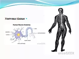
/
CNS andAnesthesiaLyon Lee DVM PhD DACVAThe Nervous SystemCentral CNS CNS andAnesthesiaLyon Lee DVM PhD DACVAThe Nervous SystemCentral CNS

CNS andAnesthesiaLyon Lee DVM PhD DACVAThe Nervous SystemCentral CNS - PDF document

reese
reese . @reese
Follow
342 views
Uploaded On 2022-08-22

CNS andAnesthesiaLyon Lee DVM PhD DACVAThe Nervous SystemCentral CNS - PPT Presentation

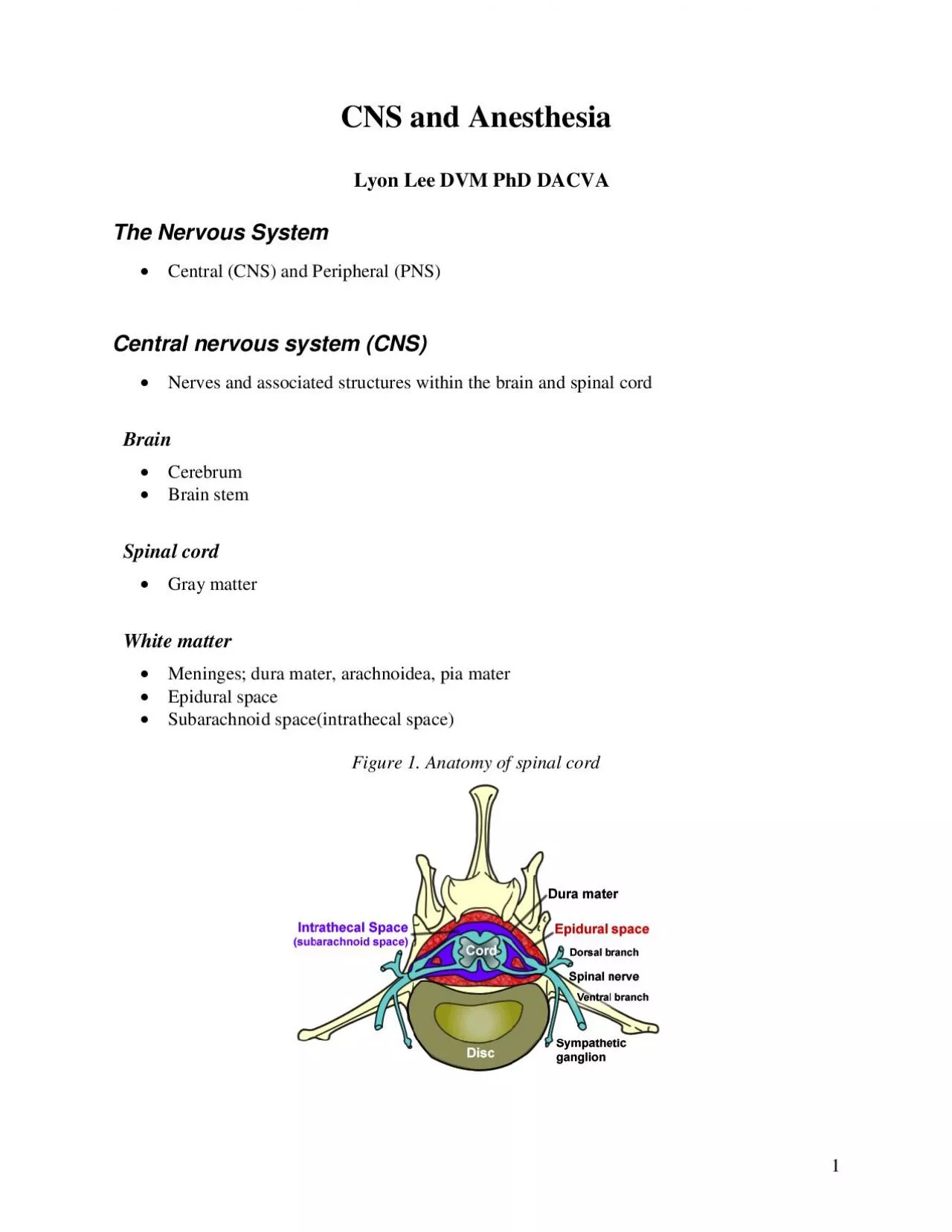
1 2 CSFFormed at choroid plexuses in the ventriclesCushioning effectNormal 10 mmHg in pressure 1002 1009 in SG 732 in pHncreased productiondecreased absorption andorobstruction of flow ofCSF a ID: 939639

nervous system sensory nerve system nervous nerve sensory motor fibers muscles spinal peripheral cns sympathetic muscle parasympathetic figure receptors

Share:

Link:

Embed:

Download Presentation from below link

Download Pdf The PPT/PDF document "CNS andAnesthesiaLyon Lee DVM PhD DACVAT..." is the property of its rightful owner. Permission is granted to download and print the materials on this web site for personal, non-commercial use only, and to display it on your personal computer provided you do not modify the materials and that you retain all copyright notices contained in the materials. By downloading content from our website, you accept the terms of this agreement.


Presentation Transcript

1 CNS andAnesthesiaLyon Lee DVM PhD DACVAThe Nervous SystemCentral (CNS) and Peripheral (PNS)Central nervous system(CNS)erves and associated structures within the brain and spinal cord CerebrumBrain stemSpinal cord Gray matterWhite matterMeningesdura mater, arachnoidea, pia materEpidural spaceSubarachnoid space(intrathecal space)Figure 1. Anatomy of spinal cord 2 CSFFormed at choroid plexuses in the ventriclesCushioning effectNormal: 10 mmHg in pressure, 1.002 1.009 in SG, 7.32 in pHncreased productiondecreased absorption, and/orobstruction of flow ofCSF all contribute to hydrocephalus symptomPeripheral nervous system(PNS)The nerves and ganglia which lie outside the brain and spinal cord. Cranial nerves and spinal nerves extend from the CNS to peripheral organs such as muscles, jointsand glands.Nerves are bundles of nerve fibers, much like muscles are bundles of muscle fibers. Ganglia are collections, or small knots, of nerve cell bodies outside the CNS.The peripheral nervous system is further subdivided into an afferent (sensory) division nd an efferent (motor) division (see figure 2)he efferent or motor di

vision is again subdivided into the somatic nervous system and the autonomic nervous system.Figure 3. ivision of the nervous system CNS PNS Motor division Sensory division Autonomic nervous system Somatic nervous system Sympathetic Parasympathetic 3 ranial nervesTable 1. Cranial nervesand their function Nerves in order Modality Function Olfactory (I) Sensory Smell Optic (II) Sensory Vision Oculomotor (III) Motor Motor Levator palpebrae, superioris, superior, medial& inferior recti muscles Parasympathetic to ciliary pupillary constrictor muscles Trochlear (IV) Motor D orsal oblique muscle Trigeminal(V) Motor Sensory Muscles of mastication Sensory for head/neck, sinuses, meninges, & external surface of tympanic membrane Abd ucens (VI) Motor Lateral rectus muscle , retractor oculi muscle Facial (VII) Motor Motor Sensory Sensory Muscles of facial expression Parasympathetic to all glands of head except the parotid he skin of external ear T aste buds of tongue Vestibulocochlear (VIII) Special SensoryHearing and Balance Glo

ssopharyngeal(IX) Motor MotorSensorySensory Sensory Stylopharyngeus muscle Parotidand zygomaticsalivary glandCarotid body and sinusSensation posterior onethirdtongue & internal surface of tympanic membrane. Taste posterior one - third tongue Vagus(X) Motor MotorSensory Sensory Muscles of pharynx , larynx and esophagus Parasympathetic to neck, thorax, abdomen Sensory from pharynx, larynx viscera Sensory from external ear Spinal Accessory (XI) Motorusclesof the neck and head Hypogloss al (XII) MotorMuscles of the pharynx, larynx and tongue 4 Spinal nervesegins at foramen magnum and terminates at L6/7 in dogsat L7/S1 in cats and L6/S1 in horses. Consists of ventral and dorsal roots(see figure The dorsal root contains sensory neurons while the ventral root contains motor neurons. Consists of white matter which forms ascending and descendingpathways and grey mater that contains cell bodiesFigure Components of a spinal nerve Anatomy and function of the nerve fibersA neuron consists of a cell body or soma, dendrites, and a nerve fiber or axon. The axon of one neuron terminates (synapses) near

the cell body ordendrites of another neuron. Neurons The larger nerve fibers are surrounded by a coat of fatty materialthe myelin sheath, while the smaller nervefibers may not be myelinated. are cells that transfer stimuli to other cells.The myelin lamellae are not continuous along the entire length of the fiber, being interrupted at more or less regular intervals (the nodes of Ranvier). Peripheral nerves are classified as A, B, and C on the basis of fiber diameter and velocity of conduction of nerve impulses (see table below). The largest diameter A fibers are subdivided into alpha, beta, gamma, and delta. alpha fibers innervate skeletal muscles. Tactile sensory receptors transmit signals in type Abeta fibers. gamma fibers are distributed to skeletal muscles. delta fibers transmit touch, temperature and fast pain. C fibers transmit slow pain, temperature and touch. A and B fibers are myelinated, whereas C fibers are unmyelinated. Nerve fibers are afferent if they transmit impulses from peripheral receptors to the spinal cord and efferent if they relay signals from the spinal cord and CNS to the periphery.

5 Table 3. Peripheral nerve classification on the basis of fiber diameter and velocity of conduction of nerve impulses fiber Anatomic location Myelinated Fiber diameter (µm) Conduction speed (m/sec) Function Sensitivity to block by local anesthetics C P ostganglionic sympathetic, sensory roots, and afferent peripheral nerves Vasomotor, visceromotor, slow pain, temperature, touch++++(highest) B Preganglionic sympatheticYesVasomotor, visceromotor++++ A Sensory roots Yes 1 - 4 12 - 30 Fast pain, temperature, touch, uscle tone, +++ Efferent to muscle spindle Yes 3 - 6 15 - 30 Fast pain, temperature, touch, m uscle tone, ++ Efferent and Afferent to muscles and joints Yes 6 - 22 30 - 70 Motor and proprioception ++ Efferent and Afferent to muscles and jointsYesMotor and proprioception(lowest) 6 Autonomicnervous system It is further subdivided into sympathetic and parasympathetic divisions(see figure 3)Because the autonomic nervous system regulates involuntary or automatic functions, it is called the in

voluntary nervous system.The ParasympatheticNervous System(craniosacral)etylcholineis transmitter both at pre postganlionic (muscarinic)neuronslong preganglionicneurons, short postganglionic neurons; ganglia are diffusly spread; allows fordiscrete, localized innervation control Vagus nerve innervatesheart, lungs, esophagus, stomach, small intestine, proximal colon, liver,gallbladder, pancreas, kidneys, upper uretersDistribution ofinnervation to the heart is to the AV node, SA node, and atria (essentiallynone to the ventricles)Sacral outflow from 2nd,3rd, and 4th sacral segments of the cord; form the pelvic nerves, and innervatethe bladder, distal colon, rectum, andsexual organsFigure 4. Parasympathetic nervous system 7 The Sympathetic Nervous System(thoracolumbar)acetylcholine istransmitter between pre postganlionic neurons; norepinephrine isneurotransmitter between the neuron effector cellsympathetic stimulatioproduces more generalized effects than parasympathetic stimulationadrenal medulla isessentially a specialized sympathetic ganglia, which functions by releasingepinephrine andnorepinephrine into the syst

emic circulation; this results inpathetic activation even in cells that do not have direct sympathetic innervation (but have sypathetic receptors)Figure 5. Sympathetic system 8 eurotransmissionA nerve impulse is anelectric current that passes along an axon to the presynaptic membrane. Uponreaching the presynaptic membrane, it causes the release of neurotransmittersinto the synaptic cleft. The neurotransmitter then interacts with receptors on effectorcells to induce a response in the effector cell.Figure 6. A nerve terminal Neuroregulators Neurotransmitters areleased into the syptic cleft in response to action potentials release isvoltage dependent and requires calcium influxNeuropeptide modulatorsare released in smaller quantities than neurotransmitters in response to actionpotentials they serve to amplify or dampen neural activity.CholinergictransmissionAcetylcholine is theneurotransmitterPrimary means ofterminating action is break down of acetylcholine into acetate choline byacetylcholine esterase (AchE), found principally in neurons neuromuscularjunctionsCholinergic receptorsare present in the parasympathe

tic nervous system, brain, ganglia of thesympathetic nervous system, and skeletal muscleTwo main types ofreceptors present Muscarinic (principallyautonomic nervous systemNicotinic (principallyskeletal muscle) 9 Adrenergic transmission:Catecholamines (dopamine,norepinephrine, epinephrine) are the neurotransmittersPrimary means ofterminating action is by neural membrane reuptake of the transmitter, althoughmetabolism bycatecholmethyltransferase (COMT) and monoamine oxidase (MAO) isimportant in some tissues.drenergic receptorsAlpha receptorsaremainly subdivided into alpha1 and alpha 2receptorsAlpharincipallyfound in peripheral vascular smooth muscleAlphoccur bothpresynaptically postsynapticallythose occurring presynaptically on sympathetic nerve terminals reduce the release of norepinephrine,thus producing a negative feedback loopalso may modulatecholinergic, serotonergic, GABAergic neuroncentral alpha2adrenergic receptor stimulation results in sedation, analgesia, decreasedsympathetic outflow, tranquilizationindirectly affectscardiac function by decreased sympathetic toneact preand postjuntionally to decrease motil

ity andsecretions in the GI tractproduces diuresis byinhibing ADH release, blocking ADH's effect in the renal tubule, increasingGFR, and inhibiting renin releasestimulate plateletaggregationBeta receptors, again, are mainly subdivided into beta1 and beta 2receptorsBetalocated in themyocardium, SA node, ventricular conduction system, and adipose tissueetavascular smoothmuscle of the skin, muscles, mesentery bronchial tree; stimulationresults in vasodilation and bronchodilationopaminergic receptors dopamine: splanchnicandrenal vasodilation 10 Figure 7. Alpha adrenergic receptor (nonadrenergicnoncholinergic)In the brain, spinal cord, and peripheral nervous system.Arginine and OIt activates guanyl cyclase to increase cGMP which leads to relaxation of smooth muscle.produce LCitrulline and NO by NO synthasesNMDA glutamate receptor activation releaseNO and turn results in excitatoryneurotransmission in the CNS.NOS inhibitor causes dosedependent MAC decreaseNeuromuscular junctionand neuromuscular blocker (NMB)It consists of presynaptic nerve terminal and postsynaptic muscular membrane.Mainly cholinergic nicotinicreceptor

s, two at postsynaptic and one presynapticThe neurotransmitter is the quaternary ammonium esteracetylcholineAcetate and choline through choline acetylase form Acetylcholineat motor nerve endingAcetylcholinesterase at cholinergic receptors is responsible for hydrolysingAch into Acetic acid and choline 11 Choline can reenter nerve terminal to again participate in the synthesis of new acetylcholineDepolarizing neuromuscular blockerSuccinylcholine (suxamethonium in Europe), mimics the action of Achby occupying postsynaptic nicotinic cholinergic receptor, thus depolarizing postsynaptic membrane. However, hydolyis of Sch is slower, so postjunctional membrane does not respond to subsequently released Ach prolonging neuromuscular blockade (Phase I)Side effects include hyperkalemia, hypertension, myalgia, cardiac arrhythmia, and increased intraocular pressure. Also known as a trigger for malignant hyperthermia in susceptible patients.Nondepolarising NMBs Some examples of drugs falling into this category are pancuronium,atracuriumdoxacurium, vecuronium and mivacurium.These agentsbind to the post synaptic nicotinic cholin

ergic receptors without causing any activation of ion channel permeability, and yet impeding normal postjunctional depolarization with less Ach availability at the receptor leading to the neuromuscular blockade.Occupation as many as 70 % does not produce neuromuscular blockade, but 80 % occupation fails neuromuscular transmission, indicating widesafety margin of the drugClinically, a peripheral nerve stimulator is employed to assess the neuromuscular blocking effect induced with the drugs.Train of , Single Twitch, Tetanic or Double Burst timulationare applied to test the degree of neuromusculartransmissionTheoriesof AnesthesiaWide range of compoundsproduce anesthesia, without any unifying chemical structure or activityWe don’t as yetunderstand how general anesthetics functionA key concept in any theory regarding anesthetic mechanisms must be the ability of the anesthetic to disrupt cellular and intercellular communication, particularly in the CNS.Many hypothesehave been proposedover the years; it appears that there is expansion and fluidization of the cellmembrane by anesthetic agents that result in depressed sy

naptic transmission,and some anesthetic agents also hyperpolarize neurons by increasing potassiumpermeabilityMeyerOverton hypothesis asserts that, anesthesia results from the presence of a certainconcentration of the anesthetic at a hydrophobic site. Evidence for this has come from the fact that potency is strongly correlated with the lipid solubility of the drugCritical volume theory asserts that anesthetic’s direct action on proteins (ion channel proteins nicotinic Ach, GABA, glycine, NMDA; signal transduction pathwayswill induce conformation change on lipoprotein (expansion beyond the critical volume) and lead to interruption of neurotransmissionby obstructing ion flux with changes of electrical conductivity in the neurons 12 The reticular activating system, a multisynaptic structureis believed to be the most important site within the central nervous system for anesthetic action.We do have anunderstanding of how certain classes of drugs work those that interact withspecific receptor sitesopioids (eg, morphine, butorphanolalpha2 receptoagonists (eg, xylazine, medetomidinebenzodiazepines (eg, diazepam, mida