/
VetScript November 2017 150 VetScript November 2017 150

VetScript November 2017 150 - PDF document

roberts
roberts . @roberts
Follow
342 views
Uploaded On 2022-10-28

VetScript November 2017 150 - PPT Presentation

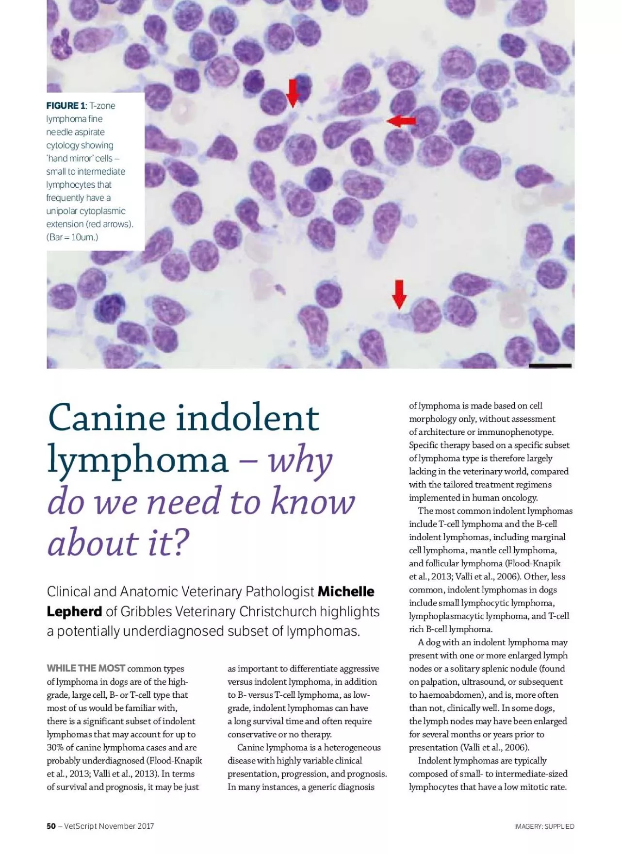
They can be mistaken for lymphoid hyperplasia or a reactive lymph node on fine needle aspirate FNA cytology as they lack the significant numbers of large lymphocytes that help to cytologically diag ID: 961508

indolent lymphoma 150 dogs lymphoma indolent dogs 150 veterinary clinical lymphomas cell 2013 knapik flood common large demodicosis zone

Share:

Link:

Embed:

Download Presentation from below link

Download Pdf The PPT/PDF document "VetScript November 2017 150" is the property of its rightful owner. Permission is granted to download and print the materials on this web site for personal, non-commercial use only, and to display it on your personal computer provided you do not modify the materials and that you retain all copyright notices contained in the materials. By downloading content from our website, you accept the terms of this agreement.


Presentation Transcript

VetScript November 2017 – They can be mistaken for lymphoid hyperplasia or a reactive lymph node on fine needle aspirate (FNA) cytology, as they lack the significant numbers of large lymphocytes that help to cytologically diagnose the more common large cell lymphomas. While the clued-up pathologist can often get a suspicion of indolent lymphoma from cytology findings and detailed clinical history, histopathology is essential for confirmation. Whole node or large wedge-shaped biopsies that incorporate capsule, cortex and medulla are required, as most indolent lymphomas have a characteristic pattern of follicular expansion and effacement. Tru-cut and narrow core biopsies are often unrewarding, as they don’t include enough architecture for diagnosis. In many cases, routine histopathology of an adequately sized biopsy will be sufficient to diagnose the subtypes of indolent lymphoma. However, in more advanced cases, where there is greater effacement of architecture and obscure residual follicles, immunophenotyping for B- and T-cell markers can be enormously helpful in recognising the follicle-related heritage and fading follicles that characterise most indolent lymphomas. The detection of clonality is a useful adjunct to histology and immunohistochemistry, particularly in those cases with an ambiguous morphology and immunophenotype.T-zone lymphoma (TZL) is the most common type of indolent lymphoma and, as the name suggests, is of T-cell origin, developing as an expansion of the paracortical areas between fading follicles. A clue to diagnosis can often be seen on FNA of affected lymph nodes, due to the presence of large numbers of small to intermediate lymphocytes that frequently have a unipolar cytoplasmic extension (hand mirror cells – Figure 1). Dogs with TZL often have generalised lymphadenopathy, and therapy does not appear to positively influence survival or quality of life (Valli et al., 2006; Valli et al., 2013). The second most common type of indolent lymphoma is marginal zone lymphoma (MZL), which is of B-cell origin, and occurs in either lymph nodes or, more commonly, the spleen. It is characterised by coalescing perifollicular proliferation of marginal cells and paracortical atrophy. Splenic MZL often has a good response to splenectomy alone (Valli et al., 2006). Interestingly, 10-50% of dogs with indolent lymphoma also have demodicosis (Flood-Knapik et al., 2013; Mizutani et al., Demodex spp. infestation is usually associated with immunosuppression and the presence of indolent lymphoma may predispose to immune dysregulation and subsequent demodicosis. Also, demodicosis itself may cause further immunosuppression. Further work-up for lymphoma may be warranted in older dogs with generalised demodicosis, and should include lymph node biopsy, rather than just FNA, to ensure indolent lymphoma can be detected. Median lymphoma-specific survival time for indolent lymphoma has been reported to be as long as 4.4 years and the median overall survival time (OST) as 21.

2 months, and is longest for TZL (median OST of 33.5 months) (Flood-Knapik et al., 2013). Dogs with solitary splenic nodules are reported to do well with splenectomy alone (Flood-Knapik et al., 2013; Valli et al., 2006). In fact, many dogs can do well without treatment, and ‘watchful waiting’ may be a reasonable approach, particularly in the earlier phases of disease. However, it appears that some indolent B-cell lymphomas can behave like, or transform into, high-grade lymphomas. There is no established consensus about when to treat dogs with indolent lymphoma. For asymptomatic TZL, Moore (2016) suggests watchful waiting with careful monitoring of peripheral nodes and haematology. Further therapy should be considered if dogs develop clinical signs, have rapid progression (defined as a tumour doubling time of less than six months), a circulating lymphocyte count greater than /L, multiple sites of lymphoma larger than 3cm in diameter or a single site greater than 7cm in diameter, development of myelosuppression due to myelophthisis, or organ dysfunction due to infiltration. Chemotherapy for indolent lymphoma ranges from prednisone alone, combination prednisone and chlorambucil, or CHOP-based chemotherapy protocols, similar to those used for aggressive lymphoma (Flood-Knapik et al., 2013). In the future, better characterisation and more precise diagnoses of lymphoma, alongside the documentation of responses to specific treatments, will hopefully allow for more accurate prognostications and lymphoma-specific therapy. REFERENCES:Flood-Knapik KE, Durham AC, Gregor TP, Sánchez MD, Durney ME, Sorenmo KU. Clinical, histopathological and immunohistochemical characterization of canine indolent lymphoma. Veterinary and Comparative OncologyMizutani, N, Goto-Hoshino Y, Takahashi M, Uchida K, Tsujimoto H. Clinical and histopathological evaluation of 16 dogs with T-zone lymphoma. Journal of Veterinary Medicine and Surgery 78, 1237–44, 2016Moore AS. Treatment of T-cell lymphoma in dogs. Veterinary Record 179, 277–81, 2016Valli VE, Vernau W, de Lorimier L-P, Graham PS, Moore PF. Canine indolent nodular lymphoma. Veterinary PathologyValli VE, Kass PH, San Myint M, Scott F. Canine lymphomas: association of classication type, disease stage, tumor subtype, mitotic rate, and treatment with survival. Veterinary Pathology 50, 738–48, 2013 WHILE THE CLUEDUP PATHOLOGIST CAN OFTEN GET A SUSPICION OF INDOLENT LYMPHOMA FROM CYTOLOGY FINDINGS AND DETAILED CLINICAL HISTORY, HISTOPATHOLOGY IS ESSENTIAL FOR CONFIRMATION. – VetScript November 2017 Canine indolent lymphoma– why do we need to know about it?Clinical and Anatomic Veterinary Pathologist Michelle Lepherd of Gribbles Veterinary Christchurch highlights T-zone lymphoma fine needle aspirate cytology showing ‘hand mirror’ cells – small to intermediate lymphocytes that frequently have a unipolar cytoplasmic extension (red arrows). WHILE THE MOST common types of lymphoma in dogs are of the high