/
Typhoid Fever Dr.  Dur  Muhammad Khan Typhoid Fever Dr.  Dur  Muhammad Khan

Typhoid Fever Dr. Dur Muhammad Khan - PowerPoint Presentation

rodriguez
rodriguez . @rodriguez
Follow
350 views
Uploaded On 2022-06-14

Typhoid Fever Dr. Dur Muhammad Khan - PPT Presentation

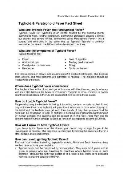
MRCP FRCP A 20years old patient presents in the OPD with a history of fever for 10 days He also complains of generalized weakness and headache There is history of dry cough and abdominal pain but no ID: 917947

week fever blood high fever week high blood weeks intestinal stage days culture treatment diagnosis typhoid perforation complications splenomegaly

Share:

Link:

Embed:

Download Presentation from below link

Download Presentation The PPT/PDF document "Typhoid Fever Dr. Dur Muhammad Khan" is the property of its rightful owner. Permission is granted to download and print the materials on this web site for personal, non-commercial use only, and to display it on your personal computer provided you do not modify the materials and that you retain all copyright notices contained in the materials. By downloading content from our website, you accept the terms of this agreement.


Presentation Transcript

Slide1

Typhoid Fever

Dr.

Dur

Muhammad Khan

(MRCP. FRCP)

Slide2

A 20years old patient presents in the OPD with a history of fever for 10 days. He also complains of generalized weakness and headache. There is history of dry cough and abdominal pain but no

diarrhoea

.

Examination reveals Temp:102 F, Pulse : 70/m, BP: 110/70.

There is mild splenomegaly. Rest of the examination is normal. What is the likely diagnosis?

Slide3

Typhoid

Definition

Etiology

Pathogenesis

EpidemiologyClinical manifestationsThe laboratory and other examinations

Complications

Diagnosis and differential diagnosis

Prognosis

Treatment

Slide4

Definition of Typhoid fever

Acute enteric infectious disease

caused by Salmonella typhi (S.Typhi).

prolonged fever, Relative bradycardia, apathetic facial expressions,

roseola, splenomegaly, hepatomegaly,

leukopenia.

intestinal perforation, intestinal hemorrhage

Slide5

Etiology

Serotype: D group of Salmonella

Gram-negative

rod

non-spore

flagella Culture characteristics

Slide6

Antigens: located in the cell capsule

H (flagellar antigen).

O (Somatic or cell wall antigen).

Vi (polysaccharide virulence)

“widel test”

Slide7

A schematic diagram of a single

Salmonella typhi

cell showing the locations of the H (flagellar), 0 (somatic), and Vi (K envelope) antigens.

Slide8

Epidemiology

continues to be a global health problem

areas with a high incidence include Asia, Africa and Latin America

affects about 6000000 people with more than 600000 deaths a year. 80% in Asia .

sporadic occur usually, sometimes have epidemic outbreaks.

Slide9

Slide10

Source of infection

Cases and chronic carriers

Cases discharge from incubation, more in 2~4 weeks after onset, a few (about 2~5%) last longer than 3 months

chronic carrier

Typhoid Mary

Slide11

Slide12

Transmission

fecal-oral route

close contact with patients or carriers

contaminated water and food

flies and cockroaches.

Slide13

Pathogenesis

gastrointestinal tract host-pathogen interactions

The amount of bacilli infection (>10

5

baeteria).

Slide14

ingested orally

Stomach barrier (some Eliminated)

enters the small intestine

Penetrate the mucus layer

enter mononuclear phagocytes of ileal peyer's patches and mesenteric lymph nodes

proliferate in mononuclear phagocytes

spread to blood. initial bacteremia (Incubation period).

Pathogenesis

Slide15

Pathogenesis

enter spleen, liver and bone marrow (reticulo-endothelial system)

further proliferation occurs

A lot of bacteria enter blood again.

(second bacteremia).

Recovery

Slide16

Pathology

essential lesion:

proliferation of RES (

reticuloendothelial system )

specific changes in lymphoid tissues

and mesenteric lymph nodes.

"typhoid nodules“

Most characteristic lesion

:

ulceration of mucosa in the region of the

Peyer’s

patches of the small intestine

Slide17

Major findings in lower ileum

Hyperplasia stage(1st week):

swelling of lymphoid tissue and proliferation of macrophages.

Necrosis stage(2nd week):

necrosis of swollen lymph nodes or solitary follicles.

Slide18

Major findings in lower ileum

Ulceration stage(3rd week):

shedding of necrosis tissue and formation of ulcer

----- intestinal hemorrhage, perforation

.

Stage of healing (from 4th week): healing of ulcer, no cicatrices and no contraction

Slide19

Clinical manifestations

Incubation period: 3

60 days(7

~14).The initial period (early stage) First week. Insidious onset.

Fever up to 39~40

0

C in 5~7 days

chills

ailment

tired

sore throat

cough ,abdominal discomfort and constipation et al.

Slide20

The fastigium satge

second and third weeks.

Sustained high fever

partly remittent fever or irregular fever. Last 10

~14 days. Gastro-intestinal symptoms: anorexia、abdominal distension or pain、

diarrhea or constipation

Neuropsychiatric manifestations: confusion

blunt respond even delirium and coma or meningism

Slide21

Circulation system

:

relative

bradycardia

.

splenomegaly

hepatomegaly

toxic hepatitis.

roseola

:30%,

maculopapular

rash

a faint pale color, slightly raised

round or lenticular, fade on pressure

2-4 mm in diameter, less than 10 in number

on the trunk, disappear in 2-3 days.

Slide22

Slide23

fatal complications:

intestinal hemorrhage

intestinal perforation

severe toxemia

Slide24

defervescence stage

fever and most symptoms resolve by the

forth week

of infection.

Fever come down, gradual improvement in all symptoms and signs, but still danger.

convalescence stage

the

fifth week

. disappearance of all symptoms, but can relapse

Slide25

Special manifestations

In children

Often atypical

sudden onset with high fever. Respiratory symptoms and diarrhea, dominant.

Convulsion common in below 3.

relative bradycardia rare.

Splenomegaly, roseola and leucopenia less common.

Slide26

In the aged

temperature not high, weakness common.

More complications.high mortality.

Slide27

Laboratory findings

Routine examinations:

white blood cell count is normal or decreased.

Leukocytopenia(specially eosinophilic leukocytopenia).

recovery with improvement of diseases decreased in relapse

Slide28

Bacteriological examinations:

Blood culture:

the most common use

80~90% positive during the first 2 weeks of illness

50% in 3rd week not easy in 4th week

re-positive when relapse and recrudesce

attention to the use of antibiotics

Slide29

The bone marrow culture

the most sensitive test

specially in patients pretreated with antibiotics.

Urine and stool cultures

increase the diagnostic yieldpositive less frequentlystool culture better in 3~4 weeks

The duodenal string test to culture bile useful for the diagnosis of carriers.

Rose spots: Not use routinely

Slide30

Complications

Intestinal hemorrhage

Commonly appear during the second-third week of illness

difference between mild and greater bleeding

often caused by unsuitable food, diarrhea et al

serious bleeding in about 2~8%

a sudden drop in temperature

rise in pulse

and signs of shock followed by dark or fresh blood in the stool.

Slide31

Intestinal

perforation:

The more serious .Incidence,1-4%

Commonly appear during 2-3 weeks. Take place at the lower end of ileum.

Before perforation,abdominal pain or

diarrhea,intestinal bleeding .

When perforation, abdominal pain, sweating, drop in temperature, and increase in pulse rate, then, rebound tenderness when press abdomen,

abdomen muscle entasia, reduce or disappear in the sonant extent of liver, leukocytosis .

Temperature rise .peritonitis appear.

celiac free air under x-ray.

Slide32

Toxic hepatitis

:

common,1-3 weeks

hepatomegaly, ALT elevated

get better with improvement of diseases in 2~3 weeks

Toxic myocarditis. seen in 2-3 weeks, usually severe toxemia.

Bronchitis, bronchopneumonia.

seen in early stage

Slide33

Other complications

:

toxic encephalopathy.

Hemolytic uremic syndrome.

acute cholecystitis

、meningitis、nephritis et al.

Slide34

Differential diagnosis

Viral infections

:

such as upper respiratory tract infection.

abrupt onset with fever, headache, leucopenia, sore throat, cough, coryza.

no rose spots, no enlargement of liver & spleen. The course of illness no more than 2 wks.

differential diagnosis depends on typical manifestations and blood culture.

Slide35

Malaria

history of exposure to malaria.

Paroxysms(often periodic) of sequential chill,high fever and sweating.

Headache, anorexia, splenomegaly, anemia, leukopenia

Characteristic parasites in erythrocytes,identified in thick or thin blood smears.

Slide36

Leptospirosis

Endemic area,contacted with urine of mice.

Abrupt fever,chills,severe headache,and myalgias, especially of the calf muscles.

Leptospires can be isolated from blood,cerebrospinal fluid.

Special agglutination titers develop after 7 days and may persist at high levels for many years.

Slide37

Tuberculosis

continuous high or low fever,fatigue,weight loss,night sweats.

Mild cough

pulmonary infiltration on chest radiograph

positive tuberculin skin test reaction(most cases)

acid-fast bacilli on smear of sputumsputum culture positive for mycobacterium tuberculosis

.

Slide38

Septicemia of Gram-negative bacilli

abrupt onset,high fever,symptom of toxemia.

Chill,sweats.

Shock.

Positive of gram-negative bacilli from blood culture.

Slide39

Prognosis:

Case fatality 0.5

1%.

but high in old ages

、infant、

and serious complications

Have immunity for ever after diseases

About 3% of patients become fecal carriers

.

Slide40

TREATMENT

General treatment

isolation and rest

good nursing care and supportive treatment

close observation T,P,R,BP,abdominal condition and stool .

suitable diet include easy digested food or half-liquid food.drink more water

intravenous injection to maintain water and acid-base and electrolyte balance

Slide41

Symptomatic treatment:

for high fever:

physical measures firstly

antipyretic drugs such as aspirin should be administrated with caution

delirium,coma or shock,2-4mg dexamethasone in addition to antibiotics reduces mortality.

Slide42

Etiologic and special treatment

1.Quinolones:

first choice

it’s highly against S.typhi

penetrate well into macrophages,and achieve high concentrations in the bowel and bile lumens

Norfloxacin (0.1

0.2 tid

qid/10

14 days).

Ofloxacin (0.2 tid 10

14days).

ciprofloxacin (0.25 tid)

caution: not in children and pregnant

Slide43

3.Cephalosporines:

Only third generation effective

Cefoperazone and Ceftazidime.

2

~4g/day .10~14 days.

4.Treatment of complication.

Intestinal bleeding:

bed rest, stop diet,close observation T,P,R,BP.

intravenous saline and blood transfusion,and attention to acid-base balances.

sometimes,operative.

Slide44

Perforation:

early diagnosis.

stop diet.

decrease down the stomach pressure.

intravenous injection to maintain electrolyte and acid-base balances.

use of antibiotics.

sometimes operative.

Slide45

Toxic myocarditis:

bed rest, cardiac muscle protection drugs,

dexamethasone, digoxin.

5.Chronic carrier:

Ofloxacin 0.2 bid or ciprofloxacin 0.5 bid, 4

6 weeks.

Ampicillin 3

6g/day tid plus probenecid 1

1.5g/day. 4

6 weeks.

TMP+SMZ

2 tabs. Bid. 1

3 months.

Cholecystitis may require cholecystectomy.

Slide46

Thank you!!!

Slide47

MCQs

Sahmonella

is

Gram +

ve

rodGram –ve cocciGram –ve rodGram +ve cocciAnaerob

Slide48

Answer: C

Slide49

Q2

In typhoid fever, splenomegaly

Is present in the early first week

Is present in the second week

Present in early third week

Is massiveCarries a poor prognosis

Slide50

Anwer: B

Slide51

Q3

Which of the following is not a complication of typhoid fever

Intestenial

haemorrhage

PneumonitisDissemeinated intravascular coagulationMeningitisPulmonary hypertension

Slide52

Answer: E