/
Nail Diseases By Dr. Salam Altemimi Nail Diseases By Dr. Salam Altemimi

Nail Diseases By Dr. Salam Altemimi - PowerPoint Presentation

roxanne
roxanne . @roxanne
Follow
343 views
Uploaded On 2022-06-18

Nail Diseases By Dr. Salam Altemimi - PPT Presentation

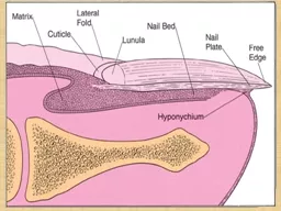
Chronic paronychia Is infection of the nail fold and matrix by candida albicans There is glazed and red swelling of the nail fold with loss of the cuticle There is mild to moderate pain Occasional bead of pus comes out from under the nail fold Ridging and furrowing of nail plate may occur due t ID: 920054

plate nail fold nails nail plate nails fold toenail paronychia treatment psoriasis onycholysis infections weeks dermatosis ridging unguium tinea

Share:

Link:

Embed:

Download Presentation from below link

Download Presentation The PPT/PDF document "Nail Diseases By Dr. Salam Altemimi" is the property of its rightful owner. Permission is granted to download and print the materials on this web site for personal, non-commercial use only, and to display it on your personal computer provided you do not modify the materials and that you retain all copyright notices contained in the materials. By downloading content from our website, you accept the terms of this agreement.


Presentation Transcript

Slide1

Nail Diseases

By

Dr. Salam Altemimi

Slide2

Slide3

Chronic paronychia

Is infection of the nail fold and matrix by candida albicans. There is glazed and red swelling of the nail fold with loss of the cuticle. There is mild to moderate pain. Occasional bead of pus comes out from under the nail fold. Ridging and furrowing of nail plate may occur due to damage of the matrix. Dark brown pigmentation of nail plate occurs in direct invasion by monilia. The disease is occupational of housewives due to wetness, which lead to maceration of the cuticle then the entrance of the microorganism.

Slide4

Slide5

Slide6

Differential diagnosis:

acute paronychia

is bacterial infection of the nail fold. There is bright red swelling of the nail fold. The inflammation is more severe, the condition is more painful.

Slide7

Acute paronychia

Slide8

Treatment of Acute paronychia

Incision to evacuate the pus.

Oral antistaphylococcal antibiotics e.g. cephalexin 250 mg four times daily for 7 days.

Slide9

Treatment of chronic paronychia

Maintain dryness.

Topical

clotrimazol

cream.

Oral choice is

fluconazole

150 mg per week for 4 weeks.

Slide10

Tinea

unguium

(

onychomycosis

)

It is the dermatophyte (ringworm) infection of the nails. Commonly caused by

trichophyton

and

epidermophyton

. The affected nail is roughing, opaque and friable. It has an accumulation of keratinous debris under it. The diagnosis can be confirmed by shaving of the nails to find fungus on 20% KOH microscopic exam. Also culture on

Sabouraud

medium can be performed.

Slide11

Slide12

Slide13

Slide14

Tinea

unguium

Slide15

Treatment of tinea

unguium

The treatment of choice is oral

terbinafine

250mg daily for 6 weeks in fingernail infections and for 12 weeks in toenail infections.

Fluconazole

150 mg once a week for 9 months.

Itraconazole

(100 mg capsule) given in pulse therapy. The pulse consists of 2 capsules twice daily for 1 week followed by 3 weeks of drug free interval. Two to 3 pulses is given for fingernail infections and 3-4 pulses for toenail infections.

Slide16

Dermatosis associated with nail involvement

Psoriasis

Pitting

Onycholysis

Discoloration

Subungual thickening

Malformed nails

Splinter hemorrhages

Slide17

Psoriasis of the nails

Slide18

Dermatosis associated with nail involvement

Lichen planus

Longitudinal grooving and ridging

Pterygium

Slide19

Lichen

planus

of the nails

Slide20

Lichen

planus

of the nail

Slide21

Alopecia areata-nails

Slide22

Alopecia areata

Pitting

Norwegian scabies

Nail plate dystrophy

Eczema and dermatitis

Ridging, thickening and discoloration

Paronychia

Ridging, thickening and discoloration

Dermatosis

associated with nail involvement

Slide23

Onycholysis

It is the separation of the nail plate from the nail bed at distal and lateral margins.

Slide24

Onycholysis

(from psoriasis)

Slide25

Etiology of onycholysis

Idiopathic

Secondary

Dermatosis

: psoriasis, fungal infections.

General medical conditions: like hyperthyroidism,

Raynaud’s

phenomenon.

Trauma: like typing, long nails.

Drugs: photo-

onycholysis

occurs with tetracycline or

psoralin

.

Slide26

Nail pitting

It is a tiny, punched out or ices pick depressions of the nail plate. Common causes are psoriasis, alopecia areata, and sometimes a normal variant.

Slide27

Koilonychia

The nail is flat or concave has spoon-shape. It is often thin and brittle. The condition associated with hypochromic iron deficiency anemia.

Slide28

Koilonychia

Slide29

Finger clubbing

It is increase in the size and curvature of nail plate with loss of the angle between the nail plate and the posterior nail fold. It is associated with many diseases e.g. carcinoma of the bronchus, heart diseases.

Slide30

Finger clubbing

Slide31

Ingrown toenail

It is the soft tissue of the side of the nail (lateral nail fold) is penetrated by the edge of the nail plate, resulting in pain, sepsis and later the formation of the granulation tissue. The great toe is often affected. The cause is compression of the toe by ill-fitting footwear and cutting of the toenail in a half-circle instead of straight across.

Slide32

Ingrowing toenail

Slide33

Treatment of ingrown toenail

Wearing wide and pliable shoes.

Antibiotics.

Cauterization of granulation tissues by silver nitrate sticks.

If yet no benefit, avulsion of nail plate or removal part of it in continuing cellulitis.

Slide34

Good luck