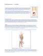
/
Figure 1 Location of the Figure 1 Location of the

Figure 1 Location of the - PDF document

roy
roy . @roy
Follow
342 views
Uploaded On 2021-10-01

Figure 1 Location of the - PPT Presentation

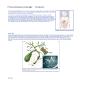
colon in the bodyHereditary Colorectal Cancer Introduction The Hereditary Colorectal Cancer Website has been sponsored by the Robert Rauschenberg FoundationWelcome to the Johns Hopkins Heredita ID: 892273

colon cancer rectum colorectal cancer colon colorectal rectum age risk hereditary doctor ileostomy rectal group figure polyps exam people

Share:

Link:

Embed:

Download Presentation from below link

Download Pdf The PPT/PDF document "Figure 1 Location of the" is the property of its rightful owner. Permission is granted to download and print the materials on this web site for personal, non-commercial use only, and to display it on your personal computer provided you do not modify the materials and that you retain all copyright notices contained in the materials. By downloading content from our website, you accept the terms of this agreement.


Presentation Transcript

1 Figure 1. Location of the colon in the
Figure 1. Location of the colon in the body. Hereditary Colorectal Cancer: Introduction The Hereditary Colorectal Cancer Website has been sponsored by the Robert Rauschenberg FoundationWelcome to the Johns Hopkins Hereditary Colorectal Cancer Website. This site is provided to educate and inform peopleabout hereditary colorectal cancer . You can find information about the basic causes of colorectal cancer , how colorectal cancer is diagnosed, what surgical therapies are available, and complications of hereditary colorectal Specific information on Familial Adenomatous Polyposis (FAP), Hereditary Nonpolyposis Colorectal Cancer (HNPCC), and a gene mutation called APC I1307K may be accessed on the navigation bar to the right. There is a section just for kids who Hereditary Colorectal Cancer Registry, which has an interactive registration page. What is Colorectal Cancer?Colorectal cancer is also called colon cancer or rectal cancer. It refers to any cancer in the colon from the beginning (at the cecum) to the end (at the rectum). Figure 2. Location of the colon in the body with correspondi There are a number of different inherited conditions that have a significant risk of colon cancer. Research into these conditions and the knowledge that researchershave gained have led to the development of screening and management guidelines for both patients and their relatives. These guidelines can lead to prevention ofcancer and other complications associated with these diseases. Figure 3. Distribution of colorectal cancers (flow chart) What is Familial Colorectal Cancer?The occurrence of colorectal cancer in more than one family member may be due to chance alone. However Figure 4. Basic family pedigree. It is estimated that 15–50% of colorectal cancers are familial. A single gene, a combination of genes, or a combination of genetic and environmental factors can causefamilial colorectal cancer. Typically these families have one or two members with a history of colorectal cancer or precancerous polyps.What is Hereditary Colorectal Cancer?We say that a fam

2 ily has hereditary colorectal cancer whe
ily has hereditary colorectal cancer when the exact gene that causes the disease is known. Several genes that cause hereditary colorectal cancerhave been identified. There may be other genes that have not yet been discovered.If more than one person in a family has colorectal cancer, it could mean that the potential for developing this form of cancer has been passed from one generation of afamily to the next generation (hereditary). In these families, relatives of people with colorectal cancer may be much more likely to develop it themselves.Inherited colorectal cancers are associated with a genetic mutation in a cancer susceptibility gene. Everyone inherits one susceptibility gene from each of theirparents, making a total of two working copies of each gene. If a mutation in one copy of a cancer susceptibility gene is passed from the parent to their child, the childis predisposed (or has the potential) to develop cancer.The genetic causes of two hereditary colorectal cancer syndromes, familial adenomatous polyposis (FAP) and hereditary nonpolyposis colorectal cancer (HNPCC)have been discovered.What is FAP?Familial adenomatous polyposis (FAP) is a disorder that leads to hundreds, even thousands, of polyps in the colon and rectum at a young age, usually as a teenageror young adult. Other names for this condition are hereditary polyposis of the colorectum, familial polyposis, and Gardner’s syndrome. This condition is inherited andprimarily affects the gastrointestinal tract, commonly the colon and less often the stomach and small intestine.What is HNPCC?Hereditary nonpolyposis colorectal cancer (HNPCC) is also known as Lynch syndrome or cancer family syndrome. It is a condition in which the tendency to developcolorectal cancer is inherited. People with HNPCC have a 50% chance of passing the HNPCC gene to each of their children. A mutation in the genes (hMLH1 andhMSH2), that when functioning normally would protect against colon cancer, is the cause of HNPCC. People affected with this type of colorectal cancer do notdevelop large numbers of polyps (only a small number may be present or none a

3 t all). In families with HNPCC, cancer u
t all). In families with HNPCC, cancer usually occurs on the right side of the colon. Itoften occurs at a younger age than colon cancer that is not inherited. Other cancers can occur in these families, including cancer of the uterus, ovaries, stomach,urinary tract, small bowel and bile ducts.What is APC I 1307K?About 6% of Jews whose ancestors came from Eastern Europe have an increased risk (18–30%) of developing colon cancer. This risk is much higher than the normal5% risk. In 1997 a Johns Hopkins research team discovered an inherited gene mutation called APC I1307K. This gene increases the susceptibility for colorectalcancer.SymptomsMany patients with colorectal cancer experience no symptoms in the early stage of their disease. In fact, many people have no symptoms until the disease is quiteadvanced. Therefore, routine colorectal screening and an appreciation of risk factors are extremely important.Since many of the symptoms of colorectal cancer are also symptoms of a variety of other colon diseases, it is important to see your physician so that the necessarytests can be performed and a diagnosis made.What are Symptoms of Colorectal Cancer?Blood in the stool Diarrhea that is not the result of diet or illness A long period of constipation Crampy pain in the abdomen Change in bowel habits Persistent decrease in the size or caliber of stool Frequent feeling of distention in the abdomen or bowel region (gas pain, bloating, fullness, with or without cramping) Weight loss with no known reason Vomiting and continual lack of energy Note: In patients with FAP, clues to early diagnosis include abnormalities in other areas of the body. These abnormalities may include bumps or lumps on the bones ofthe legs, arms, skull, and jaw; cysts of the skin; teeth that do not erupt when they should; and freckle-like spots on the inside lining of the eyes. © Copyright 2001-2013 | All Rights Reserved.600 North Wolfe Street, Baltimore, Maryland 21287 Hereditary Colorectal Cancer: Anatomy What is the colon?The colon and rectum are part of the lower digestive tract. The digestive tract is a hollow tube t

4 hat begins at the mouth and ends at the
hat begins at the mouth and ends at the anus. It has several partsincluding the esophagus, stomach, small intestine, colon, and rectum (the colon and rectum make up the large intestine). Figure 5. A, Anatomy of the colon; B, anatomy of the rectum; The intestine is about 28 feet long. The last 5–6 feet of the intestine is called the colon or large intestine. This structure has six major divisions: cecum, ascendingcolon, transverse colon, descending colon, sigmoid colon, and rectum. The last 5 or 6 inches of the large intestine is the rectum.The purpose of the digestive system is to remove nutrients (minerals, vitamins, carbohydrates, proteins, fats, and water) from the foods we eat and to store the waste.After food is digested, solid waste moves through the colon and rectum to the anus, where they are passed out of the body. © Copyright 2001-2013 | All Rights Reserved.600 North Wolfe Street, Baltimore, Maryland 21287 Hereditary Colorectal Cancer: Causes OverviewThe exact causes of colorectal cancer are not known. However, studies have shown that genetics, diet, and lifestyle can affect the risk of developing colorectalcancer.Family HistoryFamily history is one of the most significant risk factors for colorectal cancer. People who have cancer, colorectal cancer, noncancerous colon polyps, or inflammatorybowel disease are at increased risk of developing colorectal cancer. People who have been previously treated for colorectal cancer are at risk for recurrence. Womenwith uterine, ovarian, and breast cancer are also at increased risk.GeneticsPeople with FAP, HNPCC, and APC I1307K have a 50% chance of passing the condition to each of their children. Diet and LifestyleCancer of the colon or rectum (colorectal cancer) is one of the most common cancers in the United States. It occurs in both men and women, usually over the age of50. The exact causes of colorectal cancer are not known. However, studies have shown that changes in diet and lifestyle can decrease the risk of developingcolorectal cancer.The American Cancer Society recommendations for a healthy diet are consistent with

5 the USDA Food Guide Pyramid. Although n
the USDA Food Guide Pyramid. Although no diet can guarantee full protectionagainst any disease, the Food Pyramid offers the best nutritional information currently available to help reduce the risk of cancer. Figure 6. USDA Food Guide Pyramid Dietary ChangesFruits and vegetablesThe American Cancer Society recommends eating at least five servings of fruits and vegetables every day. Diets rich in raw, green, dark yellow, and cruciferous(cabbage family) vegetables are best.FiberFiber forms bulk that keeps things moving through the colon, preventing constipation. It fills you up, aids digestion, and lowers cholesterol and blood pressure. Ahigh-fiber diet includes vegetables, whole grains, and beans. High-fiber (starchy) vegetables include potatoes, lima beans, green peas, winter squash, corn, yams,and sweet potatoes. Whole grains are brown rice, barley, millet, and quinoa. Beans are lentils, chickpeas, great northern/navy/lima beans, and split and black-eyedpeas (prepared without meat or grease).MeatsThe American Cancer Society recommends no more than 6 ounces of cooked lean meat, fish, and poultry per day. Avoid red, processed, cured or fried meat withheavily browned surfaces.FatsLimit your intake of high-fat foods. Avoid foods high in saturated/animal fats. Reduce your total amount of dietary fat to less than 30% of your total daily calories. Uselow-fat cooking methods such as steaming, broiling, and baking.CalciumBe sure you are getting enough calcium in your diet. Good sources of calcium include milk, yogurt, broccoli, and grains. Take supplements if needed.FolateA daily multivitamin containing folic acid or folate may lower colorectal cancer risk.Lifestyle ChangesPhysical activityIncreased physical activity is one way people can decrease their risk for colorectal cancer. Small amounts of exercise on a regular basis can be helpful. The AmericanCancer Society recommends at least 30 minutes of physical activity three times a week.Maintain a healthy weight/low body massDecreasing obesity can be achieved by maintaining a healthy weight through a healthy eating plan using the USDA Food Pyr

6 amid and exercise.Avoid smokingThere is
amid and exercise.Avoid smokingThere is an increased risk of developing precancerous colorectal polyps in people with a long smoking history.Avoid excessive alcoholThere is an increased risk of developing colorectal cancer in people with a history of excessive alcohol intake, regardless of the type of drink. Other ChangesNSAIDs (Nonsteroidal anti-inflammatory drugs)Studies suggest that taking NSAIDs (such as aspirin and ibuprofen) reduces the risk of colorectal cancer. These medications work by reducing polyp formation. Noteveryone should take NSAIDs. Talk to your doctor about whether this is right for you and, if so, how much should you take.Estrogen replacement therapyStudies show that ERT (estrogen replacement therapy) may reduce the risk of colorectal cancer by one half in postmenopausal women. The decision to take estrogenshould be based on discussions of the benefits and risks with your doctor. © Copyright 2001-2013 | All Rights Reserved.600 North Wolfe Street, Baltimore, Maryland 21287 Hereditary Colorectal Cancer: Diagnosis Physical ExaminationDoctors should keep a detailed history on each patient including the patient’s personal and family medical history. The physician will perform a physical examinationand may order laboratory tests.Digital Rectal ExamThis is a painless examination of the rectal area. The doctor inserts a gloved, lubricated finger into the rectum to gently feel for any abnormalities.Fecal Occult Blood TestThis is a test for hidden (occult) blood in the stool. The patient is given three small cards and asked to provide samples from three consecutive bowel movements. Asmall amount of stool is placed on a special test strip on each card. This stool may be tested in the doctor’s office or sent to a laboratory to see if there is any occultblood in the specimen. Figure 7. Fecal Occult Blood Test card Endoscopic DiagnosisFlexible SigmoidoscopyThe flexible sigmoidoscopy is an examination of the rectum and the lower colon. It is done with a lighted, flexible, hollow tube, which is about the thickness of a finger.The sigmoidoscope is inserted into the anus t

7 hrough the rectum and into the large int
hrough the rectum and into the large intestine. Using the sigmoidoscope, the doctor can see whether polyps or cancerare present. At the time of sigmoidoscopy, a biopsy forceps may be inserted through a channel of the scope to remove a small sample of tissue for microscopicexamination. Sometimes it is necessary for the doctor to introduce air into the sigmoid colon to improve visibility. Most patients feel a little cramping or discomfortwhen having a flexible sigmoidoscopy. Figure 8. A, Position of the flexible sigmoidoscope in the colon; B, tip of the sigmoidoscope; C,endoscopic image. Before having a sigmoidoscopy the colon must be clear of stool so that the doctor has good visibility. The patient must undergo a preparation that may include a liquiddiet, enema, and laxatives to clear stool from the colon. The technique for bowel preparation may differ by health facility. Figure 9. Patient positioning for sigmoidoscopy and colonoscopy ColonoscopyA colonoscopy is an examination of the rectum and the entire colon. It is performed with a lighted, flexible, hollow tube, which is slightly larger in diameter than anenema tube. Colonoscopy permits the doctor to see much farther into the bowel than sigmoidoscopy.The colonoscope allows the doctor to see whether polyps or cancer are present. Figure 10. A, Position of the colonoscope in the colon; B, endoscopic view; C, colonoscope tip. A biopsy forceps may be inserted through a channel in the colonoscope to remove a small sample of tissue for microscopic examination. Sometimes it is necessaryfor the doctor to introduce air into the colon to improve visibility. Before having a colonoscopy the colon must be clear of stool so that the doctor has good visibility.The patient must undergo a preparation that may include a liquid diet, enema, and laxatives to clear stool from the colon. The technique for bowel preparation maydiffer by health facility.A sedative is given before a person undergoes colonoscopy. Many people sleep through the whole procedure and feel little or no discomfort. Occasionally, theinsertion of air during the procedure may cau

8 se the same kind of discomfort as gas pa
se the same kind of discomfort as gas pain. Barium EnemaA barium enema is a radiological examination of the rectum and the entire colon. Before a barium enema, the patient may have to undergo a preparation that includesa liquid diet, enema, or laxative to clear stool from the colon. This preparation may differ from exam to exam and from one doctor to another. Before having this exam,a barium preparation (contrast material) is administered through a rectal tube. This contrast material outlines the colon. The test allows the colon to be visualizedwhen the x-ray picture is taken. If polyps or cancer are present, they can usually be seen on the x-ray.The barium enema feels similar to an ordinary enema, causing a feeling of fullness. This test should not be performed on pregnant women because of the risk ofx-rays (radiation) on the fetus. Figure 11. Patient positioning for a barium enema x-ray. Genetic TestingGenetic testing is available for Familial Adenomatous Polyposis (FAP), Hereditary Nonpolyposis Colorectal Cancer (HNPCC) and APC I1307K.Screening Guidelines for Families with a History of Colorectal Cancer or PolypsThe following cancer-screening guidelines are for people who have a family member with colon or rectal (colorectal) cancer or adenomatous (precancerous) polyps.When one family member gets colon or rectal cancer, his or her close relatives (parents, sisters, brothers, and children) have a higher risk of developing colon orrectal cancer than the general population.These guidelines were developed to help patients and physicians. They are for people who may be at risk for developing colon or rectal cancer.Group 1People who do not have a family member with colorectal cancer or adenomatous polyps.orPeople who have a family member with colorectal cancer or adenomatous polyps but the affected family member is not their parent, sister, brother, or child (not a first-degree relative).Group 2People who have one parent, sister, brother, or child who had colon or rectal cancer after age 50 or adenomatous polyps after age 60.Group 3People who have one parent, sister, brother, or child who

9 had colon or rectal cancer before age 50
had colon or rectal cancer before age 50 or an adenomatous polyp before age 60.orPeople who have two close relatives (parent, sister, brother, child) who had colon or rectal cancer after age 50 and/or an adenomatous polyp at any age. Individualsshould consider genetic counseling and testing for genes associated with hereditary colon and rectal cancer.Group 4People who have a stronger family history than described in Group 3, should discuss the Group 4 guidelines with their doctor. For example, they would fit in Group 4 iftheir mother, her father and her sister have colon or rectal cancer and at least one of them was under age 50 when they were diagnosed. Individuals shouldconsider genetic counseling and testing for genes associated with hereditary colon and rectal cancer.Colon/RectumGroup 1Yearly test for blood in stool, start age 50.Flexible sigmoidoscopy with digital rectal exam every 5 years,start age 50 (1).Group 2See note for Group 2 (4).Group 3Yearly test for blood in stool, beginning at age of first colonoscopy.Colonoscopy with digital rectal exam every 3–5 years, beginning either at age 35–40 or 5–10 years earlier than the earliest age at which colon or rectal canceroccurred in yourfamily, whichever is younger (5).Group 4Yearly test for blood in stool, beginning at age of first colonoscopy.Colonoscopy with digital rectal exam every 1–3 years, beginning either at age 25 or 5–10 years earlier than the earliest age at which colon or rectal cancer occurredin yourfamily, whichever is younger (6).Female OrgansGroup 1Yearly pelvic exam with Pap test starting age 18, younger if sexually active (2).At menopause, endometrial biopsy if recommended by your doctor (3).Group 2 See note for Group 2 (4).Group 3Yearly pelvic exam with Pap test starting age 18, younger if sexually active (2).Group 4Yearly pelvic exam with Pap test starting age 18, younger if sexually active (2).Endometrial biopsy and/or transvaginal ultrasound beginning age 25–35 (3).BreastGroup 1Breast self-exam every month.Exam by doctor and mammography (breast x-ray) at intervals suggested by your doctor.Group 2See note for Gr

10 oup 2 (4).Group 3Breast self-exam every
oup 2 (4).Group 3Breast self-exam every month.Exam by doctor and mammography at intervals suggested by your doctor.Group 4Breast self-exam every month.Exam by doctor and mammography at intervals suggested by your doctor1 Other alternative exams include: double-contrast barium enema every 5–10 years or colonoscopy every 10 years (both with digital rectal exam). Discuss theseoptions with your doctor. After three normal exams, Pap test may be done less frequently. Discuss with your doctor. A family history of endometrial and/or colon cancer may increase your risk of endometrial cancer. Note for Group 2: Because doctors are uncertain about the best guidelines for patients in this group, we recommend that you discuss the guidelines with yourdoctor. These are broad guidelines. Discuss them with your doctor to decide what age and testing interval is right for you. Flexible sigmoidoscopy and air contrast bariumenema exam may be substituted for colonoscopy but are not preferred.6 Your doctor should decide what age and testing interval is appropriate. © Copyright 2001-2013 | All Rights Reserved.600 North Wolfe Street, Baltimore, Maryland 21287 Hereditary Colorectal Cancer: Therapy Surgical TherapyThe goal of surgery is to completely remove the cancer , alleviate symptoms and offer the best chance for a cure. Preoperative evaluations to rule out metastatic disease may include colonoscopy, barium enema , endoscopic ultrasound (EUS), CAT scan, chest x-ray, liver , and CEA (for baseline levels). Surgical removal of all orpart of the colon may be offered to reduce or eliminate the risk of cancer . Proctocolectomy and colectomy , demonstrated below, show the difference between havingboth colon and rectum removed and having just colon removed, while the rectum remains intact. Figure 12. Comparison of proctocolectomy and colectomy surgical procedures. Several different operations are currently available for treatment. The four most commonly performed operations are:1. Total proctocolectomy with Brooke ileostomy (with pouch)2. Total proctocolectomy with Koch pouch3.

11 Colectomy with ileorectostomy4. Resto
Colectomy with ileorectostomy4. Restorative proctocolectomy (ileoanal pouch procedure)Total Proctocolectomy with Brooke Ileostomy This procedure involves removal of the entire colon and rectum . The end of the small intestine is brought out as an ileostomy . Although the restorative proctocolectomy is the preferred procedure in most cases, total proctocolectomy with Brooke ileostomy is generally performed in situations when invasive cancers arepresent in the rectum , when the anal sphincter is not functioning correctly, or in elderly patients in whom strength of the sphincter muscles is diminished. Figure 13. Technique for total proctocolectomy with Brook ileostomy (front and side views). An ileostomy is an opening on the abdomen through which stool leaves the body. An ileostomy can be temporary or permanent. In most cases it is necessary to wearan appliance called an ileostomy bag to collect wastes. An ileostomy should not be considered a handicap, although it is an inconvenience. With proper care, thereshould be no odor or uncleanliness. Thousands of people of every age and of both sexes have had ileostomy surgery. After surgery, people can be just as busy,successful, and involved in daily routines as before surgery. In fact, they may be more active because of improved health.Total Proctocolectomy with Koch PouchThis operation involves complete removal of the colon and rectum with the creation of a continent ileostomy . It is similar to that of the Brooke ileostomy , but here apouch is created inside the abdominal wall with a continent ileostomy . This operation is rarely performed, but can be considered in selected cases in which transanal reconstruction is not a good option. Figure 14. Technique for total proctocolectomy with Koch pouch (front and side view) Colectomy with Ileorectostomy (Ileorectal Anastomosis In this procedure, the colon is removed but all or most of the rectum is preserved. The small intestine is attached to the upper portion of the rectum . The advantage of the ileorectostomy procedure is that

12 it is a less complicated one-stage opera
it is a less complicated one-stage operation, yet still preserves fecal continence and maintains tolerable bowel function. The mainconcern with this procedure is that the rectum is left in place, despite its potential for the development of polyps and cancer . Colectomy with ileorectal attachment isgenerally performed in patients who have few or no polyps in the rectum . In this case, the remaining rectum requires frequent surveillance and removal of premalignant polyps. Figure 15. Technique for colectomy with ileorectal anastomosis . The restorative proctocolectomy involves removal of the entire colon and all or most of the rectum . The end of the small intestine ( ileum ) is attached to the very distalrectum with the creation of an ileal pouch. Either a small portion of rectal mucosa can be left intact or the remainder of the rectal lining can be stripped. In this way,continence can be maintained and yet all or nearly all of the at-risk intestinal lining can be removed. The ileal pouch provides a reservoir for fecal storage. Typically,the operation is performed in two stages. In the first stage a temporary ileostomy is created to divert the intestinal stream during healing of the ileoanal pouch. After aperiod of time the ileostomy is reversed. Alternatively, some medical centers favor a single-stage procedure where no temporary ileostomy is used. Colectomy with Ileorectostomy (Ileorectal Anastomosis In this procedure, the colon is removed but all or most of the rectum is preserved. The small intestine is attached to the upper portion of the rectum . The advantage ofthe ileorectostomy procedure is that it is a less complicated, one-stage operation, yet still preserves fecal continence and maintains tolerable bowel function. The mainconcern with this procedure is that the rectum is left in place, despite its potential for the development of polyps and cancer . Colectomy with ileorectal attachment isgenerally performed in patients who have few or no polyps in the rectum . In this case, the remaining rectum requires frequent surveilla

13 nce and removal of premalignant polyps
nce and removal of premalignant polyps. Figure 16. Technique for restorative proctocolectomy (ileoanal pouch procedure) Restorative Proctocolectomy (Ileoanal Pouch Procedure) Figure 17. Technique for ileal pouch anal anastomosis (mucosal lining left intact) Figure 18. Technique for ileal pouch anastomosis with distal rectal mucosal stripping All operations involve removal of all or most of the colon . After a discussion of these surgical options, the patient and surgeon can decide which one is best.In some cases, after colon removal, a person may have an ileostomy . An ileostomy is an opening on the abdomen through which stool leaves the body. An ileostomy can be temporary or permanent. In most cases it is necessary to wear an appliance called an ileostomy bag to collect waste. An ileostomy should not be considered ahandicap, although it is an inconvenience. With proper care, there should be no odor or uncleanliness. Thousands of people of every age and of both sexes have had ileostomy surgery. After surgery, people can be just as busy, successful, and involved in daily routines as before surgery. In fact, they may be more active because ofimproved health. Staging of Colorectal Cancer Colorectal cancers are staged (or classified) at the time of surgery. Staging describes the extent of tumor penetration into or through the bowel wall and determineswhether the cancer has spread to lymph nodes or other organs. This is done by microscopic exam of the tumor cells removed during surgery and by radiologicalexam of the area around the colon using CAT scan or endoscopic ultrasound (EUS).There are two different methods of describing a colorectal cancer stage:1. The modified Dukes system separates colorectal cancers into four groups—A, B, C, and D.2. The TNM system separates colorectal cancer into five stages—Stages 0–IV.Both systems are described below, and are compared with each other in the table at the end of this section. Table 1. Dukes System TNM SystemThis system is recommended by the American Joint Commission on Cancer . In this classi

14 fication system three different characte
fication system three different characteristics of the tumor are evaluated andassigned a value. Figure 19. Individual layers of the wall of the colon . is used to describe the size and extent of invasion of the main tumor. Levels 1 through 4 describe the depth of tumor penetration through the bowel wall. Tis meansthat the tumor is “ in situ ” (has not gone into the bowel wall).N is used to describe whether lymph nodes have any cancer cells and the number of lymph nodes involved. N0 means no nodes are involved, N1 when one to threenodes are involved and N2-3 when more than 3 lymph nodes are positive. refers to metastasis or cancer spread to other parts of the body. For example, M0 means that there is no evidence of distant metastatic disease, M1 means there is cancer spread to another site. Table 2. Figure 20. Progression of high-grade dysplasia to cancer using the TNM staging system. Table 3. The prognosis for colorectal cancer patients depends on the extent of disease and the adequacy of the surgical procedure. Patients have a worse prognosis if the cancer has spread to lymph nodes or distant organs, has invaded blood and lymphatic vessels, or is poorly differentiated.OverviewIn addition to the risk for colorectal cancer, other complications of these hereditary conditions exist. Depending upon which form of hereditary colorectal cancer isfound in a family, certain complications may occur.Individuals with FAP often develop polyps in organs other than the colon, such as stomach and small intestine. Desmoid tumors may also develop. Affectedindividuals may also have some, or all, of the following: lumps or bumps on the bones of the legs, arms, skull and jaw; cysts of the skin; teeth that do not erupt whenthey should; and freckle-like spots on the inside lining of the eye.For families with HNPCC, the most common complication seen is the presence of cancer in organs other than the colon. This includes cancer of the uterus, ovary,stomach, urinary tract, small bowel, and bile ducts. © Copyright 2001-2013 | All Rights Reserved.600 North Wolfe Street, Baltimore, Mar