/
  Entwicklung  der  Nebennieren   Entwicklung  der  Nebennieren

  Entwicklung der Nebennieren - PowerPoint Presentation

sadie
sadie . @sadie
Follow
342 views
Uploaded On 2022-06-28

  Entwicklung der Nebennieren - PPT Presentation

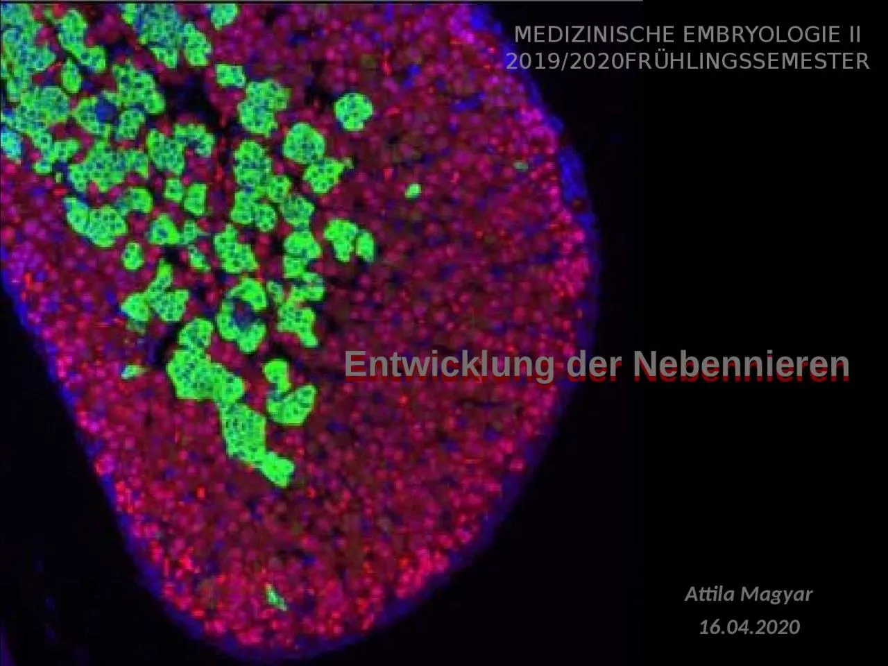
Attila Magyar 16042020 Medizinische Embryologie II 20192020Frühlingssemester The adrenals develops from the urogenital ridge on the medial and cranial ID: 928099

cells adrenal zone fetal adrenal cells fetal zone gland cortex development cell gestation zona cortisol primordium mouse human medulla

Share:

Link:

Embed:

Download Presentation from below link

Download Presentation The PPT/PDF document "  Entwicklung der Nebennieren" is the property of its rightful owner. Permission is granted to download and print the materials on this web site for personal, non-commercial use only, and to display it on your personal computer provided you do not modify the materials and that you retain all copyright notices contained in the materials. By downloading content from our website, you accept the terms of this agreement.


Presentation Transcript

Slide1


Entwicklung der Nebennieren

Attila Magyar16.04.2020

Medizinische

Embryologie

II

2019/2020Frühlingssemester

Slide2

The

adrenals develops from the urogenital ridge, on the medial and cranial part of the later mesonephric fold

Slide3

Mesonephric (m) and gonadal (g)

ridge from a mouse embryo (SEM, ed11,5). Rest of mesentery is shown by the red arrow.cranial

Slide4

medial

lateralintermedier mesoderm + coelomepithelurogenital

ridgekidneys(

mesonephric

ridge

,

metanephric

blastem

)

adrenogonadal

primordium

(

gonadal

ridge

)

adrenal

primordium

gonadalprimordium

ED 17-18

ED 24-25

ED 28-30

ED 33-35

Timetable

Early

development

of

the

adrenal

gland

Slide5

Adrenal primordium

First time it appears at the cranial and medial part of the gonadal ridgeAdrenal gland has the most complex development: its cells

derive from1. the coelomic epithel (cortex)

2.

the

intermediate

mesoderm

(

cortex

)

3.

the

mesonephric

nephrons

(

capsule)4. neural crest

cells (medulla)At first the coelomic

epithel cells migrate out of the epithel, and then

they intermingle

with mesodermal cells making cell clump: adrenal

primordium, adrenal anlage: about ED33-35

Slide6

Adrenal

gland development in humanat ED33 (a)at ED44 (b)at EW13 (c)1: aorta2: postcardinal (l) and subcardinal (m) veins (in a) or suprarenal (l) and inferior

cava veins (m; in c)3: mesonephric fold4: gonad5: sympathicoblasten

6:liver

7:

diaphragm

8:

coelom

orange

:

adrenal

anlage

brown

:

neural

crest

Slide7

Human

embryo; shortly before ED33:local thickening of the coelomic epithelium of the adreno-gonadal ridge, migration of these cells out of

the epithelial sheetin the following days:

cell

migration

to

the

cranial

pole

of

the

mesonephros

,

forming the adrenal blastem (primordium)

aorta

mesonephric ridgeroot

of mesentery

mesonephric nephron

Slide8

Adrenal primordium of a human

embryo; ED 441: adrenal primordium2: mesonephros and mesonephric duct3: stomach4: liver5: aorta6: vertebral anlage7: spinal cord8: gonadb: higher magnification of a

arrows in b: nerv fibers (left) and sympathicoblasts (right)

Neural

crest

cells

migrate

in

this

primordium

(EW5-6).

8

Slide9

Slide10

Slide11

Slide12

development of

the transitional zonemigration of trunk neural crest cellsdevelopment of the fetal and definitive

zone of the cortex;gland is encapsulated;extensive

vascularisation

adrenal

primordium

:

coelomic

and

mesodermal

cells

making

a

cell

clump

after EW22

EW 8-9

ED 33-35

Timtable

ED36-42

Embryonic and

fetal development of the adrenal gland

Slide13

12EW

fetusbetween EW8 and EW20 adrenal weight doubles weekly, from EW20 (0,5 g) up term: 8-10 gafter birth: 2 g;at the end of the puberty: 8-10 gThe rapid growth of fetal

adrenal is almost entirely due to enlargement of the FZ.

Slide14

Slide15

Structure

of fetal adrenal glandCapsule: thin

layer of connective

tissue

with

stem

cells

Cortex

:

two

regions

,

superficial

thin

layer

is

the

definitive

zone

(DZ) deeper is

the

fetal

zone

(FZ); from

the EW20 a

third

region

appears

:

the

transitional

zone

(TZ)

Medulla

as

a

discrete

strucute

exists

not

in

the

fetal

period

,

sympathicoblast

are

making

groups

,

that

lie

scattered

in

the

FZ

Proportions

of

the

different

zones

:

at

midterm

FZ

represents

75-80% of

the

gland

volume

;

at

term

FZ: 70%; DZ: 14%, TZ: 7%, „

medulla

”: 9%;

anencephalic

fetuses

:

midterm

: FZ: 57%,

at

term

FZ:

25%, DZ: 40%, TZ: 20%, „

medulla

”: 14%

Slide16

fetus

(30 EW)adult

Slide17

EW20:

fetal cortex with strangs of steroidogenic cells and wide capillaries

Slide18

EW12 (Azan

staining): 1: definitive zone 2: fetale zone3: groups of sympathicoblastEW12 (Azan staining): higher

magnification

Slide19

Cells

of the fetal cortexDefinitive

zone: smaller

,

tightly

packed

basophilic

cells

, 10-20 mm

diameter

;

many

free

ribosomes

and

glykogen

particles

;

high

proportion

of

them are

positive for

proliferation

markers, like

Ki67. At

late gestation, the cells accumulate cytoplasmic lipid

droplets and begin

to resemble

steroidogenically

active cells.

Fetal

zone

:

larger

,

acidophil

cells

20-50 mm

diameter

,

actively

synthetising

steroids

(

tubular

mitochondria

,

sER

,

lipid

drops

);

low

mitotic

activity

Differentiation

of

cortical

cells

:

During

the

entire course of pregnancy, an

intense

proliferative

activity is observed at the

periphery

of the gland, followed by

migration

of

the newly formed cells throughout the fetal

cortex. In the course of their migration, cells

differentiate, enlarge (becoming rich in steroid-filled lipid droplets) and finally die from

apoptosis in the center

of the gland

.

Slide20

Human

embryo, about ED50Definitive zone cellFetal

zone cell

Slide21

The medulla is

absent from the primate fetal adrenal as a discrete structure throughout most of gestation

except for small islands of chromaffin cells scattered through the cortex. Only after the involution of the fetal zone

during the first postnatal week

s

do the chromaffin

cells

an

aggregate

around the central vein and begin to form a

medulla

.

Fetal

medulla

EW12 (Azan

staining

):

higher

magnification

fetale zone with sympathicoblasts

Immigration of sympathicoblast

: from the EW6; TH+

Slide22

Chromaffin

cell from fetal mouse

adrenal gland

(ED17,5)

with

dendse

core

vesicles

(

arrowheads

).

Slide23

Development

of zonationThree general hypothetical models on zonation of the adrenal cortex: 1) the cell migration model; 2) the transformation field model; and 3) the zonal model. An increasing number of studies support the cell migration model of adrenal cortical cytogenesis. In this model, each zone is derived from

a common pool of progenitor cells, which then migrate and differentiate to populate the cortical zones. Centripetal migration of lipid-containing cells from the DZ to the FZ was reported in earlier studies.

Slide24

development

of zona reticularisadrenal gland at birth:fetal zone largely disappear in

several (2-4) weeks;definitive zonae glomerulosa and fasciculata

develop

;

medulla

first

appears

up

to

4.

week

from

birth

up to 4 weeks postnatal

Timtable

from 3-4 years

Postnatal development of the

adrenal gland

Slide25

Postnatal

development 1.Fetal zone: degenration

starts after

birth

(

accumulation

of

lipids

,

apoptose

,

haemorrhage

);

fetal

uzone

largely

ddisappears

up

to

6

ewwk; small

rests of fetal

zona

should remain

up

to 2.

yearFrom

the

definitve

zone

the

zonae

glomerulosa

and

fasciculata

develop

. M

orphological

studies

have identified rudimentary zonae glomerulosa and

fasciculata

(in

the

definitive

zone

)

during

late

gestation

.

The

adrenal

medulla

appears

in

the

first

few

postnatal

weeks

following

the

involution

of

the

fetal

zone

.

By

4th

week

,

the

chromaffin

cells

cluster

at

the

centre of

the

gland

.

About

12-18

month

of

age

that

the

infant

has

the

medulla

with

the

adult

type

architecture

.

Slide26

Postnatal

development 2.Zona reticularis: appears rudimental

in the 2-3

years

of

age

;

fully

developed

is

around

/

after

puberty

(11-18

years

of life).

Slide27

Steroidogenesis

in the fetal adrenal

Slide28

Early signals

for the urogenital ridgeurogenital ridge: gonad-adrenal kidney; early defects result in malformations more than one organHedgehog

signalling: DHH muation: gonadal dysgenesis,

Pallister

-Hall

syndrome

(GLI3): a

bsence

of

adrenal

gland

SALL/

Sall

signalling

:

renal abnormalities/normal

adrenals (human); kidney agenesis; absence of adrenal

gland (mouse)Fox2D: adrenal and kidney

hypoplasia (mouse)

Wnt4: unilateral absence of kidney; adrenal

not reported (human); masculinization of female

mice, compromised Sertoli

cell development, abnormal vascular pattern in gonad (female mice); decreased aldosterone

production, ectopic adrenal cells in gonadal tissue, abnormal adrenal gland vasculature

(mouse)

Pbx1: absence of gonads, kidney

hypoplasia, absence of adrenal gland

(mouse)

Slide29

Signals for

the adrenogonadal primordiumSF1/Sf1: XY sex reversal, adrenal insufficiency, adrenal hypoplasia (aplasia) (human); aplasia of

the gonads, adrenal aplasia (mouse)DAX1/Dax1:

h

ypogonadotropic

hypogonadism

,

XY

sex

reversal

,

Cytomegalic

adrenal

hypoplasia

,

persistence of fetal zone, adrenal

insufficiency (human); male infertility and XY sex reversal, lack

of X-zone regression (mouse)

Slide30

Signals for

the adrenal primordiumCited 2: KO mouse lack adrenals, but have normal gonads (adrenocortical development has a failure)

Slide31

Signals for

the fetal and neonatal adrenalDisorders of ACTH signalling: ACTH receptor mutations (MC2R, ACTHR, MRAP): adrenal insufficiency, hypoplasia with a grossly diminished or even absent ZF and are referred to as

miniature adult adrenocortical hypoplasia congenita, AHC (human);

POMC

mutations

:

Secondary

adrenal

insufficiency

(human

,

mouse

)

Slide32

Differentiation

of cortical cellsShh signalling:

secreted by

subcapsular

Sf1

+

/

SteroidogenE

-

cells

; Gli1 is

present

in

Sf1

-

/

SteroidogenE

-

subcapsular

cells

;

these

start to

migrate centripetally

and

become Sf1

+/

SteroidogenE+

and become

ZG specific

cells

(Cyp11b2

+

)

or

ZF

specific

cells

(Cyp11b1

+

)

Delta-

Notch

signalling

: DLK1 (delta

like

homolog1) is

expressed

by

subcapsular

cells

;

negatively

regulate

the

differentiation

of Gli1

+

cells

Slide33

SF1

radioactive ISH in 32 or 33 dpo (days post ovulationem) human embryosGr, gonadal ridge; m, mesonephros; asterisk, pool of SF-1 positive cells; a, adrenal cortex; g, gonad; gm, gut mesentery.

Slide34

SF1

expressioncited 2 expressionwhite arrow: gonadal

primordiumarrowhead: adrenal primordium

Slide35

white

arrow: gonadal primordium (G)arrowhead: adrenal primordium

Slide36

Slide37

?????weeks 8-10: two

different cortical cell types differentiate (fetal and definitive), a brazil cikk szerint a definitv zóna sejtjei szintetikusan inaktívak és nem osztódnak. A második trimeszter végén (22-24 hetek között) differenciálódik a tranziciós zóna. Ennek a zónának lesz 3bHSD aktivitása, ami lehetővé teszi a fetus saját kortizol termelését…. Nothing is clear about the onset of 3ß-HSD or about the factors regulating it. Surely, this event is of importance to the fetus, in that the fetal cortisol is needed for a good maturation of numerous organs (

lungs, gut, etc.).Close to birth, the definitive zone acquires 3ß-HSD but this

zone

,

being exempt

(mentes)

of cytochrome P450C17, will

secrete mineralocorticoids, which will be

needed

for

extra-uterine

life

.

Slide38

ACTH secreted by the fetal hypophysis around the 8th week of pregnancy.

At the 16th week, fetal zone cells appear to be under the control of this hormone while the definitive zone is not.However, even if ACTH is an important regulating factor of fetal adrenal functions during the entire pregnancy, it cannot explain the development of the fetal zone by itself. Indeed, anencephalic fetuses exhibit a fully constituted adrenal gland, even if reduced in the volume of the fetal zone.

Slide39

The FZ consists of large (20–50 mm) cells with ultrastructural characteristics

typical of steroidogenic cells. In the outer regions of the FZ, the cells are arranged in tightly packed cords. In the central portion, the FZ forms a reticular pattern, with cells spaced more widely and separated by numerous vascular sinusoids. Ultrastructural studies also have revealed a third zone between the DZ and FZ, which we have named the transitional zone (TZ) (11). Cells in this zone show intermediate characteristics (12). TZ cells have the capacity to synthesize cortisol, being analogous to cells of the zona fasciculata of the adult adrenal cortex. By the 30th week of gestation, the HFA cortex manifests a rudimentary form of the adult adrenal cortex; the DZ and TZ

begin to resemble the zona glomerulosa

and

the

zona

fasciculata

,

respectively

Slide40

Ishimoto: The widely

accepted concept of a key role for adrenal glucocorticoids in chromaffin cell differentiation has been challenged by a recent study in mice lacking the glucocorticoid receptor; chromaffin cells in such mice develop quite normally.

Slide41

The origin of progenitor cells during embryonic and fetal adrenal development is unclear.

Some have hypothesized that the precursors in the adrenal primordia (mouse) give rise to Sf1-negative stem cells that reside in the adrenal capsule. In response to mitogenic/morphogenic signals,

such Sf1-negative capsular stem cells would exit from the capsular niche into the subcapsular environment, where they commence Sf1 expression and proliferate.by midgestation (16–20

wk

), the FZ

clearly dominates in the gland. In contrast to the DZ, mitotic

figures in the FZ are scant. The cell number of the FZ

is not necessarily higher, but the size of it is much larger

than that of the DZ.

Apoptosis also appears to occur

in

the

HFA

,

primarily in the central portions of the FZ.(Freedman) Recent studies that used lineage tracing to

map the cell fate of Shh- and Gli1-expressing progenitor cells demonstrated radial stripes that appeared to migrate through the zG into the zF

, providing support for the model of centripetal migration.

Slide42

Slide43

the newly constituted transitional zone (TZ, medium grey) acquires the enzyme 3ß-HSD while the expression of P450C17 remains, thus allowing the production of fetal cortisol.

Near birth, cells of the definitive zone which express only 3ß-HSD, acquire the P450aldo and begin to secrete mineralocorticoids such as aldosterone. Adrenals acquire the ability to synthetise steroid hormones between weeks 6-8.Generally, the activity of steroidogenic enzymes and

regulatory proteins is higher in the fetal zone than

in

neocortex

.

Of

special

importance

are

the

studies

on

localization of the key enzymes, like 3

bHSD and 17a hydroxylase. Both are required for

the synthesis of cortizol.When 3bHSD is expressed, cell

have the

capacity to synthetize mineralocorticoids, while 17a Hase only

indicates the synhthesis of androgens.

Slide44

fetal zone:

does not express 3bHSD but 17ahydroxylase. Transitory zone (future zona fasciculata): both are expressed (from the weeks 25-30)definitive zone (future zona glomerulosa):

expresses 3bHSD from the weeks 22-24.fetal adrenal gland

secretes

cortizol

from

the

weeks

6-8: most

probably

it is

synthetised

from

placental progesterone but not from

cholesterol.Neocortex produces cortizol fetal zone DHEA and DHEA-S

What about aldosterone?CYP11A: 3-4 fold higher activity in

the fetal

zona than in the definitivequantities of secreted steroids

from tha fetal adrenals: fetal zone: DHEA and DHEA-S 200 mg/

day at

term (in both sexes); it starts at weeks 6-8aldosteron

secretion starts at weeks 20-24.

Slide45

Slide46

Slide47

Ishimoto cikk friss összefoglalása: The current data regarding the ontogeny of expression

of steroidogenic components could account for zonal differential steroidogenic activity and its onset. The HFAcortex after midgestation is composed of three functionally distinct zones, each of which expresses different combinations of steroidogenic enzymes and cofactors (Fig. 3): 1) the DZ, which is the likely site of aldosterone synthesis late in gestation because of the persistent lack of CYP17, and the eventual expression of HSD3B2, CYP11A, CYP21, and probably of CYP11B2; 2) the TZ, which appears to be the site of cortisol production late in gestation based on the persistent expression of CYP17, CYP11A, CYP21, and the eventual expression of HSD3B2, and probably of CYP11B1; and 3) the FZ, which expresses

CYP11A and CYP17 but not HSD3B2, is the site of 5-steroid production, particularly DHEA and DHEAS, throughout most of gestation. The localization and ontogeny of the steroidogenic enzymes and cofactors fit well with the concept that the DZ develops to form the zona glomerulosa, the TZ is the equivalent of the zona

fasciculata

,

and the FZ is analogous to the zona reticularis.

Slide48

steroidogenic activity in

the HFA is summarized as follows: 1) DHEAS productionappears to begin at around 8–10 wk gestation, continues thereafter, and increases considerably during the second and third trimesters, such that by term the HFA produces around 200 mg of DHEAS per day; 2) de novo cortisol production likely occurs transiently early in gestation (around 7–10 wk gestation); 3) due to the lack ofHSD3B2 expression, de novo cortisol biosynthesis appears to be suppressed until late gestation when cortisol

production escalates (76 –78); and 4) aldosterone synthesisin the HFA may be suppressed during midgestation due to the probable lack of CYP11B2 expression, but likely becomes active by term.

Slide49

Slide50

Developmental abnormalities of

theadrenal glandEctopic adrenal tissue (adrenal rest): The adrenal rests may contain cortical tissue only or other cortical and medullary tissue. Ectopic adrenal cortical tissue is found in up to 50% of neonates and usually atrophies, so that it only occurs in 1% of adults. Most ectopic

adrenal tissue is found in the vicinity of the adrenals around the coeliac axis.Adrenal heterotopia (

very rare

):

ac

c

essory

adrenal issue is incorporated into

adjacent organs such as kidney or liver.

Congenital

adrenal

hyperplasia

(CAH)

:

a group of autosomal recessive disorders characterized by enzyme defects in the pathway of cortisol synthesis

. Deficient cortisol production leads to increased ACTH production and hyperplasia of the

adrenal cortex. fetal adrenal cortex cannot synthesize adequate amounts of cortisol. The suppressed cortisol inhibits negative feedback at the fetal anterior pituitary, which

leads to a compensatory increase in ACTH secretion. The elevated ACTH causes fetal adrenal hyperplasia and increases

production of DHEAS because its biosynthesis is not affected by CYP21 deficiency. In the first trimester when sexual differentiation occurs, there is a relative lack of aromatase (CYP19) activity in contrast to high placental

aromatase activity seen later in gestation. Thus, the primary clinical manifestations of CYP21 deficiency are those of androgen excess, which are first expressed in utero, resulting in virilization

of the external genitalia of female

fetuses.