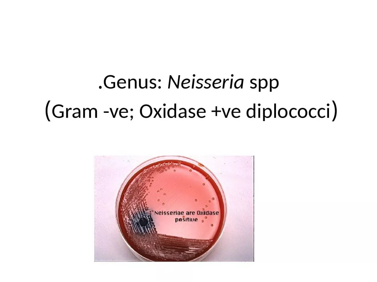
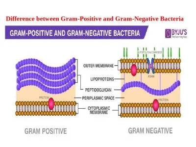
Neisseria spp Grams negative Diplococci Kidney shape with flat opposing edge Neisseria spp PATHOGENIC N gonorrhoea gonococci GC Gonorrhoeae N meningitidis ID: 1001248
Download Presentation The PPT/PDF document "Genus: Neisseria spp. (Gram -ve; Oxida..." is the property of its rightful owner. Permission is granted to download and print the materials on this web site for personal, non-commercial use only, and to display it on your personal computer provided you do not modify the materials and that you retain all copyright notices contained in the materials. By downloading content from our website, you accept the terms of this agreement.
1. Genus: Neisseria spp.(Gram -ve; Oxidase +ve diplococci)
2. Neisseria spp.Gram’s negative Diplococci Kidney shape with flat opposing edge
3. Neisseria spp.PATHOGENIC:N. gonorrhoea (gonococci, G.C) Gonorrhoeae.N. meningitidis (meningococci) meningitis. NON-PATHOGENIC:N. Catarrhalis Normal flora of upper respiratory tract
4. General characteristics of Neisseria spp.Aerobic to facultatively anaerobic Gram –ve cocci Arrange in pairs (diplococci) with adjacent sides flattened (like coffee beans)Neisseria meningitidis is encapsulated whereas Neisseria gonorrhea is not Oxidase +ve / catalase +ve Non motile / non hemolytic
5.
6. N. gonorrhoeae (gonococcus)Very delicate and fastidious, requiring increased carbon dioxide tension (3% to10%) for cultivation. Narrow optimal temperature range, between 35° and 37°C and fails to grow much above or below these temperatures. it may remain viable in dried pus for several weeks.
7. Diagnosis:Swab sample. A swab sample from the part of the body likely to be infected (cervix, urethra, penis, rectum, or throat) can be sent to a lab for testing: 1-Gram stain: A sample from the urethra or a cervix is placed on a slide and stained with Gram stain. The cells appear as Gram negative cocci form masses with adjacent cells (i.e. in diploid forms),having a bean-shape, a coffee bean-shape, or Kidney-shape. These cells are seen intracellularly (inside polymorphonuclear leucocytes), or extracellularly.N. gonorrhoeae
8.
9. Laboratory DiagnosticGram stain : G – ve, diplococci Intracellular &/or extracellular in polymrphonuclar cells.Gram’s staining slide for urethral discharge
10. 2-Urine test. Gonorrhoea in the cervix or urethra can also be diagnosed with a urine sample sent to a lab. 3-Oxidase Test - It is a hallmark test for the Neisseria. The oxidase test determines the presence or absence of cytochrome C. Organisms containing cytochrome C as part of their respiratory chain are oxidase positive and turn the reagent purple; organisms lacking cytochrome C as part of their respiratory chain do not oxidize the reagent, leaving it colorless within the time limits of the test, and are oxidase negative.
11.
12. Laboratory DiagnosticPathogenic fastidious need chocolate agar +5-10% CO2 , selective media Selective media for NeisseriaA. Thayer Martin Medium (TM) :( Chocolate agar + Enrichment element +Antibiotics : Colistin (against G – ve rods)Vancomycin (against G + ve)Nystatin (against fungi)B. Modified Thayer Martin:{same as TM + Trimethoprim (against G –ve especially swarming proteus)}.white-gray colonies(N. gonorrhoea on chocolate agar)4-Culture
13. Candle jar 5%CO2
14. Neisseria meningitidis (Meningococcus)Meningococcus is similar to the gonococcus in morphology and staining reactions. Neisseria meningitidis is Gram-negative diplococcal bacterium fastidious in growth requirements and may be cultivated on blood agar or chocolate agar on which it develops relatively large, gray, smooth, raised colonies. No hemolysis is produced on blood agar. Neisseria meningitidis is the Causative agent of meningococcal meningitis, meningococcemia, and bacterial endocarditis. It is an obligate human pathogen.
15. Diagnosis:Specimen: The gold standard of diagnosis is isolation of N. meningitidis from sterile body fluid. Blood, CSF specimen is sent to the laboratory immediately for identification of the organism. It may be isolated from nasopharynx, joint fluid, and from petechiae of the skin.Gram stain: show adjacent coffee bean-shape, Gram negative cells (diplococci) with flattened surfaces facing each other. Culture: culturing the organism on Thayer-Martin agar. Oxidase test: (all Neisseria show a positive reaction)
16. 4-Sugar fermentation tests:important in differentiation between Neisseria.carbohydrates; maltose, sucrose, and glucose test in which N. meningitidis will utilize the glucose and maltose.+ve-ve- ve+ve+ve- veCarbohydrate fermentation set for Gonococci Carbohydrate fermentation set for Meningococci
17. DIFFERENTIATION OF THE NEISSERIAEDifferent Neisseria species can be identified by the sets of sugars from which they will produce acid. For example, N. gonorrhea makes acid from only glucose, however N. meningitidis produces acid from both glucose and maltose.NeisseriaeGrowth CharacteristicsSugar fermentationGlucoseMaltoseSucrose Lactose N. GonorrheaGrowth on enriched media at 37°Cin CO2 jar; colonies similar toN. meningitides, but smaller andgrayish white + _ __ N. meningitidis Growth on enriched media at 37°Cin CO2 jar; colonies smooth,glistening, translucent, soft, orbluish gray++__
18. Laboratory Diagnostic5-Srology: determines the group of the isolated organism.If the organism reaches the circulation, then blood cultures should be drawn and processed accordingly. PCR; polymerase chain reaction tests can be used to identify the organism even after antibiotics have begun to reduce the infection
19.
20. Thank you