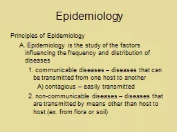
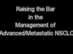
NSCLC nonsmall cell lung cancer Lung cancer incidence and mortality 1 One of the most common cancers with 2 million new cases worldwide in 2018 The most common cause of cancer death causing nearly ID: 913384
Download Presentation The PPT/PDF document "NSCLC: Epidemiology and disease charact..." is the property of its rightful owner. Permission is granted to download and print the materials on this web site for personal, non-commercial use only, and to display it on your personal computer provided you do not modify the materials and that you retain all copyright notices contained in the materials. By downloading content from our website, you accept the terms of this agreement.
Slide1
NSCLC: Epidemiology and disease characteristics
NSCLC, non-small cell lung cancer.
Slide2Lung cancer incidence and mortality1
One of the most common cancers, with
2
million new cases worldwide in 2018
The most common cause of cancer death, causing nearly one in five of all cancer deaths worldwide in 2018
Male
Female
1.
Bray F,
et al.
CA Cancer J
Clin
2018;
doi
: 10.3322/caac.21492. [
Epub
ahead of print].
Slide3Global distribution of lung cancerIn 2018, there were approximately
2,094,000 new cases
of lung cancer worldwide; incidence is estimated to
increase by 38% by 20302The geographical pattern of lung cancer incidence generally reflects historical patterns in tobacco use3
Temporal and geographical patterns of mortality mirror those of incidence owing to lung cancer’s high fatality rate (mortality-to-incidence ratio: 0.87)3,41. International Agency for Research on Cancer, World Health Organization. http://gco.iarc.fr/today/data/factsheets/cancers/15-Lung-fact-sheet.pdf (Accessed: 24 April 2019). 2. International Agency for Research on Cancer, World Health Organization. http://gco.iarc.fr/tomorrow/graphic-isotype (Accessed 24 April 2019); 3. International Agency for Research on Cancer, World Health Organization. http://gco.iarc.fr/today/data/pdf/fact-sheets/cancers/cancer-fact-sheets-11.pdf (Accessed: 24 April 2019). 4. Wong MC, et al. Sci Rep. 2017;7(1
):14300.
Global incidence and mortality rates
1
Male
Female
60
0
40
20
20
4060
Incidence
Age standardised rates (% per 100,000)
Mortality
Micronesia
Polynesia
Central and Eastern Europe
Eastern Asia
Western Europe
Southern Europe
North America
Western Asia
Northern Europe
Australia/New Zealand
South-Eastern Asia
Southern Africa
Caribbean
Melanesia
Northern Africa
South America
South-Central Asia
Central America
Middle Africa
Eastern Africa
Western Africa
Slide45-year
survival trends for various cancers
1
Breast
Colorectal
Lung
Survival rates for lung
cancer are
generally low
*Data for
2015 are predicted
based on the modelled
trend..
Percentage
(%)
Prostate
High unmet medical need
Survival
rates vary depending on stage at diagnosis.
The later the stage of diagnosis, the lower the 5-year
survival rate
2
1. National Cancer Institute Surveillance, Epidemiology, and End Results (SEER). https://seer.cancer.gov/statfacts/ (Accessed: 07 December 2018
). 2.
Ridge CA, et al.
Semin
Intervent
Radiol
2013;30(2):93–8
.
Slide5Stage of diagnosis affects 5-year survival rate
Almost 60% of patients with lung cancer
have late-stage disease at diagnosis
The 5-year survival rate is low
in patients with late-stage disease
1. National Cancer Institute Surveillance, Epidemiology, and End Results (SEER). https://seer.cancer.gov/statfacts/html/lungb.html (Accessed: 07 December 2018).
Slide6Is there a ‘typical’ lung cancer patient?EGFR, epidermal growth factor receptor; US,
United States of America
.
Lung cancer mortality in women, including young women in some countries,
has been on the rise1-4Reflecting changes in the relative incidence of lung cancer in women versus men,5 there is a recent report that incidence may now be higher in young women than young men in the US6
A higher proportion of lung cancer cases occur in never-smokers in
Asian countries than in
Western countries7
In patients with adenocarcinoma histology, mutations in
EGFR are more
prevalent in Asian populations than elsewhere8
Traditionally, the ‘typical’ lung cancer patient was thought to be an older
male with
a history of smokingHowever, recent studies have demonstrated the trend is shifting:
1.Malvezzi M, et al. Ann Oncol 2017;28(5):1117‒23. 2. Martin-Sanchez JC, et al. Cancer Epidemiol
2017;49:19–23. 3. Martin-Sanchez JC, et al. Cancer Res 2018;78(15):4436–42. 4. Levi F, et al . Int J Cancer 2007;121(2):462‒5. 5. Kozielski
J, et al. Contemp Oncol (Pozn) 2012;16(5):413–5. 6. Jemal A, et al, N Engl J Med 2018;378(21):1999–2009. 7. Toh CK, Lim WT. J Clin Pathol 2007;60(4):
337–40. 8. Midha A, et al. Am J Cancer Res. 2015;5(9):
2892‒911.
Slide7Tobacco use is the most important risk factor in lung cancer1
*Mortality rate trends over five years from 1970‒1974 to 2005‒2009 plus the year 2012 and predicted rates for 2017.
EU, European Union
.
Tobacco smoking
is the most important cause of lung cancer
Rates of lung cancer deaths attributable to smoking
vary from
>80%
in the US and France to
61%
in Asia and
40%
in sub-Saharan
Africa
1
Over the last few decades lung cancer
mortality rates
in the EU have been
decreasing in men
but
increasing in women
,
reflecting
a later
decline in smoking prevalence among
women
1,2
Trends in
lung cancer mortality rates
in a
country generally
follow trends in smoking
pre
val
ence
, with lung cancer trends lagging
by 20–30
years
1
A
ge-
standardised
EU male and female lung cancer mortality rates
2
*
1.
Islami
F, et al.
Transl
Lung Cancer Res 2015;4(4):
327–38. 2.
Malvezzi
M, et al. Ann
Oncol
2017;28(5
):
1117‒23.
Slide8There are two main types of lung cancer1. Zappa C & Mousa SA.
Transl
Lung Cancer Res 2016;5(3):288–300. 2
. Lozić AA, et al. Coll Antropol 2010;34(2):609–12.
There are
two main types
of
lung cancer
1
15%
Small cell
lung cancer
(SCLC)
85%
Non-small cell
lung cancer
(NSCLC
)
NSCLC usually
grows
and
spreads
more
slowly
than SCLC
2
Slide9NSCLC is genomically diverse
NSCLC
, non-small cell lung
cancer.
ALK
HER2
BRAF
PIK3CA
AKT1
MAP2K1
NRAS
ROS1
RET
EGFR
KRAS
Unknown
Driver mutations in
adenocarcinoma
2
Histological distribution of NSCLC
1
1. National Cancer Institute. Surveillance, Epidemiology and End Results Program. https
://seer.cancer.gov
(Accessed May 2019). 2. Li
T, et al. J
Clin
Oncol 2013;31(8):1039–49.
Driver mutations in
squamous cell
carcinoma
2
Slide10Some common NSCLC symptoms
NSCLC, non-small cell lung cancer
.
Irrespective of metastases1–3
Mortality rates are greatly improved when lung cancer is diagnosed earlyMetastatic NSCLC3,4Symptoms may vary widely and often coincide with the site of tumour metastasisWorsening long-term cough
Common symptoms
Chronic cough
Wheezing or shortness of breath
Hoarseness
Loss of appetite
Weight loss
Recurrent pneumonia
Haemoptysis
Fatigue
Constant chest pain
Recurrent bronchitis
Lumps near the surface of the body (lymph nodes), often in the neck or above the collarbone
Common symptoms
Jaundice
Dizziness
Seizures
Headaches
Bone pain
Bleeding or blood clots
Weakness or
numbness of
the
arms
or legs
1. MedlinePlus Medical Encyclopedia. http://www.nlm.nih.gov/medlineplus/ency/article/007194.htm (Accessed
: 07 December 2018).
2. Thomas KW. UpToDate. http://www.uptodate.com/contents/lung-cancer-risks-symptoms-and-diagnosis-beyond-the-basics (Accessed:
07 December
2018
).
3. WebMD. http://www.webmd.com/lung-cancer/lung-cancer-symptoms (Accessed:
07 December 2018).
4
. American Cancer Society. http://www.cancer.org/cancer/lungcancer-non-smallcell/detailedguide/non-small-cell-lung-cancer-signs-symptoms (Accessed: 07 December 2018).
Slide11Diagnostic workup of NSCLC: laboratory evaluation and imaging1
CNS, central nervous system; CT, computed tomography; FDG, fluorodeoxyglucose; MRI, magnetic resonance imaging; PET, positron emission tomography
.
Standard tests, including routine haematology, renal and hepaticfunction, and bone biochemistry
LaboratoryCT scan of chest and upper abdomen; complete assessment of liver, kidneys and adrenal glandsRadiology
CNS imaging (MRI [more sensitive] or CT scan with iodine contrast) if available; required in patients with neurological symptoms
If bone metastases suspected: PET, ideally coupled with CT, and bone scans. PET/CT is most sensitive for detecting bone metastases. MRI as needed
Assessment of mediastinal lymph nodes and distant metastases:
FDG–PET/CT scan offers highest sensitivity
1.
Planchard
D, et al. Ann
Oncol 2018;29(Suppl. 4):iv192–iv237.
Slide12NSCLC is most often diagnosed at an advanced stage
NSCLC, non-small cell lung cancer
.
Many of the symptoms that do appear with more advanced disease can be mistaken for other illnesses4
develop some symptoms3Early lung cancer may not cause any symptoms2
Early symptoms that may be difficult to notice include:
have
no symptoms
when lung cancer is diagnosed
25% 75%
Persistent
cough
Shortness
of breath
Dull and persistent pain in the chest
Repeat
infections, such as bronchitis or pneumoniaAlmost 60%
of patients with lung cancer have late-stage disease at diagnosis1
1. National Cancer Institute Surveillance, Epidemiology, and End Results (SEER). https://seer.cancer.gov/statfacts/html/lungb.html (Accessed:
07 December 2018). 2. MedlinePlus Medical Encyclopedia. http://www.nlm.nih.gov/medlineplus/ency/article/007194.htm (Accessed:
07 December 2018
). 3.
WebMD. http://www.webmd.com/lung-cancer/lung-cancer-symptoms (Accessed:
07 December 2018
). 4.
American Cancer Society. http://www.cancer.org/cancer/lungcancer-non-smallcell/moreinformation/lungcancerpreventionandearlydetection/lung-cancer-prevention-and-early-detection-early-detection (Accessed:
07 December 2018
).
Slide13Lung cancer staging and TNM classification1
The most frequently used system to stage lung cancer is the American Joint Committee on Cancer TNM system, which is based on
:
The size and extent of the primary tumour (T)Whether the cancer has spread to nearby (regional)lymph nodes (N)Whether the cancer has metastasised (M) to other organs of the bodyOnce the T, N and M categories have been defined,
this information is combined to assign an overall stage of 0, I, II, III or IVThis process is called stage groupingIt produces a range of anatomical stage or prognostic groups (right)T1mi, minimally invasive adenocarcinoma; Tis, tumour in situ; TNM, tumour, node, metastasis.
1. American Joint Committee on Cancer. Lung cancer staging.
8th ed. 2017.
https://cancerstaging.org/references-tools/Pages/Cancer-Staging-Resources.aspx (Accessed: 07 December 2018
).
Slide14Lung cancer TNM classification explained1
*There is no designation of MX. The absence of any clinical history or physical findings suggestive of metastases in a patient who has not undergone any imaging is sufficient to assign the clinical M0 category. There is no designation of pM0. Biopsy or other pathological information is required to assign the pathological M1 category. Patients with a negative biopsy of a suspected metastatic site are classified as clinical M0 (cM0).
cM
, clinical metastasis; pM, pathological metastasis; TNM, tumour, node, metastasis.
1. American Joint Committee on Cancer. Lung cancer staging. 8th ed. 2017. https://cancerstaging.org/references-tools/Pages/Cancer-Staging-Resources.aspx (Accessed: 07 December 2018).
Slide15SummaryLung cancer is one of the most common cancers, with ~2 million new cases worldwide in 20181There are two main types of lung cancer: SCLC and NSCLC
2,3
NSCLC usually grows and spreads more slowly than
SCLCNSCLC comprises adenocarcinoma and squamous cell carcinoma4Major driver mutations in adenocarcinoma include EGFR and KRASThe majority of patients with lung cancer have late-stage disease at time of diagnosis5–7The
TNM system, is used for staging, taking into account the size and extend of the primary tumour (T), whether it has spread via the lymph nodes (N) or metastasised (M)The 5-year lung cancer survival rate is low in patients with late-stage diseaseNSCLC, non-small cell lung cancer; SCLC, small cell lung cancer.1. Bray F, et al. CA Cancer J Clin 2018; doi: 10.3322/caac.21492. [Epub ahead of print]. 2. Zappa C & Mousa SA. Transl Lung Cancer Res 2016;5(3):288–300. 3. Lozić AA, et al.
Coll Antropol 2010;34(2):609–12. 4. Li T, et al. J Clin Oncol 2013;31(8):
1039–49. 5. National Cancer Institute Surveillance, Epidemiology, and End Results (SEER). https://seer.cancer.gov/statfacts/html/lungb.html (Accessed: 07 December
2018). 6. American Joint Committee on Cancer. Lung cancer staging. 8th ed. 2017. https://cancerstaging.org/references-tools/Pages/Cancer-Staging-Resources.aspx (Accessed: 07 December 2018
). 7. Ridge CA, et al. Semin Intervent
Radiol 2013;30(2):93–8.