/
H.RIAHI, B.SOUISSI, I.OMRI, K.BOUAZIZ, O.AZAIZ, M.BEN MESSAOUD, R.ALLANI, H.RIAHI, B.SOUISSI, I.OMRI, K.BOUAZIZ, O.AZAIZ, M.BEN MESSAOUD, R.ALLANI,

H.RIAHI, B.SOUISSI, I.OMRI, K.BOUAZIZ, O.AZAIZ, M.BEN MESSAOUD, R.ALLANI, - PowerPoint Presentation

sophia
sophia . @sophia
Follow
64 views
Uploaded On 2023-12-30

H.RIAHI, B.SOUISSI, I.OMRI, K.BOUAZIZ, O.AZAIZ, M.BEN MESSAOUD, R.ALLANI, - PPT Presentation

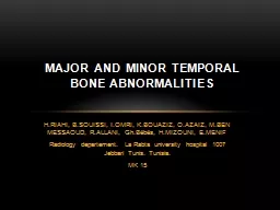
GhBébés HMIZOUNI EMENIF Radiology departement La Rabta university hospital 1007 Jebbari Tunis Tunisia MK 15 MAJOR AND MINOR TEMPORAL BONE ABNORMALITIES INTRODUCTION In contrast to anatomical variants congenital malformations CM synonyms are abnormalities or ID: 1036190

canal ear auditory vestibular ear canal vestibular auditory patients week cavity cochlear external cochlea scc abnormalities malformations middle vestibule

Share:

Link:

Embed:

Download Presentation from below link

Download Presentation The PPT/PDF document "H.RIAHI, B.SOUISSI, I.OMRI, K.BOUAZIZ, O..." is the property of its rightful owner. Permission is granted to download and print the materials on this web site for personal, non-commercial use only, and to display it on your personal computer provided you do not modify the materials and that you retain all copyright notices contained in the materials. By downloading content from our website, you accept the terms of this agreement.


Presentation Transcript

1. H.RIAHI, B.SOUISSI, I.OMRI, K.BOUAZIZ, O.AZAIZ, M.BEN MESSAOUD, R.ALLANI, Gh.Bébés, H.MIZOUNI, E.MENIFRadiology departement. La Rabta university hospital 1007 Jebbari Tunis. Tunisia.MK 15MAJOR AND MINOR TEMPORAL BONE ABNORMALITIES

2. INTRODUCTION In contrast to anatomical variants, congenital malformations (CM) – synonyms are abnormalities or dysplasias – are not only characterized by a deviation from normal anatomical development, but also from regular function. They can result from a developmental arrest, irregular embryogenesis or from both due to spontaneous genetic mutations – this is the case in the majority of CM of the ear –, genetic transmission, and exogenic factors—in about 10% of cases. CM of the ear may be found in syndromes—the most known of them are the Franceschetti (Treacher-Collins), Goldenhar and Klippel-Feil syndrome.

3. Materials and patientsRetrospective study during 48 months from July 2008 to June 2011 .50 patients were explored with MDCT including 26 women 24 menAll of them consulted for sensorineural hearing loss.

4. Exploration method CT examinations of the temporal bone are performed at our institution by using a GE 64 multislices CT without injection of iodinated contrast: 50 patients so 100 ears Acquisition sub-millimeter volume Axial reconstruction: lateral semicircular canal plan Perpendicular coronal reconstructions Oblique reconstructions: Reconstructed double-oblique coronal image shows normal anatomy of the long axis of the incudal body , double-oblique orientation of the axial reconstruction plane enables the optimal display of both the crura and the stapedial footplate at the oval window, Oblique coronal planes located along the long axes of malleus and incus

5. The following structures were evaluated in 100 temporal bones: The external auditory canal: stenosis or atresia of the cartilaginous part of the auditory canal, stenosis or atresia of the bony part, The extent of the middle ear cavity, form of tegmen.Diminution, dysplasia, rotation, fusion, ectopia, tympanic wall adherenceOssicular chain dysplasia, fusion, absenceLabyrinthine windows open or closed; fistula.Cochlear turns, vestibule, semicircular canals, aqueducts.The internal auditory canal Facial nerve canal: aberration, dehiscence, hypoplasia, thickening, splitting.

6. resultsExternal and medial ear deformity:structurenombreExternal auditory canal9 (in 9 patients)Tympanic cavity3 (3 patients)Oval/ round windows24 (15 patients)malleus24 (19 patients)incus31 (23 patients)stapes34 (24patients)

7. Inner ear deformitystructurenombreMichel4(2patients)Cochlear dysplasia8 (8 patients)Vetibule dysplasia22 (12patients)Semicircular canal60 (41 patients)Vestibular aqueduct10 (6 patients)Internal auditory canal17 (10patients)

8. EMBRYOLOGYIn the 3rd week of gestation: formation of the otic placode, an ectodermal thickening in the neighborhood of the myelencephalon. Invagination of the otic placode leads to an otic pit and to fusion of their external lips to the otic vesicle. In the 8th week, the otic capsule is formed, providing the stapes footplate and the ligament of the oval window. By the 12th week, the labyrinth is well differentiated. The facial nerve grows to reach its destinations between the 4th and 5th week.

9. The middle and external ear develops from the mesodermal first and second branchial arch and the endodermal first pharyngeal pouch between the fourth and 30th weeks. Developmental anomalies of the first pharyngeal pouch lead to disturbances of the eustachian tube and of the tympanic and mastoid pneumatization. Failure of differentiation of the first branchial arch leads to malformations of the incudomalleal joint, tensor tympani muscle, and mandible. Failure of differentiation of the second branchial arch affects the facial nerve canal, the stapedius muscle, the lower part of the ossicular chain, and the styloid process.

10. Disorders of the first and second branchial arches also result in dysplasia of the auricular cartilage (leading to microtia in the seventh to eighth week, the earlier the more severe, or to anotia in the seventh week).The external auditory canal arises from deepening of the first branchial groove in the 9th week. Opening of the bony part of the external auditory canal starts only in the 30th week, after complete differentiation of the inner, middle, and outer ear. Failure of the epithelial cells of the first branchial groove to split causes stenosis or atresia of the external auditory canal, which might be isolated in an otherwise normal temporal bone. Anomalies of the internal carotid artery are thought to be caused by maldevelopment of the third branchial arch during the 4th week

11. DISCUSSIONMost inner ear malformations arise when formation of the membranous labyrinth is interrupted during the first trimester of pregnancy.Only approximately 20% of patients who present with congenital SNHL have imaging manifestations.Malformations of the inner ear may be associated with various other embryological abnormalities: cervical fistula, renal abnormalities in the brachio-oto-renal syndrom.INNER EAR DEFORMITY

12. Schematic representation of different stages of developmental arrest in the inner ear development. The letters a and b represent sections through the internal auditory canal and round window, respectively.

13. A: cochlear aplasia/deformityNormal cochleaMichel deformity: complete labyrinthine aplasia.Vestibule and semicircular canal dysplasiaCochlear aplasiaEnlarged vestibular aqueduct.Common cavity.Incomplete partitionCochlear hypoplasia

14. Michel deformityComplete absence of all cochlear and vestibular elements.Rarely unilateral.Associated lesions:Aberrant facial nerve Absent stapesAbnormality of the skull baseAberrant internal carotidstapedial artery

15. Cochlear aplasiaThis is seen as dense otic bone on the anterior part of the IAC. Because of the absence of the cochlea, the course of the labyrinthine segment of the facial canal is more anterior at the usual location of the cochlea.It is important to differentiate this malformation from cochlear ossification in which the basal turn of the cochlea produces the characteristic bulging in the middle ear(the promontory).

16. Common cavity.cochlea and vestibule form a common cavity, which represents both of these structures without any differentiation.Usually, the internal auditory canal enters the common cavity at its center.

17. Incomplete partitionCystic dilated vestibule accompanied the cystic, empty cochlea.the dimensions of the cochlea and vestibule are normal but the internal architecture is missing. There is no modiolus in the cochlea, giving it the shape of an empty cystic structure.Usually, the cribriform area between the cochlea and the IAC is also defective.

18. Cochlear hypoplasia Cochlear and vestibular structures are separate from each other.In hypoplasia the cochlea is smaller than normal.Only the basal turn is developed, the other turns form one commun cavity.The accompanying vestibular malformation is usually absence or hypoplasia.

19. VESTIBULE and semicircular canal dysplasiaSCC malformations also frequently occur in association with other inner ear anomalies.The SCCs begin as disk-shaped evaginations arising from the vestibular appendage in the 6th gestational week.

20. The superior SCC is the first to form, followed by the posterior and then the lateral SCC. Therefore, superior and posterior SCC anomalies are almost invariably associated with anomalies of the lateral SCC, whereas abnormalities of the lateral SCC can occur in isolation.On axial CT images, subtle SCC abnormalities may be indicated by a small or enlarged lateral SCC bony island. Normally, the transverse diameter of this bony island measures between 2.6 and 4.8 mm

21. Large vestibuleThere appears to be a reciprocal relationship between the size of semi circulars canals particularly the lateral canal and the size of the vestibule.An enlarged vestibule is associated with underdeveloped lateral semi-circular canal

22. Enlarged vestibular aqueductVestibular aqueduct enlargement (also referred to as enlarged vestibular aqueduct syndrome) is the single most common inner ear anomaly seen in individuals with SNHL.It is frequently seen in association with other inner ear anomalies( vestibular enlargement, and SCC dysplasias)On CT, vestibular aqueduct enlargement is diagnosed when the aqueduct is 1.5 mm in width (roughly the diameter of the simultaneously visualized posterior SCC) at the midpoint between the common crus and its external aperture. MR imaging additionally demonstrates enlargement of the endolymphatic duct and sac.

23. Small internal auditory canalThis deformity is characterized by a decrease in the diameter of the IAC Presumably, this hypoplastic changes are secondary to hypooplasia or aplasia of the cochlear nerve

24. EXTERNAL AND MIDDLE EAR DEFORMITYMany classificationsModerate malformationSevere malformationElementary lesions:External acoustic canalMiddle earossicular chain

25. External auditory canal Failures of the canalisation result in EAC malformations including membranous and/or bony atresia. The classification of malformations of the EAC according to Weerda EAC stenosis type A: a marked narrowing of the EAC with an intact skin layer. EAC stenosis type B: a partial development of the EAC with an atresia plate at the medial part.Type C: complete bony EAC atresia.

26. Middle ear Middle ear malformations can affect the normal development of the tympanic cavity as well as ossicles and form a continuum with varying degrees of involvement. This is linked with a change in configuration or size of middle ear spaces:Mastoid pneumatization may be reduced or absent. Tympanic cavity: may be hypoplasia: the cavity is shorter and or smaller than normal; split-like deformed: then often opacified and filled with non-resorbed embryonic or fibrosing tissue aplasia:absence extracavitation: an additional cavity on a wrong place There may be abnormalities of the oval window and, rarely, of the round window.

27. ossicular chainMalleus: fused malleus and incus fixation of malleus and/or incus on the tympanic wall aplasia of the head of the malleus

28. Incus:No or hypoplasic incus long processIncus fused to medial attic wallNo connexion with malleusincus long process fused to promontriumFused incus and stapes

29. Stapes:Stapes without anterior crus or stapes suprastructure Stapes with monopolar crusStapes footplate fixationNo stapes footplate

30. CONCLUSIONWell-established temporal bone abnormalities such as cochlear and vestibular abnormalities and a grossly enlarged vestibular aqueduct are significantly found in patients with sensorineural hearing loss. CT and MRI allow labelling from isolated malformations or from simple morphological considerations to guide to an abnormal type geyser, a syndrome branchio-oto-renal or load before any result would then specify the genetic disorder in question.