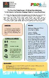
/
Department of Surgical Oncology Suleyman Demirel University Faculty o Department of Surgical Oncology Suleyman Demirel University Faculty o

Department of Surgical Oncology Suleyman Demirel University Faculty o - PDF document

sophie
sophie . @sophie
Follow
342 views
Uploaded On 2022-08-24

Department of Surgical Oncology Suleyman Demirel University Faculty o - PPT Presentation

Ulus Travma Acil Cerrahi Derg January 2016 Vol 22 No 1 Address for correspondenceOktay Karaköse MDSüleyman Demirel Üniversitesi T28p Fakültesi Cerrahi Onkoloji Bilim Dal28 32260 I ID: 941020

bile perforation duct spontaneous perforation bile spontaneous duct 150 cerrahi abdomen case acute diagnosis common determined hepatic tube acil

Share:

Link:

Embed:

Download Presentation from below link

Download Pdf The PPT/PDF document "Department of Surgical Oncology Suleyman..." is the property of its rightful owner. Permission is granted to download and print the materials on this web site for personal, non-commercial use only, and to display it on your personal computer provided you do not modify the materials and that you retain all copyright notices contained in the materials. By downloading content from our website, you accept the terms of this agreement.


Presentation Transcript

Department of Surgical Oncology, Suleyman Demirel University Faculty of Medicine, Isparta, TurkeyDepartment of General Surgery, Suleyman Demirel University Faculty of Medicine, Isparta, TurkeyABSTRACTSpontaneous extrahepatic bile duct perforation is generally seen in infants. Although rarely seen in adults, it may be seen with fatal bile peritonitis. Therefore, for a patient presenting with acute abdominal symptoms, differential diagnosis must be made with radiological imaging such as abdominal ultrasonography or computed tomography, without any loss of time. In these imaging tests, in cases of gallstone disease together with perihepatic free �uid or choledocus which can not be monitored, it should be considered in the differential Ulus Travma Acil Cerrahi Derg, January 2016, Vol. 22, No. 1 Address for correspondence:Oktay Karaköse, M.D.Süleyman Demirel Üniversitesi Tp Fakültesi, Cerrahi Onkoloji Bilim Dal, 32260 Isparta, TurkeyTel: +90 246 - 211 20 00/2224 E-mail: oktaykarakose@gmail.com Qucik Response CodeUlus Travma Acil Cerrahi Dergdoi: 10.5505/tjtes.2015.95142Copyright 2016 Pülat et al. A rare cause of acute abdomen: spontaneous common hepatic duct perforationthe pancreas parenchyma consistent with ischaemia or necrosis.The patient was admitted for surgery with an initial diagnosis of bile duct perforation. In the exploration, there was widespread bile within the abdomen. Following dissection of adhesions in the subhepatic area, it was seen that the hydropic and wall thickness of the gallbladder had increased. Millimetric stones were palpated in the gallbladder neck. In the continuation of the exploration, a 2 cm ischaemic area was determined in the anterior of the common hepatic canal and it was here that the perforation had occurred (Fig. 1c). There was slight oedema of the head of the pancreas, but no haemorrhagic or necrotising appearance in the pancreas. After cleaning within the abdomen, and cholecystectomy, the distal choledocus and proximal bile ducts were checked with choleducus coils. As no additional pathology, such as choledocus stone, papillary stenosis or choledocal cyst was determined, a T-tube drain was placed from the perforation area to the common hepatic canal and the operation was terminated. On the postoperative twenty-rst day, the passage to the duodenum was seen to be normal on cholangiography applied from the T-tube, and the T-tube was removed. Pathological examination of cholecystectomy determined ulcerous cholesistitis and cholelithiasis. During a one-year follow-up period, no complications were seen. DISCUSSIONIn the majority of SEBDP cases, perforation location is deterer Very few cases have been reported in the common hepatic cannal, especially in adults.dults. As it is rarely seen, prevalence, morbidity and mortality rates are not certain. However, when diagnosis is delayed, fatal bile peritonitis may develop.. While 80% of cases with SEBDP are seen to be clinically subacute, acute abdominal ndings occur in the remainder.. The case presented here had ndings of acute abdomen.In adults, the etiological factors held responsible are viral infections, stone and/or erosion caused by a stone, biliary sludge, congenital weakness of the wall because of a connective tissue defect, bile c

anal ischaemia, intramural infection, cyst or diverticulum and pancreas uid reux.. In the current case, there were many millimetric stones within the gallbladder lumen. However, they were not of a size to create pressure on the extrahepatic bile ducts. In the extrahepatic bile ducts, no gallstones, sludge or sphincter which would increase pressure in the lumen were determined. However, the wall of the common hepatic canal was extremely thin and weak in structure. As the patient had recently had pancreatitis, the etiological reasons could be considered to be pancreatic uid reux, wall weakness and stones in the gallbladder lumen determined in the operation and necrosis of Advanced methods such as scintigraphy and choledoscopy can be useful in preoperative diagnosis but are dicult to apply under emergency conditions.y under emergency conditions. Even though it is said in the literature that generally radiological imaging methods such as abdominal US and CT are insucient for preoperative diagnosis,e diagnosis, after careful examination of these tests, bile duct perforation can be determined. Therefore, in patients presenting with acute abdomen, SEBDP should be considered in the dierential diagnosis in cases of gallstone disease determined together with perihepatic free uid or peritonitis or when the chholedocus can not be monitored.In the treatment of SEBDP, there are choices such as symptomatic, medical, interventional, and surgical methods.entional, and surgical methods. From surgical treatment choices, the most frequently recommended ones are simple peritoneal drainage when it is seen that there is no obstruction in the distal, and T-tube drainage applied to the perforation area together with cholecystectomy.. The T-tube should be removed after remaining in place for free drainage for up to two-three weeks.eeks. In the case presented here, no problems were experienced during drainage or after removing the T-tube.Possible complications of SEBDP are cholangitis, portal vein thrombosis and bile leakage.. However, with early diagnosis and appropriate surgical intervention as in the current case, the frequency of complications is reduced.Ulus Travma Acil Cerrahi Derg, January 2016, Vol. 22, No. 1 Figure 1. (a) Axial coronal CT scan of the abdomen showing edematous and in�amed head of pancreas and collection of peripancreatic Axial coronal CT scan of the abdomen showing compressing the duodenum and multiple lobulated collection of �uid. Intraoperative appearance of T- tube in the perforation area. Pülat et al. A rare cause of acute abdomen: spontaneous common hepatic duct perforationIn conclusion, in a patient with a recent history of pancreaticobiliary pathology presenting with acute abdomen, abdominal US and multidetector CT must be taken without losing any time. In cases of gallstone pathology and determination of pericholecystic, perihepatic free uid and when the choledocus can not be monitored on these imaging tests, extrahepatic bile duct perforation should be considered in the dierential diagnosis. Emergency surgery must be planned in the shortest time possible to reduce and protect against Conict of interest: None declared.REFERENCESÇeleb F, Balk AA, Yldrgan

M, Demirci N, Ören D. Spontaneous perforation of the choledoc (Case report). T Klin J Med Sci 2002;22:56–8.Sözütek A, Karabua T, Derici H, Bozda AD. A rare cause of acute abdomen: spontaneous choledochus perforation. Akademik gastroenteroloji dergisi 2010;9:32–3.Sert , Salimolu S. Spontaneous perforation of the left hepatic duct: Case report. Turkiye Klinikleri J Case Rep 2013;21:53–5.Sen M, Turan M, Kurt A, Er Y. Spontaneous perforation of the ductus choledochus: case report. Cumhuriyet Med J 2009;31:75–7.Ozdemir T, Akgül AK, Arpaz Y, Arikan A. Spontaneous bile duct perforation: a rare cause of acute abdominal pain during childhood. [Article in Turkish] Ulus Travma Acil Cerrahi Derg 2008;14:211–5.Rege SA, Lambe S, Sethi H, Gandhi A, Rohondia O. Spontaneous common bile duct perforation in adult: a case report and review. Int Surg Aydin U, Yazici P, Coker A. Spontaneous rupture of intrahepatic biliary ducts with biliary peritonitis. Indian J Gastroenterol 2007;26:188–9.Imanieh MH, Mowla A, Zohouri D, Forootan HR, Karimi M. Spontaneous perforation of the common bile duct with eosinophilia in an 18-month-old girl: a case report and review of literature. Med Sci Monit 2006;12:CS31–3. Kang SB, Han HS, Min SK, Lee HK. Nontraumatic perforation of the bile duct in adults. Arch Surg 2004;139:1083–7.Megison SM, Votteler TP. Management of common bile duct obstruction associated with spontaneous perforation of the biliary tree. Surgery Fitzgerald RJ, Parbhoo K, Guiney EJ. Spontaneous perforation of bile ducts in neonates. Surgery 1978;83:303–5.Evans K, Marsden N, Desai A. Spontaneous perforation of the bile duct in infancy and childhood: a systematic review. J Pediatr Gastroenterol Nutr 2010;50:677–81.Ulus Travma Acil Cerrahi Derg, January 2016, Vol. 22, No. 1 Nadir bir akut karn nedeni: Spontan ortak hepatik kanal perforasyonuDr. Hüseyin Pülat, Dr. Oktay Karaköse, Dr. Mehmet Fatih Benzin, Dr. Mehmet Zafer Sabuncuoğlu, Dr. Recep ÇetinSüleyman Demirel Üniversitesi Tıp Fakültesi, Cerrahi Onkoloji Bilim Dalı, IspartaSüleyman Demirel Üniversitesi Tıp Fakültesi, Genel Cerrahi Anabilim Dalı, IspartaSpontan ekstrahepatik safra yollarnn perforasyonu, genellikle bebeklerde görülür. Erikinlerde nadir görülmesine ramen ölümcül safra peritonitiyle seyredebilir. Bundan dolay akut karn tablosu ile bavuran bir hastaya zaman kaybetmeden karn ultrasonograsi ve bilgisayarl tomogra gibi radyolojik görüntüleme yöntemleri ile ayrc tan yaplmaldr. Bu görüntüleme incelemelerinde safra ta hastal ile birlikte perihepatik serbest sv veya koledoun takip edilememesi durumunda ayrc tanda düünülmelidir. Ciddi komplikasyonlarndan korunmak için acil cerrahi giriim planlanmaldr. Bu yazda erikin bir hastada spontan ortak hepatik kanal perforasyonuna bal gelien nadir bir akut karn sebebini sunmay amaçladk.Anahtar sözcükler: Akut karn; cerrahi; ortak hepatik kanal; spontan perforasyon.Ulus Travma Acil Cerrahi Derg 2016;22(1):103–105 doi: 10.5505/tjtes.2015.9514