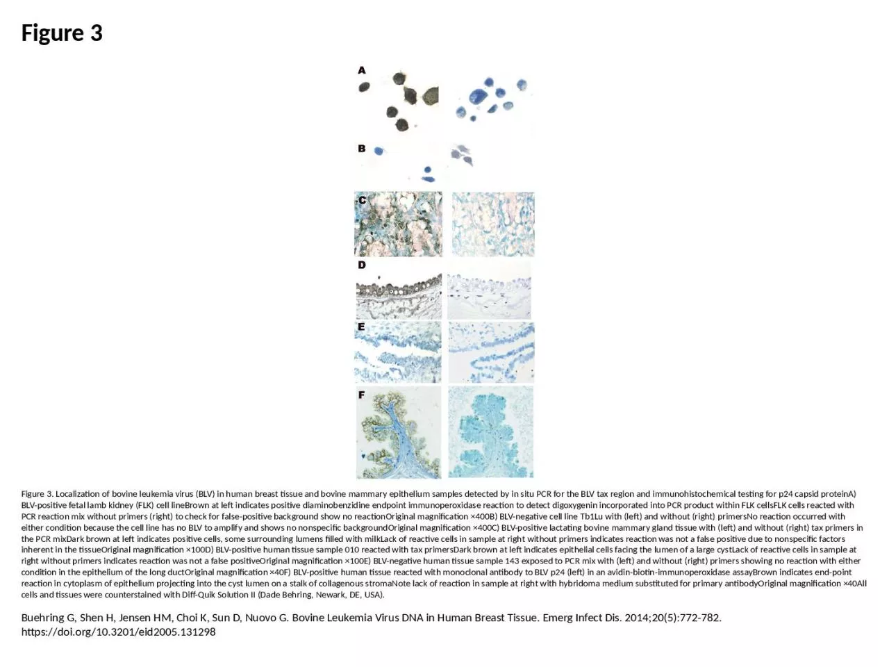
/
Figure 3 Figure 3. Localization of bovine leukemia virus (BLV) in human breast tissue Figure 3 Figure 3. Localization of bovine leukemia virus (BLV) in human breast tissue

Figure 3 Figure 3. Localization of bovine leukemia virus (BLV) in human breast tissue - PowerPoint Presentation

sophie
sophie . @sophie
Follow
66 views
Uploaded On 2023-05-29

Figure 3 Figure 3. Localization of bovine leukemia virus (BLV) in human breast tissue - PPT Presentation

Buehring G Shen H Jensen HM Choi K Sun D Nuovo G Bovine Leukemia Virus DNA in Human Breast Tissue Emerg Infect Dis 2014205772782 httpsdoiorg103201eid2005131298 ID: 999916

positive blv left reaction blv positive reaction left cells magnification tissue primers sample human pcr bovine tax cell reacted

Share:

Link:

Embed:

Download Presentation from below link

Download Presentation The PPT/PDF document "Figure 3 Figure 3. Localization of bovin..." is the property of its rightful owner. Permission is granted to download and print the materials on this web site for personal, non-commercial use only, and to display it on your personal computer provided you do not modify the materials and that you retain all copyright notices contained in the materials. By downloading content from our website, you accept the terms of this agreement.


Presentation Transcript

1. Figure 3Figure 3. Localization of bovine leukemia virus (BLV) in human breast tissue and bovine mammary epithelium samples detected by in situ PCR for the BLV tax region and immunohistochemical testing for p24 capsid proteinA) BLV-positive fetal lamb kidney (FLK) cell lineBrown at left indicates positive diaminobenzidine endpoint immunoperoxidase reaction to detect digoxygenin incorporated into PCR product within FLK cellsFLK cells reacted with PCR reaction mix without primers (right) to check for false-positive background show no reactionOriginal magnification ×400B) BLV-negative cell line Tb1Lu with (left) and without (right) primersNo reaction occurred with either condition because the cell line has no BLV to amplify and shows no nonspecific backgroundOriginal magnification ×400C) BLV-positive lactating bovine mammary gland tissue with (left) and without (right) tax primers in the PCR mixDark brown at left indicates positive cells, some surrounding lumens filled with milkLack of reactive cells in sample at right without primers indicates reaction was not a false positive due to nonspecific factors inherent in the tissueOriginal magnification ×100D) BLV-positive human tissue sample 010 reacted with tax primersDark brown at left indicates epithelial cells facing the lumen of a large cystLack of reactive cells in sample at right without primers indicates reaction was not a false positiveOriginal magnification ×100E) BLV-negative human tissue sample 143 exposed to PCR mix with (left) and without (right) primers showing no reaction with either condition in the epithelium of the long ductOriginal magnification ×40F) BLV-positive human tissue reacted with monoclonal antibody to BLV p24 (left) in an avidin-biotin-immunoperoxidase assayBrown indicates end-point reaction in cytoplasm of epithelium projecting into the cyst lumen on a stalk of collagenous stromaNote lack of reaction in sample at right with hybridoma medium substituted for primary antibodyOriginal magnification ×40All cells and tissues were counterstained with Diff-Quik Solution II (Dade Behring, Newark, DE, USA).Buehring G, Shen H, Jensen HM, Choi K, Sun D, Nuovo G. Bovine Leukemia Virus DNA in Human Breast Tissue. Emerg Infect Dis. 2014;20(5):772-782. https://doi.org/10.3201/eid2005.131298