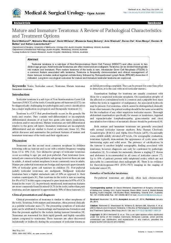
/
David Wetherell*, Mahesha Weerakoon, David Williams, Ania SliwinskiDep David Wetherell*, Mahesha Weerakoon, David Williams, Ania SliwinskiDep

David Wetherell*, Mahesha Weerakoon, David Williams, Ania SliwinskiDep - PDF document

stefany-barnette
stefany-barnette . @stefany-barnette
Follow
503 views
Uploaded On 2016-06-13

David Wetherell*, Mahesha Weerakoon, David Williams, Ania SliwinskiDep - PPT Presentation

Corresponding author Dr David Wetherell Department of Surgery University of Melbourne Urology Unit Austin Hospital Room 8244 Level 8 HaroldStokes Building Austin Hospital 145 Studley Road ID: 360949

*Corresponding author: David Wetherell

Share:

Link:

Embed:

Download Presentation from below link

Download Pdf The PPT/PDF document "David Wetherell*, Mahesha Weerakoon, Dav..." is the property of its rightful owner. Permission is granted to download and print the materials on this web site for personal, non-commercial use only, and to display it on your personal computer provided you do not modify the materials and that you retain all copyright notices contained in the materials. By downloading content from our website, you accept the terms of this agreement.


Presentation Transcript

David Wetherell*, Mahesha Weerakoon, David Williams, Ania SliwinskiDepartment of Surgery, University of Melbourne, Urology Unit, Austin Hospital, Heidelberg, Victoria, AustraliaDepartment of Anatomical Pathology, Austin Hospital, Heidelberg, Victoria, Australia Ludwig Institute for Cancer Research, Austin Hospital, Heidelberg, Victoria, Australia *Corresponding author: Dr David Wetherell, Department of Surgery, University of Melbourne, Urology Unit, Austin Hospital, Room 8244, Level 8, Harold-Stokes Building, Austin Hospital, 145 Studley Road, Heidelberg, Victoria 3141, Australia, Tel: +61 3 9496 5458; Fax: +61 3 9496 3617; E-mail: November 13, 2013; Wetherell D, Weerakoon M, Williams D, Beharry BK, Sliwinski A, et al. (2014) Mature and Immature Teratoma: A Review of Pathological Characteristics Wetherell D, et al This is an open-access article distributed unrestricted use, distribution, and reproduction in any medium, provided the Keywords: Testis; Testicular cancer; Teratoma; Mature teratoma; Immature teratomaIntroduction Testicular teratoma is a sub-type of Non-Seminomatous Germ Cell Tumours (NSGCT) of the testis. Gonadal germ cell tumours (GCT) can be diagnostically challenging for pathologists and correct classication has a major implication on prognosis and therapeutic treatment [1].Teratoma is a GCT that predominantly occurs in the gonads: the testis and ovaries. ey contain well-dierentiated or incompletely dierentiated elements of at least two germ cells layers (endoderm, ectoderm and/or mesoderm). Mature teratomas are well dierentiated relative to the germ cell layers. Immature teratomas are incompletely dierentiated and are similar to foetal or embryonic tissue [2]. is article discusses and summaries the pertinent features of mature and immature teratomas of the testis and their signicance. DiscussionTeratomas are the second most common neoplasm in children following yolk sac tumour and occur with a relative frequency ranging from 13 to 19% [3,4]. Two distinctive groups of testicular teratomas occur according to age: pre and post-pubertal. Pure teratomas (non-mixed) are common in the paediatric sub-group, however these are rare in adults. A mixed variant neoplasm is more commonly seen in adults. Mature pre-pubertal teratomas are benign and represent approximately 30% of testicular germ cell tumours in children [5]. Post-pubertal (adult) testicular teratomas are malignant. Malignant testicular teratomas have a higher metastasis rate of 20% as opposed to their ovarian counterparts [6]. Pure teratoma in the testis is rare accounting for 4% of GCT in this organ compared to pure teratoma in 95% of GCTs found in the ovary [1]. As previously mentioned, teratomatous features are more commonly found in mixed GCTs in the testis, rather than pure teratoma, and are apparent in approximately 50% of these tumours [1]. Clinical presentation and diagnosisClinical presentation of teratoma is similar to other neoplasms of the testis. Teratoma, both mature and immature, oen present clinically as a painful testicular mass [7]. e presenting symptom of testicular pain can be attributed to haemorrhage and haematoma formation and thus the pressure applied to the testicle during tumour formation [8]. Teratomas are renowned for their rapid growth and highly vasculitic nature compared to seminoma. ese tumours are oen discovered incidentally or indirectly during the assessment of testicular trauma as an initial presenting complaint. ey can be present for some time prior to detection, as is the case with most testicular masses. Examination ndings for teratoma are usually consistent with those for a suspected testicular neoplasm. On examination atrophy of the aected or contralateral testis is common and a palpable rm mass within the testis is suggestive of malignancy. An associated hydrocele may be present. For teratomas, which cannot be distinguished clinically from other tumours, the patient workup should be the same as would be for the evaluation of any other suspicious testicular masses. orough abdominal examination specically for masses or tenderness, inguinal and supraclavicular lymphadenopathy, gynecomastia and chest auscultation for evidence of metastatic disease should be performed [9]. Both mature and immature teratomas are generally associated with normal testicular tumour markers: Beta Human Chorionic Gonadotropin (B-hCG) and Alpha Feto-Protein (aFP). Occasionally some exhibit mildly elevated aFP levels. On sonographic evaluation, teratomas typically demonstrate the appearance of cystic areas with intervening septa and solid areas. e presence of calcications in the tumour is another helpful sonographic nding associated with teratomas, however, diagnosis can only be conrmed by pathologic evaluation [4]. To evaluate for metastatic disease a staging CT thorax and abdomen is recommended in all cases of testicular cancer [7]. Up to 10% of patients present with subpleural nodes, which are not detectable by conventional chest radiograph [8]. ere is no evidence for uorodeoxygenase-PET (FDG-PET) imaging in the staging oftesticular cancer [9].Genetics of testicular teratomas Pre-pubertal teratomas are diploid, oen lack chromosomal Testicular teratoma is a sub-type of Non-Seminomatous Germ Cell Tumour (NSGCT) and often occurs in two distinct age groups. Adult testicular teratomas are often mixed and are malignant. Teratoma can be divided histologically into mature and immature. Pure mature teratoma of the testis is rare. Intratubular Germ Cell Neoplasia (ITGCN) is a common feature associated with teratoma. Teratoma is frequently chemoresistant and clinical management of these tumours includes radical inguinal orchidectomy followed by Retroperitoneal Lymph Node (RPLND) dissection if Med Surg Urol Medical & Surgical Urology- Wetherell et al-, Med Surg Urol 1003, 290http9..dx-doi-org.00-3061.1057,8746-0000013 Review Article Wetherell D, Weerakoon , Sliwinski A, et al.(2014) Mature and Immature Teratoma: A Review of Pathological Characteristics and Treatment Options. Med Surg Urol 3: 124. doi:10.4172/2168-9857.1000124 cytological atypia, which are microscopic features of mature post-pubertal teratomas [18]. Lipogranuloatous reaction in the testicular parenchyma and pilosebaceous cyst arrangement is diagnostically characteristic of testicular dermoid cysts [1]. Epidermoid cysts are similarly not associated with ITGCN and lack atypia and mitotic activity [14] (Figures 1-3).previously discussed, mature pre-pubertal teratoma has a benign benign testicular tumours (GCTs) in adults is radical inguinal orchidectomy. Retroperitoneal metastasis has been identied in approximately 20 to metastasis has been identied in approximately 20 to stage I pure teratoma. e adjuvant treatment options remain RPLND or surveillance, however as previously stated around a quarter of these patients will have retroperitoneal metastasis and increased to 75% for imbalances and do not exhibit isochromosome formation (i(12p)) [10,11]. In contrast, adult teratomas, most of which are mixed type, are hypotriploid and are associated with chromosomal abnormalities. A pertinent genetic feature of testicular GCTs is the acquisition and over representation of 12p sequences. Gain of one or more of these genes on 12p is crucial in the development of testicular GCTs [12]. Other genetic changes found in a quarter of these tumours include: partial loss of chromosome 13 (particularly q31) and gain of chromosome 7 (particularly q11), chromosome 8 and the X chromosome [13]. Macroscopic and microscopic features Mature post-pubertal teratomas have a macroscopic appearance of solid testicular tumours. Microscopically mature teratomas have a disordered arrangement and demonstrate cytological atypia [1]. Adjacent seminiferous tubules oen display carcinoma in situ or intratubular germ cell neoplasia (ITGCN) in up to 90%, which is associated with malignant potential. Pre-pubertal teratomas, which are benign tumours, are seldom associated with ITGCN. ere is frequently widespread testicular atrophy and absent spermatogenesis [14]. As teratomas are germ cell tumours they can host a variety of non-tumour native tissues. For instance, in mature ovarian teratomas choroid plexus, thyroid and pituitary tissue can be present, with the latter manifesting systemically as prolactinomas [15]. In mature testicular tumours however, this non-tumour native tissue is seen less frequently. Pure adult testicular teratomas are rare and around a third are mixed GCTs. e occurrence of mixed teratoma GCTs can be attributed to the pathogenesis whereby malignant germ cells (ITGCN) dierentiate into non-teratomatous cells prior to the formation of teratomatous elements [1]. is accounts for the association of ITGCN seen in pure teratomas, as well as metastases of non-teratomatous cell types. e diagnosis of immaturity for a teratoma requires tissue resembling embryonic origin, in particular the appearance of neuroepithelium [1]. However, the presence of this tissue or ‘immaturity’ of a post-pubertal testicular teratoma is not signicant. Some pathologists believe that the teratomas should not be classied as mature or immature due to they having the same genetic changes and biological potential, in both pre and post-pubertal populations [15]. As mentioned previously, the pathogenesis of testicular teratoma is that malignant transformation occurs prior to teratomatous dierentiation. Hence the further immature dierentiation of teratomatous tissue is irrelevant, as the malignant process has occurred earlier [1]. Primitive Neuro ectodermal Tumour (PNET) is diagnosed in the presence of an overgrowth of immature neural elements in teratomas [1]. Extra testicular PNETs from GCTs as a result of metastatic spread are frequently resistant to chemotherapy and are associated with a high rate of mortality [16]. Despite testicular teratomas having malignant behaviour in the absence of excess neural tissue (i.e. PNET), the presence of this tissue does have a negative prognostic value [1].Associated tumours: dermoid and epidermoid cysts Testicular dermoid cysts are rare lesions and whether these should be classed as a variant of mature teratoma is still under discussion [17]. Epidermoid cysts represent 1% of all testicular tumours but whether they are true neoplasms still remains a topic of debate [18]. Testicular dermoid cysts are synonymous with the features of ovarian dermoid cysts. eir macroscopic appearance is of a cystic tumour oen containing grossly identiable hair [19]. In contrast to mature post-pubertal teratomas, these lesions oen occur in a testis with normal spermatogenesis and lack of atrophy. ey lack adjacent ITGCN and Figure 1: Mature teratoma (H&E, 40x) showing squamous epithelium (left upper) and enteric glandular epithelium (bottom right). Figure 2: Mixed mature (right) and immature (left) teratoma (H&E 40x), Wetherell D, Weerakoon , Sliwinski A, et al.(2014) Mature and Immature Teratoma: A Review of Pathological Characteristics and Treatment Options. Med Surg Urol 3: 124. doi:10.4172/2168-9857.1000124 adjuvant treatment option used following RPLND for other viable NSGCT elements [21].Advanced stage (Stage IIA/B)teratoma, present with advanced metastatic disease. is particular group of men oen has regression of other NSGCT elements in the the dependent upon histology of primary tumour and the prognosis as dened as per the International Germ Cell Consensus Classication cases [22]. ere is a general consensus that patients with advanced with advanced initial indication; with PEB (cisplatin, etoposide, bleomycin) being the chemotherapy of choice as per the IGCCCG guidelines [7,23,24]. with adjuvant PEB (2 cycles) in the case of metastatic disease. e outcomes post RPLND and chemotherapy are equivocal with 98% cure cure discussion needs to be taken between patient and clinician with regards to treatment options. Seminomas are radiosensitive hence Radiotherapy (RT) is an combination with surgical excision of residual mass (e.g. teratoma) teratoma) Advanced stage (Stage IIC and III) Around 37% of patients with pure teratoma present with advanced disease [20]. All advanced (stage III) NSGCTs, including mature and immature teratoma, should be assessed and stratied on a clinical basis into either favourable-risk or intermediate/poor risk group. Recommended treatment for the favourable risk group is at least three cycles of adjuvant chemotherapy (PEB). ese patients should all undergo restaging following primary chemotherapy and if there is complete remission resection is not indicated [26]. e extent of resection of residual masses (e.g. RPLND or wedge lung resection) should also take into account individual patient and quality of life factors [7]. e histological diagnosis should not inuence the treatment course in stage III disease and in general all advanced stage NSGCTs, including pure teratoma, should be treated according to the same pathway as described above [7,20].e [7,20].impact of teratomatous elements in orchidectomy specimens has been investigated to detail. Currently, evidence shows a correlation between teratoma within the orchidectomy specimen with an increased likelihood of teratoma in the post chemotherapy RPLND specimen with a current rate of 20% of teratomatous elements found in primary RPLND and 40% of teratomatous elements in patients undergoing RPLND post chemotherapy [19-21]. On the contrary, the absence of teratoma in the retroperitoneum is not suggested if teratoma is not is not Mature teratoma is found in approximately 35 to 40% of primary chemotherapy RPLND (PC-RPLND) specimens for advanced NSGCT [27]. One study by Steyerberg et al. analysed 556 RPLND specimens post-chemotherapy for NSGCTs and found mature teratoma in 42% and that a teratoma negative primary tumour was a strong predictor of necrosis in the PC-RPLND specimen [28]. Currently there is no imaging modality that accurately distinguish between necrosis, viable viable Retroperitoneal teratoma in NSCGT treated with primary chemotherapy ()Serologic and radiographic complete response to rst line chemotherapy (dened as a residual transverse axial lesion on CT less than 1 cm) is achieved in 26 to 64% of patients with advanced NSCGT [27]. ese patients are considered to be in a low–risk group, however, some centers still recommend RPLND following primary chemotherapy from a rationale that radiographic estimation of the size of residual nodal tissue following primary chemotherapy is unreliable. is remains a controversial topic and in particular the lack of imaging criteria and consensus on nodal size criteria [27]. A study by Oldenburg et al. of 87 RPLND patients post-chemotherapy showed that 30% with lymph nodes ad viable tumour present (20% teratoma and 10% malignant tumour) [30]. e European Germ Cell Cancer Consensus group and recent literature recommends that patients who achieve remission, dened as residual retroperitoneal lesion an be safely observed [31-34]. Retroperitoneal lymph-node dissection should be performed for radiographic mass cm;&#x, c-;0es 1cm post-chemotherapy for NSGCTs [27]. Loehrer et al. reported on 51 patients who had surgical resections of teratoma aer cisplatin-based chemotherapy. In this study twenty patients (39%) experienced relapse with either histologically proven teratoma (10 patients) or viable GCT (10 patients) [35]. Sonneveld Immature teratoma (H&E 200x) showing immature neuroepithelium Wetherell D, Weerakoon , Sliwinski A, et al.(2014) Mature and Immature Teratoma: A Review of Pathological Characteristics and Treatment Options. Med Surg Urol 3: 124. doi:10.4172/2168-9857.1000124 et al. reported on 51 patients with retroperitoneal teratoma aer aer experienced a relapse, with growing mature teratoma in 56%, teratoma with malignant transformation in 33%, and viable GCT in 11%. Another contemporary cohort of 210 patients by Carver et al. with only pure teratomatous elements at post chemotherapy RPLND revealed mature teratoma in 178 patients (85%), immature teratoma in 15 patients (7%) and teratoma with malignant transformation in 17 patients (8%) [21]. Of the 193 patients with mature or immature teratoma, 24 patients (12%) experienced relapse aer post chemotherapy RPLND and two patients experienced relapse with teratoma with malignant transformation. For men relapsing with only teratomatous histology, in seven and four patients respectively. Other recurrence sites include include malignant transformation are known to have a poorer prognosis and attempted resection of the primary site of recurrence remains the treatment of choice due to the poor sensitivity of these tumours to chemotherapy [21]. Oncological outcomesAs previously discussed, the oncological outcomes for teratoma pre and post chemotherapy RPLND are determined by the IGCCCG staging and grading of severity. Signicant predictors for increased risk of disease recurrence include higher pre and post chemotherapy nodal size, intermediate or poor IGCCG risk classication and evidence of malignant transformation [21]. Of particular note, histological ndings of immature teratoma, when compared to mature teratoma did not increase the risk of disease recurrence [37]. However, mature teratoma in the PC-RPLND specimen is commonly associated with late relapse [36,38]. Overall, the oncological outcomes for immature and mature teratoma remain equivocal as per IGCCCG staging and degree of severity. Conclusion In adults mixed GCTs with teratomatous elements are more common than pure teratoma. Mature and immature teratomas in the post-pubertal setting and oen exhibit malignant behavior. Distinction between mature and immature teratoma is made histologically, with the latter closely resembling foetal or embryonic tissue. e biological behaviour of immature teratoma is identical to that of mature teratoma. RPLND for patients with clinical stage I pure teratoma is still controversial. Treatment of metastatic testicular teratomas is complex but is oen primary chemotherapy followed by RPLND or surveillance and is guided clinically. However, teratomas are resistant and therefore oen respond poorly to chemotherapy. Long-term oncological outcomes are equivocal for both mature and immature teratoma. TM (2005) Germ cell tumors of the gonads: a selective review emphasizing problems in differential diagnosis, newly appreciated, and WS, Wein AJ, Kavoussi LR, Novick AC, Partin AW, et al. (2012) Brosman SA (1979) Testicular tumors in prepubertal children. Urology 13: 581-588.4. Simmonds PD, Lee AH, Theaker JM, Tung K, Smart CJ, et al. (1996) Primary 5. Grady RW, Ross JH, Kay R (1997) Epidemiological features of testicular teratoma in a prepubertal population. J Urol 158: 1191-1192.6. BS, Al-Ahmadie H, Sheinfeld J (2007) Adult and pediatric testicular F, Bokemeyer C, Cohn-Cedermark G, et al. (2011) EAU guidelines on testicular cancer: 2011 update. Eur Urol 60: 304-319. Chest staging in testis cancer patients: imaging modality selection based upon risk assessment as determined by abdominal M, Brenner W, Hartmann M, Kotzerke J, Hellwig D, et al. (2008) [18F]-FDG-PET in clinical stage I/II non-seminomatous germ cell tumours: results of SA, Wiley JM, Perlman EJ (1994) DNA ploidy analysis of pediatric germ 11. Comparative genomic and in situ hybridization of germ cell tumors of the MC, Verkerk AJ, van de Pol M, Heighway J, Marynen P, et al. (1998) Identi�cation of the critical region of 12p over-representation in testicular germ D, Suijkerbuijk RF, Geurts van Kessel A, et al. (1996) Comparative genomic hybridization of germ cell tumors of the adult testis: con�rmation of karyotypic �ndings and identi�cation of a Reinberg Y, Niehans GA, Fraley EE (1989) Intratubular germ cell neoplasia in testicular teratomas and epidermoid cysts. Correlation with IA, Davis CJ Jr (2004) Pathology of germ cell tumors of the testis. Ulbright TM, Foster RS, Miller KD (1997) Primitive neuroectodermal tumors arising in testicular germ cell neoplasms. Am J Surg TM, Srigley JR (2001) Dermoid cyst of the testis: a study of �ve postpubertal cases, including a pilomatrixoma-like variant, with evidence supporting its separate classi�cation from mature testicular teratoma. Am J EB Jr (1969) Epidermoid cysts of the testis: a clinical and pathologic F, Farivar-Mohseni H, Leon A, Motzer RJ, Bosl GJ, et al. (2003) Clinical outcome after retroperitoneal lymphadenectomy of patients with pure I, Foster RS, Ulbright TM, Donohue JP (1995) Adult primary pure n B, Serio A, Motzer RJ, Bosl GJ, et al. (2007) Long-term clinical outcome after postchemotherapy retroperitoneal lymph node dissection GM, Stenning SP (1997) The International Germ Cell Consensus Classi�cation: a new prognostic factor-based staging classi�cation for Hendry WF (1985) Clinical stage II non-seminomatous germ cell testicular tumours. Results of management by primary chemotherapy. Br J Urol A, Norman A, Fisher C, Hendry WF, Nicholls J, et al. (1994) Primary chemotherapy for stage II nonseminomatous germ cell tumors of the testis. J Kim HJ (2013) Current management of testicular cancer. Korean J Rivoire M (2001) Advanced testis cancer. Curr Treat Options Oncol S, Albers P, Fosså SD, Heidenreich A, Kollmannsberger C, et al. (2012) Contemporary management of postchemotherapy testis cancer. Eur EW, Keizer HJ, Fossa SD, Sleijfer DT, Toner GC, et al. (1995) Prediction of residual retroperitoneal mass histology after chemotherapy for metastatic nonseminomatous germ cell tumor: Multivariate analysis of S, Djaladat H, Nichols C (2011) Management of residual mass in nonseminomatous germ cell tumors following chemotherapy. Ther Adv Urol Wetherell D, Weerakoon , Sliwinski A, et al.(2014) Mature and Immature Teratoma: A Review of Pathological Characteristics and Treatment Options. Med Surg Urol 3: 124. doi:10.4172/2168-9857.1000124 30. Alfsen GC, Lien HH, Aass N, Waehre H, et al. (2003) Postchemotherapy retroperitoneal surgery remains necessary in patients with nonseminomatous testicular cancer and minimal residual tumor masses. J Clin Brames MJ, Beck SD, Foster RS, Einhorn LH (2010) Long-term follow-up of Cisplatin combination chemotherapy in patients with disseminated nonseminomatous germ cell tumors: is a postchemotherapy retroperitoneal lymph node dissection needed after complete remission? J Clinical Oncol 28: consensus conference on diagnosis and treatment of germ cell cancer: a report of the second meeting of the European Germ Cell Cancer Consensus group C, Daneshmand S, So A, Chi KN, Murray N, et al. (2010) Management of disseminated nonseminomatous germ cell tumors with risk-based chemotherapy followed by response-guided postchemotherapy surgery. Kakiashvili D, Jewett MA (2009) Surveillance in stage I Hui S, Clark S, Seal M, Einhorn LH, et al. (1986) Teratoma following cisplatin-based combination chemotherapy for nonseminomatous DJ, Sleijfer DT, Koops HS, Keemers-Gels ME, Molenaar WM, et al. (1998) Mature teratoma identi�ed after postchemotherapy surgery in patients with disseminated nonseminomatous testicular germ cell tumors: a plea for an Richie JP, Nucci MR, Renshaw AA (1998) Prognostic features of teratomas with malignant transformation: a clinicopathological study Ehrlich Y, Baniel J (2007) Late relapse of testis cancer. Urol Clin North Am 34: 253-258. Submit your next manuscript and get advantages of OMICS Unique features:• User website-translation your world’s languages• Audio Version • Digital articles exploreSpecial features:• 350 Journals• 30,000 • 21 days rapid review process• Quality quick review processing• Indexing (partial), Scopus, EBSCO, Index Copernicus Scholar • Sharing Networking • Authors, Reviewers rewarded • Better for your articles your .omicsonline.org0submission0 Wetherell D, Weerakoon M, Williams D, Beharry BK, Sliwinski A, et al. (2014) Mature and Immature Teratoma: A Review of Pathological Characteristics and Treatment Options. Med Surg Urol 3: 124. doi: Med Surg Urol Med Surg Urol Wetherell et Med Surg 1003, 290 Review Article Med Surg Urol Wetherell D, Weerakoon , Sliwinski A, et al.(2014) Mature and Immature Teratoma: A Review of Pathological Characteristics and Treatment Options. Med Surg Urol 3: 124. doi:10.4172/2168-9857.1000124 30. Oldenburg J, Alfsen GC, Lien HH, Aass N, Waehre H, et al. (2003)Postchemotherapy retroperitoneal surgery remains necessary in patients withnonseminomatous testicular cancer and minimal residual tumor masses. J Clin Ehrlich Y, Brames MJ, Beck SD, Foster RS, Einhorn LH (2010) Long-termof Cisplatin combination chemotherapy in patients with disseminated nonseminomatous germ cell tumors: is a postchemotherapy retroperitoneallymph node dissection needed after complete remission? J Clinical Oncol 28: Krege S, Beyer J, Souchon R, Albers P, Albrecht W, et al. (2008) Europeanof the second meeting of the European Germ Cell Cancer Consensus group Kollmannsberger C, Daneshmand S, So A, Chi KN, Murray N, et al. (2010)disseminated nonseminomatous germ cell tumors with risk-based chemotherapy followed by response-guided postchemotherapy surgery. D, Jewett MA (2009) Surveillance in stage I Loehrer PJ Sr, Hui S, Clark S, Seal M, Einhorn LH, et al. (1986) Teratomafollowing cisplatin-based combination chemotherapy for nonseminomatous Sonneveld DJ, Sleijfer DT, Koops HS, Keemers-Gels ME, Molenaar WM, et al. (1998) Mature teratoma identi�ed after postchemotherapy surgery in patients with disseminated nonseminomatous testicular germ cell tumors: a plea for an Comiter CV, Kibel AS, Richie JP, Nucci MR, Renshaw AA (1998) Prognosticteratomas with malignant transformation: a clinicopathological study Ehrlich Y, Baniel J (2007) Late relapse of testis cancer. Urol Clin North Am 34: 253-258. David Wetherell*, Mahesha Weerakoon, David Williams, Ania SliwinskiDepartment of Surgery, University of Melbourne, Urology Unit, Austin Hospital, Heidelberg, Victoria, AustraliaDepartment of Anatomical Pathology, Austin Hospital, Heidelberg, Victoria, Australia Ludwig Institute for Cancer Research, Austin Hospital, Heidelberg, Victoria, Australia *Corresponding author: Dr David Wetherell, Department of Surgery, Universityof Melbourne, Urology Unit, Austin Hospital, Room 8244, Level 8, Harold-StokesBuilding, Austin Hospital, 145 Studley Road, Heidelberg, Victoria 3141, Australia,Tel: +61 3 9496 5458; Fax: +61 3 9496 3617; E-mail: November 13, 2013; Wetherell D, Weerakoon M, Williams D, Beharry BK, Sliwinski A, et al. (2014) Mature and Immature Teratoma: A Review of Pathological Characteristics Wetherell D, et al This is an open-access article distributed unrestricted use, distribution, and reproduction in any medium, provided the Keywords: Testis; Testicular cancer; Teratoma; Mature teratoma;Immature teratomaIntroduction Testicular teratoma is a sub-type of Non-Seminomatous Germ Cell Tumours (NSGCT) of the testis. Gonadal germ cell tumours (GCT) can be diagnostically challenging for pathologists and correct classication has a major implication on prognosis and therapeutic treatment [1].Teratoma is a GCT that predominantly occurs in the gonads: the testis and ovaries. ey contain well-dierentiated or incompletely dierentiated elements of at least two germ cells layers (endoderm, ectoderm and/or mesoderm). Mature teratomas are well dierentiated relative to the germ cell layers. Immature teratomas are incompletely dierentiated and are similar to foetal or embryonic tissue [2]. is article discusses and summaries the pertinent features of mature and immature teratomas of the testis and their signicance. DiscussionTeratomas are the second most common neoplasm in children following yolk sac tumour and occur with a relative frequency ranging from 13 to 19% [3,4]. Two distinctive groups of testicular teratomas occur according to age: pre and post-pubertal. Pure teratomas (non-mixed) are common in the paediatric sub-group, however these are rare in adults. A mixed variant neoplasm is more commonly seen in adults. Mature pre-pubertal teratomas are benign and represent approximately 30% of testicular germ cell tumours in children [5]. Post-pubertal (adult) testicular teratomas are malignant. Malignant testicular teratomas have a higher metastasis rate of 20% as opposed to their ovarian counterparts [6]. Pure teratoma in the testis is rare accounting for 4% of GCT in this organ compared to pure teratoma in 95% of GCTs found in the ovary [1]. As previously mentioned, teratomatous features are more commonly found in mixed GCTs in the testis, rather than pure teratoma, and are apparent in approximately 50% of these tumours [1]. Clinical presentation and diagnosisClinical presentation of teratoma is similar to other neoplasms of the testis. Teratoma, both mature and immature, oen present clinically as a painful testicular mass [7]. e presenting symptom of testicular pain can be attributed to haemorrhage and haematoma formation and thus the pressure applied to the testicle during tumour formation [8]. Teratomas are renowned for their rapid growth and highly vasculitic nature compared to seminoma. ese tumours are oen discovered incidentally or indirectly during the assessment of testicular trauma as an initial presenting complaint. ey can be present for some time prior to detection, as is the case with most testicular masses. Examination ndings for teratoma are usually consistent with those for a suspected testicular neoplasm. On examination atrophy of the aected or contralateral testis is common and a palpable rm mass within the testis is suggestive of malignancy. An associated hydrocele may be present. For teratomas, which cannot be distinguished clinically from other tumours, the patient workup should be the same as would be for the evaluation of any other suspicious testicular masses. orough abdominal examination specically for masses or tenderness, inguinal and supraclavicular lymphadenopathy, gynecomastia and chest auscultation for evidence of metastatic disease should be performed [9]. Both mature and immature teratomas are generally associated with normal testicular tumour markers: Beta Human Chorionic Gonadotropin (B-hCG) and Alpha Feto-Protein (aFP). Occasionally some exhibit mildly elevated aFP levels. On sonographic evaluation, teratomas typically demonstrate the appearance of cystic areas with intervening septa and solid areas. e presence of calcications in the tumour is another helpful sonographic nding associated with teratomas, however, diagnosis can only be conrmed by pathologic evaluation [4]. To evaluate for metastatic disease a staging CT thorax and abdomen is recommended in all cases of testicular cancer [7]. Up to 10% of patients present with subpleural nodes, which are not detectable by conventional chest radiograph [8]. ere is no evidence for uorodeoxygenase-PET (FDG-PET) imaging in the staging oftesticular cancer [9].Genetics of testicular teratomas Pre-pubertal teratomas are diploid, oen lack chromosomal Testicular teratoma is a sub-type of Non-Seminomatous Germ Cell Tumour (NSGCT) and often occurs in two distinct age groups. Adult testicular teratomas are often mixed and are malignant. Teratoma can be divided histologically into mature and immature. Pure mature teratoma of the testis is rare. Intratubular Germ Cell Neoplasia (ITGCN) is a common feature associated with teratoma. Teratoma is frequently chemoresistant and clinical management of these tumours includes radical inguinal orchidectomy followed by Retroperitoneal Lymph Node (RPLND) dissection if Medical & Surgical Urology