/
CHROMOSOME ABNORMALITOES AND GENETIC COUNSELINGS CHROMOSOME ABNORMALITOES AND GENETIC COUNSELINGS

CHROMOSOME ABNORMALITOES AND GENETIC COUNSELINGS - PowerPoint Presentation

summer
summer . @summer
Follow
28 views
Uploaded On 2024-02-02

CHROMOSOME ABNORMALITOES AND GENETIC COUNSELINGS - PPT Presentation

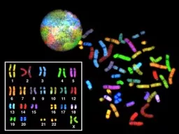
Presenter R2 張家甄 Supervisor Fellow 楊思婷 醫師 Outline PART ONE BASIC CONCEPTS 2 Chromosome Analysis INTRODUCTION CLASSICAL CYTOGENETIC ANALYSIS MICROARRAY ID: 1043770

chromosome microarray classical cytogenetic microarray chromosome cytogenetic classical chromosomes number genetic dna snp cgh copy chromosomal genomic detect amp

Share:

Link:

Embed:

Download Presentation from below link

Download Presentation The PPT/PDF document "CHROMOSOME ABNORMALITOES AND GENETIC COU..." is the property of its rightful owner. Permission is granted to download and print the materials on this web site for personal, non-commercial use only, and to display it on your personal computer provided you do not modify the materials and that you retain all copyright notices contained in the materials. By downloading content from our website, you accept the terms of this agreement.


Presentation Transcript

1. CHROMOSOME ABNORMALITOES AND GENETIC COUNSELINGSPresenter: R2 張家甄Supervisor: Fellow 楊思婷醫師

2.

3. OutlinePART ONE: BASIC CONCEPTS2. Chromosome AnalysisINTRODUCTIONCLASSICAL CYTOGENETIC ANALYSISMICROARRAY ANALYSISGENETIC COUNSELING CONSIDERATIONS

4. 01 INTRODUCTION

5. INTRODUCTION: Clinical cytogeneticsMolecular methodologiesThe first Half-century MicroscopyThis century Modern cytogenomic reportssophisticated documents

6. 02 CLASSICAL CYTOGENETIC ANALYSIS

7. CLASSICAL CYTOGENETIC ANALYSISPlain staining (“solid staining”)Early 1970s: the only stains availableGiemsa, orcein, and Leishman

8. CLASSICAL CYTOGENETIC ANALYSISGiemsa or G-bandingMain staining method: routine classical cytogeneticsTrypsin (protein digestion) stepThe detection and delineation of chromosomal structural abnormalitiesArrest the chromosome in its more elongated state at early metaphase or prometaphase

9. CLASSICAL CYTOGENETIC ANALYSISGiemsa or G-bandingMain staining method: routine classical cytogeneticsTrypsin (protein digestion) stepThe detection and delineation of chromosomal structural abnormalitiesArrest the chromosome in its more elongated state at early metaphase or prometaphaseThe same morphology are quinacrine or Q-banding, and reverse or R-banding

10. CLASSICAL CYTOGENETIC ANALYSISGiemsa or G-bandingThe same morphology are quinacrine or Q-banding, and reverse or R-bandingFIGURE 2–1 Chromosome pairs 1, 6, 15, 16, and Y and X stained by varioustechniques

11. CLASSICAL CYTOGENETIC ANALYSISConstitutive or C-bandingConstitutive heterochromatin: mainly the centromeric heterochromatinthe short arms of the acrocentric chromosomesthe distal part of the long arm of the Y chromosomeFIGURE 2–1 Chromosome pairs 1, 6, 15, 16, and Y and X stained by varioustechniques

12.

13. CLASSICAL CYTOGENETIC ANALYSISReplication bandingIdentify inactive X chromatinAdd a nucleotide analog (BrdU): Distinction of chromatin that replicates early from lateA banding pattern similar to that of R-bandingFIGURE 2–1 Chromosome pairs 1, 6, 15, 16, and Y and X stained by varioustechniques

14. CLASSICAL CYTOGENETIC ANALYSISNOR (silver) stainingNucleolar organizing regions (NOR)Multiple copies of genes coding for rRNASited on the satellite stalks of the acrocentric chromosomesFIGURE 2–1 Chromosome pairs 1, 6, 15, 16, and Y and X stained by varioustechniques

15. CLASSICAL CYTOGENETIC ANALYSISDistamycin A/DAPI stainingFluorescent stain: Identified heterochromatin of chromosomes 1, 9, 15, 16, and YInverted duplication 15 chromosome ↔ other small marker chromosomeshttps://thebiotechnotes.files.wordpress.com/2019/04/ezy-watermark_26-04-2019_03-50-58pm.jpg?w=616&h=413

16. CLASSICAL CYTOGENETIC ANALYSISFluorescence in situ hybridization (FISH)1990s: major cytogenetic advance Identify specific chromosomes, and parts of chromosomes → in situ hybridization with labeled probesWidely used: detect submicroscopic deletions and more obvious chromosome anomaliesHybridization methodDirect: attachment of a detectable molecule (e.g. a fluorophore)Indirect: the probe with a hapten detectable (biotin–avidin and digoxigenin systems)Assessment of the structural nature of imbalances revealed by microarray analysis

17.

18. CLASSICAL CYTOGENETIC ANALYSISComparative genomic hybridization (CGH)Differentially labeled, fluorophore-tagged DNA from the patient and a normal controlA metaphase slide prepared from a “standard” normal personRelative excesses and deficiencies of patient DNA bind competitivelySmall imbalances may be identifiable by this approach, ~10 Mb or greater

19. https://www.wikilectures.eu/images/3/34/CGH.gif

20.

21. Disadvantages of CGHInaccuracies in certain regions of chromosomes (in regions with high amounts of repeat sequences, centromeric regions of acrocentric chromosomes, in the telomeres of most chromosomes)Copy number changes can only be spotted if more than 50% of cells analysed contain a chromosomal gain or lossNot been able to identify chromosomal abnormalities that are balancedDecreased sensitivity due to contamination of the test cells with normal cells

22.

23. 03MICROARRAY ANALYSIS

24. MICROARRAY ANALYSIS2010s: Microarray has become the first-tier clinical diagnostic test Developmental disabilities or congenital anomaliesMicroarray techniques in use:CGH approachSingle nucleotide polymorphisms (SNP): the number of alleles

25. MICROARRAY ANALYSIS2010s: Microarray has become the first-tier clinical diagnostic test Developmental disabilities or congenital anomaliesMicroarray techniques in use:CGH approachSingle nucleotide polymorphisms (SNP): the number of alleles most microarrays comprise thousands of spots of reference DNA sequences

26. MICROARRAY ANALYSISNot that classical cytogenetics is likely to fade altogether from view:Not all array results can give a definitive constructionThe array cannot detect balanced rearrangements, and recognition of the carrier state will continue to need an old-fashioned chromosome test.The molecular cytogeneticist/cytogenomicist will not lose the intuitive understanding of what chromosomes are really like.

27. MICROARRAY ANALYSISBalanced Chromosomal RearrangementsAll of the genetic information is present in the correct amount But it is in the wrong location or in a different order

28. MICROARRAY ANALYSISBalanced Chromosomal RearrangementsAll of the genetic information is present in the correct amount But it is in the wrong location or in a different orderhttp://viafet.ae/media/files/balanced_inversions_translocation_viafet.jpg

29. MICROARRAY ANALYSISThe fundamental principle is essentially the same as in chromosomal CGH, but using the array, rather than the metaphase spread, as substrate.The fluorescent intensities of each dye are measuredExcess at a locus(duplication or aneuploidy) → more reflect the dye of the patient’s DNADeficiency at a locus(deletion or unbalanced translocation) → more reflect the dye of the control DNAThe fluorescent intensities are presented as a log ratio of each of the dyesComparative Genomic Hybridization

30. https://www.wikilectures.eu/images/3/34/CGH.gif

31.

32. MICROARRAY ANALYSISConstruction of Microarray-based CGHBacterial artificial chromosomes (BACs) or oligonucleotidesAn array with 3,000 BAC spots could detect unbalanced rearrangements at a 1 Mb resolution across the entire genome A study from Finland Approximately 20% of 150 patients with mental retardationwhose G-banded karyotypes → normalMicroarray → A presumed pathogenic imbalanceComparative Genomic HybridizationSiggberg L, Ala-Mello S, Jaakkola E, et al. Array CGH in molecular diagnosis ofmental retardation—A study of 150 Finnish patients. Am J Med Genet 152A:1398–1410, 2010.

33. MICROARRAY ANALYSISCharacteristics of Microarray-based CGHEfficiently detect copy number variations (CNVs) across the whole genomeEfficiently detect the amplifications and deletions on a specific chromosomeCannot detect the inversions and balanced reciprocal rearrangementsWidely used to diagnose postnatal cases with developmental delay (DD), autism, and/or congenital abnormalities.Comparative Genomic Hybridization

34. MICROARRAY ANALYSISDetect the number of alleles in a specimenProvide two types of informationIntensity of the signal arising from each SNP → A log ratio ↑signal intensity: Copy number gain↓signal intensity: Deletion SNP arrays produce genotyping informationHeterozygosityHomozygosity: a loss of DNA, such as a deletionThree allele: a gain of DNA copy number, such as a duplication or trisomySingle Nucleotide Polymorphism (SNP)

35.

36.

37. MICROARRAY ANALYSISAdded advantage Detecting uniparental disomy (UPD)The exceptional inheritance of the two chromosomes of a pair from the same parentNot affect number and structure of chromosomes → escapes cytogenetic detectionSingle Nucleotide Polymorphism (SNP)

38. Genes 2020, 11, 1454; doi:10.3390/genes11121454

39. MICROARRAY ANALYSISDisadvantages Probe spacing and genome coverage are limited by the nonrandom distribution of SNPs in the genome. Regions of segmental duplications are usually poorly or not at all covered.Single Nucleotide Polymorphism (SNP)

40. MICROARRAY ANALYSISAn efficient and cost-effective molecular tool to copy or amplify small segments of DNA or RNAUse specific primers to amplify segments of DNA to determine copy number and identify deletions or aneuploidyQuantitative fluorescent polymerase chain reaction (QF-PCR)Multiplex ligation-dependent probe amplification (MLPA)POLYMERASE CHAIN REACTION-BASED APPLICATIONS

41.

42. MICROARRAY ANALYSISApplicationsIdentification and characterization of infectious agentsGenetic fingerprinting (forensic application/paternity testing)Detection of mutation (investigation of genetic diseases)Cloning genesPCR sequencingPOLYMERASE CHAIN REACTION-BASED APPLICATIONS

43. MICROARRAY ANALYSISBased on massively parallel genomic sequencingWith the entire expressed genetic complement, the “exome,” and even the whole genomeAs a highly accurate molecular counting tool, sequencing cell-free DNA circulating in the maternal plasma → mapping each sequence read back to its chromosome of originNEXT-GENERATION SEQUENCING

44. MICROARRAY ANALYSISNEXT-GENERATION SEQUENCINGhttps://irepertoire.com/wp-content/uploads/2020/03/Sequencing-Workflow-1-1024x614.jpge.g. Illumina (Solexa)

45.

46. MICROARRAY ANALYSISCharacteristicsLow-coverage genome sequencingwith 100% sensitivity, copy number variants diagnosed by microarrayThe additional benefit: detecting balanced chromosome rearrangementsHigher levels of coverageConsiderable diagnostic yield → for the diagnosis of sequence-level mutationsDisadvantagesMore expensiveNEXT-GENERATION SEQUENCING

47. MICROARRAY ANALYSISCoverage is variable within a sample and typical coverage ranges from 30 or less to >1000 reads for typical human genetic and cancer applications, respectively.NEXT-GENERATION SEQUENCINGhttps://irepertoire.com/wp-content/uploads/2020/05/NGS-Considerations-Sequencing-Workflow-1024x768.png

48. MICROARRAY ANALYSISIntuitive pictorial formMay also include a listing of Presumed significant genes in the regionA comment upon imprintingThe likelihood of benign versus causative genomic changesCYTOGENETIC (OR CYTOGENOMIC) REPORTS

49. MICROARRAY ANALYSISIntuitive pictorial formCYTOGENETIC (OR CYTOGENOMIC) REPORTS

50. 04GENETIC COUNSELING CONSIDERATIONS

51. GENETIC COUNSELING CONSIDERATIONSIn the majority of cases,The abnormalities found → clear clinical relevance for the patient and familyUncover DNA changes of unclear clinical significance →Testing of additional family membersThe possibility of findings of unclear clinical significance should be discussed when ordering the test.molecular-based tests have the ability to interrogate the entire genomethe pretest genetic counseling should include information about uncovering unwanted informationThe higher resolution potential of these new technologies will increase the detection rate of chromosome abnormalities → improve our ability to make diagnoses

52. Take Home MessageTomac, Višnja & Pušeljić, Silvija & Škrlec, Ivana & Andelic, Mirna & Kos, Martina & Wagner, Jasenka. (2017). Etiology and the Genetic Basis of Intellectual Disability in the Pediatric Population. SEEMEDJ. 1. 144-153. 10.26332/seemedj.v1i1.28.

53. Thank You!