/
GENETIC  DISORDERS DISEASES GENETIC  DISORDERS DISEASES

GENETIC DISORDERS DISEASES - PowerPoint Presentation

natalie
natalie . @natalie
Follow
342 views
Uploaded On 2022-06-20

GENETIC DISORDERS DISEASES - PPT Presentation

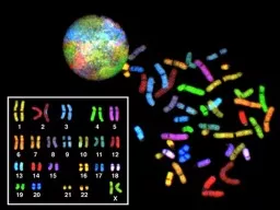
GENETIC ENVIRONMENTAL BOTH CONGENITAL HEREDITARY FAMILAL MUTATIONS PERMANENT change in DNA GENE MUTATION may and often result in a single base error CHROMOSOME MUTATION visible chromosome changepart of chromosome ID: 921018

gene dna genetic chromosome dna gene chromosome genetic protein autosomal snps mutation syndrome single sex disease genome storage proteins

Share:

Link:

Embed:

Download Presentation from below link

Download Presentation The PPT/PDF document "GENETIC DISORDERS DISEASES" is the property of its rightful owner. Permission is granted to download and print the materials on this web site for personal, non-commercial use only, and to display it on your personal computer provided you do not modify the materials and that you retain all copyright notices contained in the materials. By downloading content from our website, you accept the terms of this agreement.


Presentation Transcript

Slide1

GENETIC

DISORDERS

Slide2

DISEASESGENETICENVIRONMENTAL

BOTH

Slide3

CONGENITALHEREDITARYFAMILAL

Slide4

MUTATIONSPERMANENT change in DNA

GENE MUTATION: (may, and often, result in a single base error)CHROMOSOME MUTATION: (visible chromosome change)part of chromosomeTranslocationinversions

GENOME

MUTATION: (whole chromosome)

Base pair

 triplet gene chromosome segment whole chromosome genome

Slide5

COMPLEX MULTIGENIC DISORDERInteraction btw varient forms of genes and environmental factorsGene variation---polymorphism----multigenic or polygenic( atherosclerosis, diabetes, hypertension, ht &WT

Slide6

CONSTITUTIONAL - germ cells inherited disorder—in all cellsSOMATIC → cancer ↘congenital malformation

in specific cellsPENETRATION

Slide7

GENE MUTATIONDELETION OF A SINGLE BASE

SUBSTITUTION OF A SINGLE BASE

Slide8

POINT MUTATION---WITHIN CODING SEQUENCE--single base/different base---Results with replacement of one amino acid with another1. Missence mutation conservative nonconservative

2.Nonsense mutation---AA change—stop codon—Beta thal

Slide9

POINT MUTATION

Slide10

MUTTIONS in NON-coding sequences

 defective transcription, regulation, apop.DELETIONS/INSERTIONS 1.Multiple of three2.Premature stop codon

3.“frameshift”

mutation, involvement is NOT a multiple of 3

Tri-nucleotide

REPEATS

, e.g., CGG repeats many times in fragile X syndrome, CAG in others

Slide11

SEQUENCE AND COPY NUMBER VARIATIONS (POLYMORPHISMS)any two individuals share greaterthan 99.5% of their DNA sequencesdiversity of humans is encoded in less than 0.5% of our

, this 0.5% represents about 15 million basepairs. two most common forms of DNA variations(polymorphisms) in the human genome are singlenucleotidepolymorphisms (SNPs) and copy numbervariations (CNVs).• SNPs represent variation at single isolated nucleotide

Slide12

Two most common forms of DNA variations(polymorphisms) in the human genome are 1.Singlenucleotide polymorphisms (SNPs)

SNPs represent variation at single isolated nucleotide2.Copy number variations (CNVs).•

Slide13

positions and are almost always biallelic (i.e., one of onlytwo choices exist at a given site within the population,such as A or T). Much effort has been devoted to makingSNP maps of the human genome. These efforts haveidentified over 6 million SNPs in the human population,

many of which show wide variation in frequency in differentpopulations. SNPs may occur anywhere in thegenome—within exons, introns, or intergenic regions—but less than 1% of SNPs occurs in coding regions

Slide14

CNVs are a recently identified form of genetic variationconsisting of different numbers of large contiguousstretches of DNA from 1000 base pairs to millions ofbase pairs. In some instances these loci are, like SNPs,biallelic and simply duplicated or deleted in a subset of

Nature of Genetic Abnormalities Contributing to Human Disease 217pathologic conditions, such as cancer. Andrew Fire andCraig Mello were awarded the Nobel prize in physiologyor medicine in 2006 for their work on miRNAs.By current estimates, there are approximately 1000genes in humans that encode miRNAs. Transcription ofmiRNA genes produces primary miRNA transcript (primiRNA),which is processed within the nucleus to formanother structure called pre-miRNA (Fig. 6–1). With the

the population. In

Slide15

Epigenetic ChangesEpigenetic changes are those involving modulation of gene or protein expression in the absence of alterations in DNA sequence (i.e., mutation) Epigenetic regulation is of critical importance during development, as well as in homeostasis of fully developed tissues.

Slide16

alterations in the methylation of cytosine residues at gene promoters—heavily methylated promoters become inaccessible to RNA polymerase, leading to transcriptionalsilencing. Promoter methylation and silencing of tumor suppressor genes leads to unchecked cell growth –cancer

Slide17

Another major player in epigenetic arehistone proteins, which are components of structures called nucleosomes, around which DNA is coiled. Histone proteins undergo a variety of reversible modifications (e.g., methylation,acetylation) that affect secondary and tertiary DNA structure, and hence gene

Slide18

Alterations in Non-Coding RNAs—so-called “non-coding RNAs (ncRNAs)”—play important regulatory functions.Although many distinct families of ncRNAs existtwo imortant are microRNAs (miRNAs), and long non-coding RNAs

Slide19

The miRNAs, unlike messenger RNAs, donot encode proteins but instead inhibit the translation of target mRNAs into their corresponding proteins. Posttranscriptionalsilencing of gene expression by miRNA is preserved in all living forms from plants to humans and is therefore a fundamental mechanism of gene regulation

Slide20

In addition to alterations in DNA sequence, coding genes also can undergo structural variations, such as copy number changes (amplifications or deletions), or translocations,resulting in aberrant gain or loss of protein function. with mutations, Philadelphia chromosome— translocation t(9;22) between the BCR and ABL genes inchronic myelogenous leukemia

Slide21

GENE MUTATIONSINTERFERE with protein synthesis

SUPPRESS transcription, DNARNAPRODUCE abnormal mRNA

DEFECTS carried over into

TRANSLATION

ABNORMAL proteins

WITHOUT impairing syntheses

Slide22

GENETIC DISORDERSSINGLE gene mutations, following classical MENDELIAN inheritance patterns the most

MULTIFACTORIAL inheritanceCHROMOSOMAL disordersNON-MENDELIAN disorders

Slide23

MENDELIAN inheritance patternsAUTOSOMAL DOMINANT

AUTOSOMAL RECESSIVESEX-LINKED (recessive), involving “X” chromosome

Slide24

• Suspected sex chromosome abnormality (e.g., Turner syndrome)• Suspected fragile X syndrome• Infertility (to rule out sex chromosome abnormality)• Multiple spontaneous abortions (to rule out the parents as carriers of balanced translocation;

Slide25

AUTOSOMAL DOMINANTDisease is in HETEROZYGOTES

NEITHER parent may have the disease (NEW mut.)REDUCED PENETRANCE (environment?, other genes?)VARIABLE EXPRESSIVITY (environment?, other genes?)May have a DELAYED ONSET

Usually result in a

REDUCED PRODUCTION

or INACTIVE protein

Slide26

AUTOSOMAL DOMINANTHUNTINGTON DISEASE

NEUROFIBROMATOSISMYOTONIC DYSTROPHYTUBEROUS SCLEROSISPOLYCYSTIC KIDNEY

HEREDITARY SPHEROCYTOSIS

VON WILLEBRAND DISEASE

MARFAN SYNDROME

EHLERS-DANLOS SYNDROMES (some)

OSTEOGENESIS IMPERFECTA

ACHONDROPLASIA

FAMILIAL HYPERCHOLESTEROLEMIA

ACUTE INTERMITTENT PORPHYRIA

Slide27

AUTOSOMAL DOMINANT PEDIGREE

1) BOTH SEXES INVOLVED

2) GENERATIONS

NOT

SKIPPED

Slide28

AUTOSOMAL RECESSIVEDisease is in HOMOZYGOTES

More UNIFORM expression than ADOften COMPLETE PENETRANCEOnset usually EARLY in lifeNEW mutations rarely detected clinically

Proteins show

LOSS of FUNCTION and compensated in heterozygote form

Include ALL inborn errors of metabolism

MUCH more common that autosomal dominant

Slide29

AUTOSOMAL RECESSIVE

CFPKUGALACTOSEMIA

HOMOCYSTINURIA

LYSOSOMAL STORAGE

Α

-1 ANTITRYPSIN

WILSON DISEASE

HEMOCHROMATOSIS

GLYCOGEN STORAGE DISEASES

Hgb S

THALASSEMIAS

CONG. ADRENAL HYPERPLASIA

EHLERS-DANLOS (some)

ALKAPTONURIA

NEUROGENIC MUSC. ATROPHIES

FRIEDREICH ATAXIA

SPINAL MUSCULAR ATROPHY

Slide30

AUTOSOMAL RECESSIVE PEDIGREE

1) BOTH SEXES INVOLVED

2) GENERATIONS

SKIPPED

Slide31

SEX (“X”) LINKEDMALES ONLY

HIS SONS are OK, right?ALL his DAUGHTERS are CARRIERSThe “Y” chromosome is NOT homologous to the “X”, i.e., the classic concept of dominant/recessive has no meaning hereHETEROZYGOUS FEMALES have no phenotypic expression (carriers)….usually, this means autosomal “recessive”, right?

Slide32

SEX (“X”) LINKEDDUCHENNE MUSCULAR DYSTROPHYHEMOPHILIA , A and B

G6PD DEFICIENCYAGAMMAGLOBULINEMIAWISKOTT-ALDRICH SYNDROMEDIABETES INSIPIDUSLESCH-NYHAN SYNDROMEFRAGILE-X SYNDROME

Slide33

SEX LINKED PEDIGREE

1) MALES ONLY, sons of affected males are OK

2) GENERATION SKIPPING DOESN’T MATTER

Slide34

SINGLE GENE DISORDERSENZYME

DEFECT (Most of them, e.g., PKU)Accumulation of substrateLack of productFailure to inactivate a protein which causes damage

RECEPTOR/TRANSPORT PROTEIN

DEFECT

(Familial Hypercholesterolemia)

STRUCTURAL PROTEIN

DEFECT

(Marfan, Ehl-Dan)

Structure

Function

Quantity

ENZYME DEFECT WHICH INCREASES DRUG SUSCEPTIBILITY

: G6PD

Primaquine

Slide35

STRUCTURAL PROTEIN DEFECTSMarfan

SyndromeFibrillin-1 defect (not -2 or -3)Tall, dislocated lens, aortic arch aneurysms, etc.Abraham Lincoln?, Osama bin-Laden?

Ehlers-

Danlos

Syndromes (AD, AR)

Multiple (6?) different types

Classical,

Hypermob

.,

Vasc

.,

KyphoSc

.,

ArthChal

.,

Derm

Various collagen defects

Hyperelastic

skin,

hyperextensible

joints

Slide36

RECEPTOR PROTEIN DEFECTSFAMILIAL HYPERCHOLESTEROLEMIA

LDL RECEPTOR defectCholesterol TRANSPORT across liver cell impairedergo, CHOLESTEROL BUILDUP IN BLOOD“Scavenger System” for CHOL kicks in, i.e., MACROPHAGES

YOU NOW KNOW THE REST OF THE STORY

YOU NOW KNOW WHY MACROPHAGES are “FOAMY”

Slide37

ENZYME DEFICIENCIESBY FAR, THE LARGEST KNOWN CATEGORY

SUBSTRATE BUILDUPPRODUCT LACKSUBSTRATE could be HARMFULLYSOSOMAL STORAGE DISEASES comprise MOST of them

Slide38

LYSOSOMAL STORAGE DISEASESGLYCOGEN STORAGE DISEASES

SPHINGOLIPIDOSES (Gangliosides)SULFATIDOSESMUCOPOLYSACCHARIDOSESMUCOLIPIDOSESOTHERFucosidosis, Mannosidosis, Aspartylglycosaminuria

WOLMAN, Acid phosphate deficiency

Slide39

GLYCOGEN STORAGE DISEASESMANY TYPES (at least 13)

Type 2 Pompe (acid-α-glucosidase) , von

Gierke

(

Glu-6P-ase

),

McArdle

(

phosphorylase

), most studied and discussed, and referred to

Storage sites:

Liver

, Striated Muscle (

Skel

+

Ht

)

Slide40

SPHINGOLIPIDOSESMANY types,

Tay-Sachs most often referred toGANGLIOSIDES are ACCUMULATEDAshkenazi Jews (1/30 are carriers)CNS neurons a site of accumulation

CHERRY RED

spot in Macula

Usually fatal by age 4

Slide41

SULFATIDOSESMANY types, but the metachromatic leukodystrophies (CNS), Krabbe, Fabry, Gaucher, and Niemann-Pick (A and B) are most commonly referred to

SULFATIDES, CEREBROSIDES, SPHINGOMYELIN are the accumulations

Slide42

NIEMANN-PICKTYPES A, B, C

SPHINGOMYELIN BUILDUPSphingomyelinase (ASM), is the missing enzyme

MASSIVE SPLENOMEGALY

ALSO in ASHKANAZI JEWS

OFTEN FATAL in EARLY LIFE, CNS, ORGANOMEGALY

Slide43

GAUCHER DISEASEGLUCOCEREBROSIDE BUILDUP99% are type I, NO CNS involvement

ALL MACROPHAGES, liv, spl, nodes, marrow

Slide44

MUCOPOLYSACCHARIDOSESHURLER/HUNTER, for I and II, respectively, 14 types

DERMATAN sulfate, HEPARAN sulfate buildup, respectivelycoarse facial featuresclouding of the cornea

joint stiffness

mental retardation

URINARY EXCRETION of SULFATES COMMON

Slide45

OTHER LYSOSOMAL STORAGE DIS.FUCOSIDOSISMANNOSIDOSIS

ASPARTYLGLYCOSAMINURIAWOLMAN (CHOL., TRIGLYCERIDES)ACID PHOSPHATE DEFICIENCY (PHOS. ESTERS)

Slide46

ALCAPTONURIANOT a LYSOSOMAL ENZYME DISEASE

FIRST ONE TO BE DESCRIBEDHOMOGENTISIC ACIDHOMOGENTISIC ACID OXIDASEBLACK URINEBLACK NAILS (OCHRONOSIS), SKIN

BLACK

JOINT CARTILAGE (SEVERE ARTHRITIS)

Slide47

Slide48

Slide49

NEUROFIBROMATOSIS1 and 2

1-von Recklinghausen2- “acoustic” neurofibromatosis1Neurofibromas, café-au-lait, Lisch nodules

Slide50

NEUROFIBROMATOSIS1 and 2

1-von Recklinghausen2- “acoustic” neurofibromatosis2Bilateral acoustic neuromas and multiple meningiomas

Slide51

MULTIFACTORIAL INHERITANCEMulti-”FACTORIAL”, not just multi-GENIC

“SOIL” theoryCommon phenotypic expressions governed by “multifactorial” inheritanceHair colorEye colorSkin colorHeight

Intelligence

Diabetes, type II

Slide52

FEATURES ofmultifactorial inheritance

Expression determined by NUMBER of genesOverall 5% chance of 1st degree relatives having itIdentical twins >>>5%, but WAY less than 100%This 5% is increased if more children have it

Expression of

CONTINUOUS

traits (e.g., height) vs. DISCONTINUOUS traits (e.g., diabetes)

Slide53

“MULTIFACTORIAL” DISORDERSCleft lip, palate

Congenital heart diseaseCoronary heart diseaseHypertensionGoutDiabetesPyloric stenosis

MANY, MANY, MANY, MANY MORE…..

Slide54

KARYOTYPINGDefined as the study of CHROMOSOMES

46 = (22x2) + X + YConventional notation is “46,XY” or “46,XX”G(iemsa)-banding, 500 bands per haploid recognizableShort (“p”-etit) arm = p, other (long) arm = q

Slide55

Slide56

More KARYOTYPING infoA,B,C,D,E,F,G depends on chromosome lengthA longest

G shortestGroups within these letters depend on the p/q ratioARMREGIONBANDSub-BAND, numbering from the centromere progressing distad

Slide57

Slide58

F.I.S.H. (gene “probes”)greatly enhances G-banding

Fluorescent In-Situ

H

ybridization

Uses fluorescent labelled DNA fragments, ~10,000 base pairs, to bind (or not bind) to its complement

Slide59

FISHSUBTLE MICRODELETIONSCOMPLEX TRANSLOCATIONS

AND TELOMERE ALTERATIONS

Slide60

TRIPLE CHROMOSOME #20

A DELETION in

CHROMOSOME #22

Slide61

SPECTRAL KARYOTYPING

Slide62

CYTOGENETIC DISORDERSDEFINITIONS:EUPLOID (46XX or 46XY)

ANEUPLOID (NOT AN EXACT MULTIPLE OF 23)MONOSOMY, AUTOSOME OR SEXTRISOMY, AUTOSOME OR SEXDELETIONBREAKAGE

Slide63

MORE DEFINITIONS

Slide64

COMMON CYTOGENETIC DISEASESAUTOSOMES

TRISOMY-21 (DOWN SYNDROME)8, 9, 13 (Patau), 18 (Edwards), 2222q.11.2 deletionSEX CHROMOSOMESKLINEFELTER

: XXY, XXXY, etc.

TURNER

: XO

Slide65

TRISOMY-21

Slide66

TRISOMY-21Most trisomies (monosomies, aneuploidy) are from maternal non-disjunction(non-disjunction or anaphase lag are BOTH possible)

#1 cause of mental retardationMaternal age relatedCongenital Heart Defects, risk for acute leukemias, GI atresiasMost LOVABLE of all God’s children

Slide67

Slide68

Chromosome 22q11.2 Deletion Syndrome

Because of a DELETION, this cannot be detected by standard karyotyping and needs FISHCardiac defects, DiGeorge syndrome, velocardiofacial, CATCH*

Slide69

Slide70

SEX CHROMOSOME DISORDERSProblems related to sexual development and fertilityDiscovered at time of puberty

Retardation related to the number of X chromosomesIf you have at least ONE “Y” chromosome, you are male

Slide71

KLINEFELTER (XXY, XXXY, etc.)Hypogonadism found at puberty#1 cause of male infertility

NO retardation unless more X’s47, XXY 82% of the timeL----O----N----G legs, atrophic testes, small penis

Slide72

Slide73

TURNER (XO)45, X is the “proper” designation

Mosaics commonOften, the WHOLE chromosome is not missing, but just partNECK “WEBBING”EDEMA of HAND DORSUMCONGENITAL HEART DEFECTS most FEARED“STREAK” OVARIES

Slide74

Slide75

HERMAPHRODITESGENETIC SEX is determined by the PRESENCE or ABSENCE of a “Y” chromosome, but there is also,

GONADAL (phenotypic), and DUCTAL sexTRUE HERMAPHRODITE: OVARIES AND TESTES, often on opposite sides (VERY RARE)

PSEUDO-HERMAPHRODITE:

MALE: TESTES with female characteristics (Y-)

FEMALE: OVARIES with male characteristics (XX)

Slide76

SINGLE GENE, NON-MendelianTriplet repeats

Fragile X (CGG)Others: ataxias, myotonic dystrophyMitochondrial Mutations: (maternal) (LEBER HEREDITARY OPTIC NEUROPATHY)Genomic “IMPRINTING”: (Inactivation of maternal or paternal allele, contradicts Mendel)

Gonadal “MOSAICISM”:

(only gametes have mutated cells)

Slide77

MOLECULAR DX by DNA PROBESBIRTH DEFECTS, PRE- or POST- NATALTUMOR CELLS

CLASSIFICATIONS of TUMORSIDENTIFICATION of PATHOGENSDONOR COMPATIBILITYPATERNITYFORENSIC

Slide78

H&E tissue structures

Immuno- Antigen Proteins

GENES that

MAKE those

PROTEINS

Slide79

METHODS OF DNA ANALYSISFluorescence in Situ Hybridization (FISH)FISH utilizes DNA probes that recognize sequences specific to chromosomal regions of greater than 100 kilobases in size, which defines the limit of resolution with this technique for identifying chromosomal changes.

Such probes are labeled with fluorescent dyes and applied to metaphase spreads or interphase nuclei.

Slide80

The probe hybridizes to its complementary sequence on the chromosome and thuslabels the specific chromosomal region that can then be visualized under a fluorescence microscope.

Slide81

Array-Based Genomic HybridizationFISH requires previous knowledge of the one or few specific chromosomal regions However, chromosomal abnormalities may also be detected without previous knowledge global strategy known as array based CGH. Test DNA and a reference (normal) DNA are labeled with two different fluorescent dyes ( Cy5 and Cy3, which fluoresce red and green, ). The differentially labeled samples are then

hybridized to an array of segments of genomic DNA

Slide82

Amplifications and deletions in the test sample produce an increase or decrease in signal relative to the normal DNA that can be detected down to a 10-kilobase (kb) resolutionNewer generations of microarrays usingsingle-nucleotide polymorphisms (SNPs) provide even higher resolution

Slide83

Polymerase Chain Reaction (PCR) AnalysisDirect Detection of DNA Mutations by Polymerase Chain Reaction (PCR) Analysis PCR analysis, which involves exponential amplification of DNA, is now widely used in molecular diagnosis.If RNA is used as the substrate, it is first reverse-transcribed to obtain cDNA and then amplified by PCR. This method involving reverse transcription (RT) often is abbreviated as RT-PCR.

Slide84

One prerequisite for direct detection is that the sequence of the normal gene must be known. To detect the mutant gene, two primers that bind to the 3′ and 5′ ends ofthe normal sequence are designed. By utilizing appropriate DNA polymerases and thermal cycling, the target DNA is greatly amplified, producing millions of copies of the DNA

Slide85

Linkage Analysis and Genome-Wide Association StudiesDirect diagnosis of mutations is possible only if the gene responsible for a genetic disorder is known and its sequence has been identified. In several diseases that have a genetic

basis, including some common disorders, direct genetic diagnosis is not possible, either because the causal gene has not been identified or because the disease is multifactorial (polygenic) and no single gene is involved.

Slide86

Two types of analyses can be performed for unbiased identification of disease-associated gene(s): linkage analysis genome-wide association studies (GWASs).

In both surrogate markers in the genome, marker loci, must be used to localize the chromosomal regions of interest, based on their linkage to one regions

Slide87

Prenatal genetic analysis should be offered to all patients who are at risk of having cytogenetically abnormal progeny.It can be performed on cells obtained by amniocentesis, on chorionic villus biopsy material, or on umbilical cord blood . Indications are the following:

Advanced maternal age (beyond 34 Years),which is associated with greater risk of trisomies

Slide88

Confirmed carrier status for a balanced reciprocal translocation, Robertsonian translocation, or inversion (inPrenatal genetic analysis should be offered to all patients

Slide89

• A chromosomal abnormality affecting a previous child• Determination of fetal sex when the patient or partner is a confirmed carrier of an X-linked genetic disorder

Slide90

Postnatal genetic analysis usually is performed on peripheral blood lymphocytes.• Multiple congenital anomalies• Unexplained mental retardation and/or developmental delay• Suspected aneuploidy (e.g., features of Down syndrome)

• Suspected unbalanced autosome (e.g., Prader-Willi syndrome)