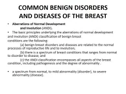
Aberrations of Normal Development and Involution ANDI The basic principles underlying the aberrations of normal development and involution ANDI classification of benign breast conditions ID: 913096
Download Presentation The PPT/PDF document "COMMON BENIGN DISORDERS AND DISEASES OF ..." is the property of its rightful owner. Permission is granted to download and print the materials on this web site for personal, non-commercial use only, and to display it on your personal computer provided you do not modify the materials and that you retain all copyright notices contained in the materials. By downloading content from our website, you accept the terms of this agreement.
Slide1
COMMON BENIGN DISORDERSAND DISEASES OF THE BREAST
Aberrations of Normal
Development
and Involution
(ANDI
)
.
The basic principles underlying the aberrations of normal development
and
involution (ANDI) classification of benign breast
conditions
are the following:
(
a) benign breast disorders
and diseases
are related to the normal processes of reproductive
life and
to involution;
(
b) there is a spectrum of breast
conditions that
ranges from normal to disorder to disease; and
(
c) the
ANDI classification
encompasses all aspects of the breast
condition, including
pathogenesis and the degree of abnormality
.
a
spectrum from normal, to mild abnormality (disorder
),
to
severe abnormality (disease
).
Slide2Aberrations of Normal Developmentand Involution
Early
Reproductive Years
.
Fibroadenomas
:
women
aged 15 to 25 years
- Small
fibroadenomas
(≤1 cm in size) are considered normal, whereas larger
fibroadenomas
(≤3 cm) are disorders and giant
fibroadenomas
(>3 cm) are disease.
-
Similarly, multiple
fibroadenomas
(more than five lesions in one breast) are very uncommon and are considered disease.
A spectrum of changes
from limited
to massive stromal hyperplasia (
gigantomastia
) is
seen.
Nipple
inversion is a disorder of development of the
major ducts.
Mammary duct
fistulas arise when nipple inversion predisposes
to major
duct obstruction, leading to recurrent
subareolar
abscess and
mammary duct fistula.
Slide3Later
Reproductive Years
.
Cyclical
mastalgia
and
nodularity usually
are associated with premenstrual enlargement of
the breast
and are regarded as normal
.
Painful nodularity
that persists
for >1 week of the menstrual cycle is considered a disorder.
papillary projections
sometimes give rise to bilateral bloody nipple discharge.
Slide4Involution.
Involution of lobular epithelium is
dependent on
the specialized stroma around it
.
When the
stroma involutes
too quickly, alveoli remain and form
microcysts
, which
are precursors of
macrocysts
. The
macrocysts
are
common, often
subclinical, and do not require specific treatment
.
Sclerosing
adenosis
is considered a disorder of both the
proliferative and
the
involutional
phases of the breast cycle
.
Duct
ectasia (dilated ducts) and
periductal
mastitis are other
important components
of the ANDI
classification
About 60% of women ≥70 years of age exhibit
some degree
of epithelial
hyperplasia
Atypical
proliferative diseases
include ductal and lobular hyperplasia, both
of which
display some features of carcinoma in situ.
Slide5Table 17-2 ANDI
classification of benign breast disorders
Normal
Disorder
Disease
Early reproductive years
(age 15–25 y)
Lobular development
Fibroadenoma
Giant
fibroadenoma
Stromal development
Adolescent hypertrophy
Gigantomastia
Gigantomastia
Nipple inversion
Subareolar
abscess
Mammary duct fistula
Later reproductive years
(age 25–40 y
)
Cyclical changes of menstruation
Cyclical
mastalgia
Incapacitating
mastalgia
Nodularity
Epithelial hyperplasia of
pregnancy
Bloody nipple discharge
Involution (age 35–55 y)
Lobular involution
Macrocysts
Sclerosing
lesions
Duct involution
Dilatation
Duct ectasia
Periductal
mastitis
Sclerosis
Nipple retraction
Epithelial turnover
Epithelial hyperplasia
ANDI = aberrations of normal development and involution.
Slide6Table 17-4
Classification of benign breast disorders
Nonproliferative
disorders of the breast
Cysts and apocrine metaplasia
Duct ectasia
Mild ductal epithelial hyperplasia
Calcifications
Fibroadenoma
and related lesions
Proliferative breast disorders without atypia
Sclerosing
adenosis
Radial and complex
sclerosing
lesions
Ductal epithelial hyperplasia
Intraductal
papillomas
Atypical proliferative lesions
Atypical lobular hyperplasia
Atypical ductal
hyperplasia
Fibrocystic Disease.
Slide7Abnormality
Relative Risk
Nonproliferative
lesions of the
breast
No increased risk
Sclerosing
adenosis
No increased risk
Intraductal
papilloma
No increased risk
Florid hyperplasia1.5 to 2-foldAtypical lobular hyperplasia4-foldAtypical ductal hyperplasia4-foldDuctal involvement by cells ofatypical ductal hyperplasia7-foldLobular carcinoma in situ10-foldDuctal carcinoma in situ10-fold
Table 17-3
Cancer risk associated with benign
breast disorders and in
situ carcinoma of the breast
Slide8Pathology
of
Nonproliferative
Disorders
Of paramount importance for the optimal management
of benign
breast disorders and diseases is the histologic
differentiation of
benign, atypical, and malignant changes.32,33
Determining the
clinical significance of these changes is a problem
that is
compounded by inconsistent nomenclature.The classificationsystem originally developed by Page separates the various types of benign breast disorders and diseases into three clinically relevant groups: nonproliferative disorders, proliferative disorders without atypia, and proliferative disorders with atypiaThis category includes:- cysts, duct ectasia, periductal mastitis, calcifications, fibroadenomas, and related disorders. Adenomas, Hamartomas, Adenolipomas, Fibrocystic Disease. The term fibrocystic disease is nonspecific.- and carry no increased risk for the development of breast cancer.
Slide9Pathology
of Proliferative
Disorders Without Atypia:
include
sclerosing
,
adenosis
, radial scars, complex
sclerosing
lesions, ductal epithelial
hyperplasia
, and
intraductal
papillomas.
Slide10Pathology of Atypical Proliferative Diseases:
The atypical proliferative diseases have some of the features of carcinoma in situ but either lack a major defining feature of carcinoma in situ or have the features in less than fully developed form.
Atypical ductal hyperplasia (ADH) appears similar to low grade ductal carcinoma in situ (DCIS) histologically and is composed of monotonous round, cuboidal, or polygonal cells enclosed by basement membrane with rare mitoses.
A
lesion will
be considered to be ADH if it is up to 2 or 3 mm in size
but would
be called DCIS if it is larger than 3 mm. The
diagnosis can
be difficult to establish with core needle biopsy
specimen alone
and most cases will require excisional biopsy
specimen for
classification.35Atypical lobular hyperplasia (ALH) results in minimal distention of lobular units with cells that are similar to those seen inlobular carcinoma in situ (LCIS).
Slide11Treatment of Selected Benign BreastDisorders and Diseases
Cysts.
Because needle biopsy of breast masses may
produce artifacts
that make mammography assessment more
difficult, many
multidisciplinary teams prefer to image breast
masses before
performing either fine needle aspiration or core
needle biopsy.36,37
In practice, however, the first investigation of
palpable breast.
In practice, however, the first investigation of
palpable breast masses may be a needle biopsy, which allows for the early diagnosis of cysts.A 21-gauge needle attached to a 10-mL syringe is placed directly into the mass, which is fixed by fingers of the nondominant hand. The volume of a typical cyst is 5 to 10 mL, but it may be 75 mL or more.If a mass was noted on initial ultrasound or there is a residual mass post-aspiration then a tissue specimen is obtained usually by core biopsy.When cystic fluid is bloodstained, fluid can be sent for cytologic examination
Slide12Fibroadenomas
.
Most
fibroadenomas
are self-limiting
and many
go undiagnosed, so a more conservative approach
is reasonable.
Careful ultrasound examination with
core-needle biopsy
will provide for an accurate diagnosis
.
Ultrasonography may reveal specific features that are pathognomonic for fibroadenoma and in a young woman (e.g., under 25 years) where the risk of breast cancer is already very low a core-needle biopsy may not be necessary.Cryoablation and ultrasound-guided vacuum assisted biopsy are approved treatments for fibroadenomas of the breast, especially lesions <3 cm. Larger lesions are often still best removed by excision.Larger lesions are often still best removed by excision.With short-term follow-up a significant percentage of fibroadenomas will decrease in size and will no longer be palpable.However, many will remain palpable, especially those larger than 2 cm.women should be counseled that the options for treatment include surgical removal, cryoablation, vacuum assisted biopsy, or observation.
Slide13Treatment of Selected Benign BreastDisorders and Diseases (cont.)
Sclerosing
Disorders:
The clinical significance of
sclerosing
adenosis
lies in its imitation of cancer
.
Excisional biopsy and
histologic examination are frequently necessary.The diagnostic work-up for radial scars and complex sclerosing lesions frequently involves stereotactic biopsy.It usually is not possible to differentiate these lesions with certainty from cancer by mammographic features, so a larger tissue biopsy is recommended either by way of vacuum assisted biopsy or an open surgical excisional biopsy.
Slide14Treatment of Selected Benign BreastDisorders and Diseases (cont.)
Periductal
Mastitis:
Painful and tender masses behind
the nipple-areola
complex are aspirated with a 21-gauge
needle attached
to a 10-mL syringe. Any fluid obtained is
submitted for
culture using a transport medium appropriate for the
detection of
anaerobic organisms
.In the absence of pus, women are started on a combination of metronidazole and dicloxacillin while awaiting the results of culture. Antibiotics are then continued based on sensitivity tests. Then continued based on sensitivity tests.when considerable purulent material is present, repeated ultrasound guided aspiration is performed and ultimately in a proportion of cases surgical treatment is required.In a woman of childbearing age, simple drainage is preferred, but if there is an anaerobic infection, recurrent infection frequently develops. Recurrent abscess with fistula is a difficult problem.Treatment of periductal fistula was initially recommended to be opening up of the fistulous track and allowing it to granulate.However, nowadays the preferred initial surgical treatment is by fistulectomy and primary closure with antibiotic coverage.Excision of all the major ducts is an alternative option depending on the circumstances.However, when subareolar sepsis is diffuse rather than localized to one segment or when more than one fistula is present, total duct excision is the most expeditious
Slide15Treatment of Selected Benign BreastDisorders and Diseases (cont.)
Nipple Inversion:
More women request correction of
congenital nipple
inversion than request correction for the
nipple inversion
that occurs secondary to duct ectasia
.
Although
the results
are usually satisfactory, women seeking correction
for cosmetic
reasons should always be made aware of the
surgical complications of altered nipple sensation, nipple necrosis, and postoperative fibrosis with nipple retraction.Because nipple inversion is a result of shortening of the subareolar ducts, a complete division of these ducts is necessary for permanent correction of the disorder.
Slide16Slide17Gynecomastia:
Gynecomastia
refers to an enlarged breast in the male
.
Physiologic gynecomastia
usually occurs during three phases of
life: the
neonatal period, adolescence, and senescence
.
However, the
hypoandrogenic
state
of Klinefelter’s syndrome (XXY), in which gynecomastia is usually evident, is associated with an increased risk of breast cancer.Neonatal gynecomastia is caused by the action of placental estrogens on neonatal breast tissues, whereas in adolescence, there is an excess of estradiol relative to testosterone, and with senescence, the circulating testosterone level falls, which results in relative hyperestrinism.Treatment: medical or surgical.I
Slide18INFECTIOUS AND
INFLAMMATORY DISORDERS
OF THE
BREAST :
Bacterial
Infection
Mycotic
Infections:
Fungal infections of the breast are rare
.
Hidradenitis Suppurativa: Hidradenitis suppurativa of the nipple-areola complex or axilla is a chronic inflammatory condition that originates within the accessory areolar glands of Montgomery or within the axillary sebaceous glands.When located in and about the nipple-areola complex, this disease may mimic other chronic inflammatory states, Paget’s disease of the nipple, or invasive breast cancer. Treatmebnt: a.biotics, Sugery, Surgery with graft.
Slide19Mondor’s
Disease:
Mondor’s
disease is a variant
of thrombophlebitis
that
involves the
superficial veins of the anterior chest wall and
breast.
Slide20Embryology:
In most mammals, paired
breasts develop
along these ridges, which extend from the base of
the forelimb
(future axilla) to the region of the hind limb (
inguinal area).
These ridges are not prominent in the
human embryo and disappear
after a short
time.
Accessory breasts (
polymastia
) or accessory nipples (polythelia) may occur along the milk line.Absence of the breast (amastia) is rare and results from an arrest in mammary ridge development that occurs during the sixth fetal week.Accessory axillary breast tissue is uncommon and usually is bilateral.
Slide21Slide22DIAGNOSIS OF BREAST CANCER
History.
Examination.
Imaging
Techniques:
Mammography.
Ductography
.
The primary indication for
ductography
is nipple
discharge, particularly when the fluid contains
blood. Radiopaque contrast media is injected into one or more of the major ducts and mammography is performed.Ultrasonography.Magnetic Resonance Imaging.
Slide23DIAGNOSIS OF BREAST CANCER (cont.)
Breast
Biopsy:
-
Nonpalpable
Lesions.
Image-guided breast biopsy:
Ultrasound localization
techniques.
The
combination of
diagnostic mammography, ultrasound
or
stereotactic.fine-needle aspiration (FNA) biopsy achieves almost 100% accuracy in the preoperative diagnosis of breast cancer.core-needle permits the analysis of breast tissu architecture and allows the pathologist to determine whether invasive cancer is present.Core-needle biopsy is preferred over open biopsy for nonpalpable breast lesions because a single surgical procedure can be planned based on the results of the core biopsy. - Palpable Lesions.FNA or core biopsy of a palpable breast mass can usually be performed in an outpatient setting.A 1.5-in, 22-gauge needle attached to a 10-mL syringe or a 14 gauge core biopsy needle is used. Both air-dried and 95% ethanol–fixed microscopic sections are prepared for analysis.Core-needle biopsy of palpable breast masses is performed using a 14-gauge needle, such as the Tru-Cut needle. If the target lesion was microcalcifications, the specimen should be radiographed to confirm appropriate sampling..Tissue specimens are placed in formalin and then processed to paraffin blocks. -
Slide24