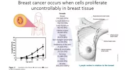
Azra Tabassum Facts about Breast cancer Worldwide increasing incidence gt 1 million newly diagnosed cases each yr 2 nd ID: 584580
Download Presentation The PPT/PDF document "investigations of the breast lump" is the property of its rightful owner. Permission is granted to download and print the materials on this web site for personal, non-commercial use only, and to display it on your personal computer provided you do not modify the materials and that you retain all copyright notices contained in the materials. By downloading content from our website, you accept the terms of this agreement.
Slide1
investigations of the breast lump
Azra Tabassum
Slide2
Facts about Breast cancer
Worldwide increasing incidence -> 1 million newly diagnosed cases each yr
2
nd
leading cause of death and 2
nd
most common cancer in women
Mortality highest in UK ---28
th
deaths/100,000women per
anum
Incidence increasing with age
All women are at risk
Most
masses are benign; that is, they are not cancerous, do not grow uncontrollably or spread, and are not life-threatening
.
• Some breast cancers are called in situ because they are confined within the ducts (
ductal
carcinoma in situ or DCIS) or lobules (lobular carcinoma in situ or LCIS) where they originated.
The
majority of in situ breast cancers are DCIS, which accounted for about 83% of in situ cases diagnosed during 2004-2008
.Slide3
Australian facts & figures
Breast cancer is a major cause of illness and death for females in Australia
On average, one in eight Australian females will develop breast cancer and one in 37 females will die from it before the age of 85 years.
In 2008 in Australia:
• Breast cancer was by far the most commonly diagnosed invasive cancer in females , accounting for 28% of all cancers in females
• A total of 13,567 breast cancers were diagnosed in Australian females.
More than 69% of breast cancers were diagnosed in those aged 40–69
The risk that a female would be diagnosed with breast cancer before the age of 85 was 1 in 8. Slide4Slide5
How to detect breast lump and further investigate Slide6
Triple assessment
Diagnostic tests – all breast lumps or suspected carcinoma
History and clinical examination
Radiological assessments
Cytological assessment
Staging investigation
Liver US
Staging CT
Chest x ray
Bone scan
Sensitivity of triple assessment with regard to histopathology is 100, Specificity is 99.3% and concordance is 99.3%. Slide7
Breast
lump
characteristics
Changes
in size over time
Change
relative to menstrual cycle
Duration
of mass
Pain
or swelling
Redness
, fever, or discharge
Diet
and medications
Current medications
Relevant History in Women with Palpable Breast MassesSlide8
Well-established risk factors
Probable risk factors
Age
50 or
older
Benign
breast disease, especially
cystic
disease, proliferative types of hyperplasia, and atypical
hyperplasia
Exposure
to ionizing
radiation
First
childbirth after age
20 Higher socioeconomic status History of breast cancer History of breast cancer in a first-degree relative Hormone therapy Nulliparity Obesity (i.e., BMI ≥ 30 kg per m2)*
Alcohol consumption
Did not breastfeed Elevated endogenous estrogen levels High BMI*Hormonal contraception therapy Increased mammographic density of breast tissue Menarche before age 12 Menopause after age 45 Mutations in BRCA 1 and BRCA 2 genes
Risk Factors for Breast CancerSlide9Slide10
Palpable lesions are always imaged before a biopsy is done.Slide11
Breast imaging studies
Ultrasound
Film screen mammography
( Screening and diagnostic)
Digital Mammography
Nuclear medicine
MRI
CT
Galactography
Slide12
Ultrasound
Ultrasound has become a
valuable tool in assessing
breast masses, as it is
widely available, quick to
perform, non-invasive and
less expensive than other
imaging modalities
.Slide13
The specificity of ultrasound in
detecting cystic lesions is 98%, and cysts ≥2 mm can be detected.Slide14
Ultrasound fcats
Ultrasound has a higher sensitivity than mammography in detecting lesions in women
with dense breast tissue
In this setting, its use as an adjunct to mammography may increase the accuracy by up to 7.4%
With regard to clinically palpable solid lesions, the specificity of ultrasound is superior
to mammography: 97% versus 87%
Complementary modality to an equivocal CBE and a normal mammogram in determining whether a mass is present
The evaluation of non palpable lesions detected on screening mammography,
image-guided biopsy of lesions and follow-up of benign lesions such as
fibroadenomas
.
It is an operator dependent technique with a lower sensitivity than mammography.Slide15Slide16Slide17Slide18Slide19Slide20Slide21Slide22Slide23Slide24Slide25Slide26Slide27Slide28Slide29Slide30Slide31Slide32
MammogramSlide33Slide34
Facts
The sensitivity of diagnostic mammography is around 90% (90% in fatty replaced breast and 65% in dense breast ) and
and
the specificity up to 88%.
Bilateral synchronous cancers are reported in 3% of cases; approximately 65% of these are detected only by mammography.
The known false negative rate of mammography is between 8% and 10%.
The sensitivity of mammography is decreased by dense breast tissue obscuring a
lesion.
False-negative results arise with poor technique and inadequate views that do not
include the mass, or when the findings are misinterpreted by the radiologist, notably
when there is overlap in the mammographic features of benign and malignant masses Slide35
MammographySlide36Slide37
Digital vs
film screen mammography
Advantages of digital mammography include better image quality, fewer artefacts, fewer patient recalls and
telemammography
.Slide38Slide39
Mammographic view
Standard views
Mediolateral
View – MLO views
Craniocaudal
views CC ViewsSlide40Slide41
Craniocaudal
Views
abnormality demonstrated are seen medical or lateral to the nipple
for lateral pathologies is more clearly demonstrated by rotating the breast medically
and
for medical pathology rotating
the breast laterally
Slide42
Mediolateral
Views
Demonstrate the maximum amount of the breast tissue.
A technically adequate exam has the nipple in profile, allows visualization of the
Axillary tail
pectoral muscle
inframammary
fold Slide43
Mammographic view
Additional views – additional information
Extended CC view -- medical or
lateal
rotation
Extended CC VIEWS – Cleopatra views
True lateral
Axillary view – Axillary tail view
Cleavage views– valley views
Paddle compression views
Spot views Magnification views Eklund views – for breast implant Slide44Slide45
An axillary
view
(also known as a "
Cleopatra view
“) is a type of supplementary mammographic view
.
It is an
exaggerated
craniocaudal
view
for better imaging of the lateral portion of the breast to the axillary tail. This projection is performed whenever we want to show a lesion seen only in the axillary tail on the MLO view. An optimal axillary view require to be clearly displayed the most lateral portion of the breast including the Axillary tail, as well the pectoral muscle and the nipple in profile.Slide46
A
magnification view
is performed to evaluate and count
microcalcifications
and its extension (as well the assessment of the borders and the tissue structures of a suspicious area or a mass) by using a magnification device which brings the breast away from the film plate and closer to the x-ray source. Slide47Slide48
Spot magnification views
are a combination of spot compression (small paddle) and magnification (
mag
stand and settings) techniques. Spot magnification is often used to evaluate the margins of a mass.
Any time you are doing a spot or spot
mag
view, it is critical that your image includes the lesion in question. Slide49
The cleavage view is another attempt at maximizing visualization of medial breast tissue when some of the breast being imaged may get pulled or left out of the compression field, or when a finding in the
mediolateral
-oblique view cannot be found on the cranial-caudal view. Slide50
The
ML view
is best for lesions located in the central or lateral breast.
LM views
are extremely useful in determining the exact location of an abnormality in the breast.
The lateral view is an additional view obtained at virtually every diagnostic evaluation. A lateral view may be obtained as a
mediolateral
(ML)
or lateralmedial (LM) view depending on where the imaging tube and detector are located.Slide51
Rolled views (rolled medial and rolled lateral) are used in the workup of an asymmetry on the CC view.
Prior to compressing, the technologist rolls the superior breast either medial or lateral, while simultaneously rolling the inferior breast in the
contralateral
direction
This motion separates the
tissues;superimposed
tissue will spread out, while a true lesion will persist. Slide52Slide53
Primary signs of cancer on Mammography
Major Signs:
Stellate
mass
Calcification -- Macro or Micro – calcification - which has
charctersotc
suspicion for malignancy
Breast Density
Locali
z
ed stromal distortion or asymmetry of parenchyma with out previous surgery Minor Signs: Skin and nipple changes Changes in vascularity Asymmetry of the duct pattern particularly affecting one segment
Enlarged , dense Axillary lymph nodes Slide54
The mass itself is typically then described according to
three
features
the shape or contour
the margin
the density.
shape
, if it is round, oval, or slightly
lobular
, the mass is probably benign. If the mass has a multi-lobular contour, or an irregular shape, then it is suggestive of malignancy. ‘Margin‘ refers to the characteristics of the border of the mass image. When the margin is circumscribed and well-defined the mass is probably benign. If the margin is obscured more than
75% by adjacent tissue, it is moderately suspicious of malignancy..Slide55
Density
‘ is usually classified as either fatty, low,
iso
-dense, or high. The mass is probably benign for fatty and low densities
moderately suspicious of malignancy for an
iso
-density
, and
highly suspicious
of malignancy at high densities
Dense breasts are not abnormal but thety are linked to higher risk of breast cancer Breast density is based on : how much fibrous and glandular tissue : how is the distribution within the breast tissue : How is breast made up of fatty tissure Slide56Slide57
Microcalcifications
Microcalcifications
are on of the main ways breast cancer is
mammographically
detected when it is in the very early stages.(DCIS)
Microcalcifications
are actually tiny specks of mineral deposits (such as calcium) They can be distributed in various ways.
Sometimes
microcalcifications
are found scattered throughout the breast tissue, and they often occur in clusters.
Most of the time, microcalcification deposits are due to benign causes. However, certain features and presentations of microcalcifications are more likely to be associated with malignant breast cancer.Slide58
Benign/probably benign
suspicious/malignant
Morphology
Popcorn egg shell with a dense center
x (adenofibroma)
curvilinear egg shell with a clear center
x (calcified cyst,
cytosteatonecrosis)
intra and peri-ductal bilateral
x (secretory disease) smudgy on CC and fluid level on lateralx (milk of calcium) powderish/BIRADS: amourphous,indistint
x
crushed stone/BIRADS: pleomorphic, heterogeneous x casting/BIRADS:linear, branching xDistributiondiffuse, scatteredx regionalx
segmental/within a major system, wedge shaped, nipple oriented
x
clustered/usually grouped in less than 2 cubic centimetres
x
linear, branching within the galactophores
x
Density
homogeneous
x
heterogenous
x
Size and number
size in mm
x ( >1 )
x ( < 0.2 to 1 )
number per cubic centimetre
x ( < 3.5 )
x ( > 3-5 )
Benign and malignant indicators for micro calcificationsSlide59
If the
microcalcifications
are
‘
Powderish
’, with either a fine,
indescernible
, or ‘cotton ball’ appearance, then the probability of DCIS is about 47%, and most frequently results in a ‘low-grade’ cancer.
Crushed Stone’ characteristic, appearing either as coarse, granular, angular, broken-needle-tip, arrowhead, or a spearhead shape, then the probability of DCIS is about 61%, the breast cancer classified as low to intermediate-grade.
Casting’ appearance, then the probability of DCIS is about 96%, the breast cancer classified as high-grade.
Casting microcalcifications typically appear in two variations. Variant A is called ‘dense casting, with linear and branched, fragmented, or irregular features.
Variant B is called ‘dotted casting’, with granular and branched, dotted, or snakeskin-like features.Slide60
An
axillary
lymph node that seems enlarged on a mammogram could contain cancer
However,
mammographical
features of benign and malignant
lymphadenopathy
are quite often indistinguishable.
Sometimes the presence of
intranodal
calcifications can be more suggestive of malignancy as well.As a rule of thumb, an axillary lymph node is suspicious if its size is greater than 2 cm and with no fatty hilum. More precicely, when a lymph node has a fatty hilum
visible, the outer cortex should be 5 millimeters thick at most, and usually less. When the cortex is 6mm or thicker, chances of cancer spread into the lymph node are significant. Slide61
Mass with extensive
spiculated
marginsSlide62
Malignant calcification , increased
parenchymal
density
Reticular calcification in
intraductal
cancerSlide63
Extensive branching ,coalescent calcification
Multifocal carcinomaSlide64
Mammographic features of benign lesions
Outline and shapes
well defined s masses are usually fibro adenoma in young breast or cyst in peri menopausal women
Well circumscribed borders
Halo sign : complete and partial ring surrounding the periphery of a breast mass
A lesion containing material of fat density has high probability of benign lesion
such as
lipoma
, traumatic oil cyst
In general benign lesion are of low density
Slide65
Fibroadenoma
Cyst with Halo signSlide66
Well defined , homogenous lesion --
Lipoma
Slide67
Magnetic resonance imaging (MRI)
High-resolution contrast-enhanced MRI has recently emerged as a sensitive imaging modality for the detection of breast cancer.
The high sensitivity, which approaches 98%, makes MRI useful in specific clinical situations,
Evaluating patients with breast implants
detecting local recurrence
After breast-conserving therapy,
detecting multifocal/
multicentric
disease.
MRI avoids exposure to radiation,
Technique is cumbersome and expensive, not readily available, Does not detect microcalcifications
However, the moderately low specificity of 47 - 67% requiresComputed tomography (CT) scanningThis modality has no established place in the evaluation of palpable breast masses. Slide68Slide69Slide70Slide71Slide72Slide73Slide74Slide75