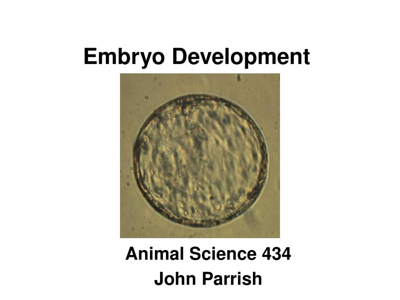
/
Embryo Development Animal Science 434 Embryo Development Animal Science 434

Embryo Development Animal Science 434 - PowerPoint Presentation

summer
summer . @summer
Follow
342 views
Uploaded On 2022-06-01

Embryo Development Animal Science 434 - PPT Presentation

John Parrish Errors in Fertilization Polyspermy polyandry Multiple sperm penetration Invertebrates excess sperm eliminated because sperm centriole contributes to first embryonic cleavage spindle ID: 912753

cleavage cell control gene cell cleavage gene control blastocyst embryonic cycle early translation day hours pregnancy embryo formation sperm

Share:

Link:

Embed:

Download Presentation from below link

Download Presentation The PPT/PDF document "Embryo Development Animal Science 434" is the property of its rightful owner. Permission is granted to download and print the materials on this web site for personal, non-commercial use only, and to display it on your personal computer provided you do not modify the materials and that you retain all copyright notices contained in the materials. By downloading content from our website, you accept the terms of this agreement.


Presentation Transcript

Slide1

Embryo Development

Animal Science 434John Parrish

Slide2

Errors in Fertilization

Polyspermy - polyandryMultiple sperm penetration

Invertebrates

excess sperm eliminated because sperm centriole contributes to first embryonic cleavage spindle

Mammals

Sperm centriole not essential so development continues but fails early to midpregnancy due to multiploidy

Occurs most often in aged oocytes due to failure of zona block to polyspermy

Slide3

Errors in Fertilization (cont.)

PolygynyMultiple maternal pronuclei + 1 paternal pronuclei

Artificially created only

Suppress extrusion of the PBII

Androgenote

Union of 2 paternal pronuclei

Artificially created only

From pronuclear exchange

Slide4

Errors in Fertilization (cont.)

GynogenoteUnion of 2 maternal pronuclei

Artificially created

Induced oocyte activation and supression of PBII extrusion

Parthenogenesis

Activation of the oocyte without a sperm

Embryo is either haploid or gynogenesis occurs to form diploid

Platties - sperm activates but then gynogenesis occurs and sperm extruded from embryo

Slide5

Oocyte Development and Fertilization

MPF

MPF

MPF

MPF

Primary Oocyte

GV-Intact

GVBD

(8 hr)

Metaphase I

PB-1

Secondary Oocyte

Metaphase II

(21 hr)

Ovulation

(29 hr)

LH Surge

(0 hr)

Slide6

Zona Pellucida

Perivitelline

Space

Oocyte

Ca

2+

Sperm Penetration

of the

Zona Pellucida

and

Fusion with the Oocyte

(30 hr)

Slide7

Embryo Development in the Bovine

2 cell

(62 hr, day 2)

4 cell

(75 hr, day 3)

8 cell

(90 hr, day 3)

16 cell

(120 hr,

day 4)

32 cell

Morula

(day 5-6)

Zygote

(34 hr, day 1)

Tight

Morula

(day 6-7)

Early Blastocyst

(day 7-8)

Blastocyst

(day 7-9)

Expanded

Blastocyst

(day 8-10)

Hatched

Blastocyst

(day 9-11)

Compaction

Blastocoel

Inner Cell

Mass

Trophectoderm

Slide8

Fertilization to Cleavage

Polar Body

Zona Pellucida

Pronuclei

Perivitelline Space

Zygote

Blastomere

Slide9

Fertilization to Cleavage

Imprinting

Maternal Gene Control

Long Cell Cycle

Slide10

Imprinting

Egg Pronucleus

Sperm

Pronucleus

Slide11

Imprinting

Egg Pronucleus

Sperm

Pronucleus

Androgenote

Gynogenote

Slide12

Imprinting

Controls

Egg

Pronucleus

Sperm Pronucleus

Androgenote

Gynogenote

Slide13

Imprinting

Androgenote

Gynogenote

Controls

No Inner Cell Mass

Normal Placenta

Fails during embryo

development

Normal Fetus

Small Placenta

Fails Midpregnancy

Normal Fetus

Normal Placenta

Normal Pregnancy

Maternal and Paternal Genomes Are Expressed Differently in the Embryo and Fetus

Slide14

Gene Control of Development

Oocyte

Growth

LH

Surge

Fertilization

Cleavage

Transciption

Translation

Transcription

Translation

Post

-Translation

Translation

Post-Translation

No transcription

Translation

Post-Translation

Maternal Gene Control

Embryonic

Gene

Control

Slide15

Fertilization to Cleavage

Maternal Gene Control

Slide16

Fertilization to Cleavage

Long Cell Cycle

Penetration to Cleavage

32 hour

(Bovine)

Slide17

Precompaction Cleavage

Cell size decreases

Cell cycle

Embryonic gene control

Asynchrony of cell divisions

Movement into Uterus

Early pregnancy factor

Slide18

Precompaction Cleavage

Cell size decreases

Cell cycle

Asynchrony of cell divisions

Embryonic gene control

Movement into Uterus

Early pregnancy factor

Slide19

Precompaction Cleavage

Cell size decreases

Cell cycle

Embryonic gene control

Asynchrony of cell divisions

Movement into Uterus

Early pregnancy factor

13 hours

15 hours

30 hours

32 hours

Slide20

Cell Cycle Lengths

1st Cell Cycle

(zygote 2 cell)

2nd Cell Cycle

(2 cell 4 cell)

G1

S

G2 + M

Total = 32 hr

Total = 13 hr

8 hr

8 hr

16 hr

<1 hr

2 hr

8 hr

Slide21

Precompaction Cleavage

Cell size decreases

Cell cycle

Embryonic gene control

Asynchrony of cell divisions

Movement into Uterus

Early pregnancy factor

13 hours

15 hours

30 hours

32 hours

Short G1 and G2

Short G1 and G2

Slide22

Precompaction Cleavage

Cell size decreases

Cell cycle

Asynchrony of cell divisions

Embryonic gene control

Movement into Uterus

Early pregnancy factor

Faster dividing

blastomeres

go

to center of embryo

Slide23

Asynchronous Cleavage

- Inside Outside Theory

If a marked blastomere

is placed into the interior of a 8-cell embryo, it and its progeny become part of the ICM.

If a marked blastomere is placed on the outside of a 8-cell embryo, it and its progeny become part of the trophectoderm.

Trophectoderm

Inner Cell Mass

Slide24

Asynchronous Cleavage Use

Create embryos from different speciesPlacenta from one species

Host mother

Embryo from some other species

Donor mother

Slide25

Precompaction Cleavage

Cell size decreases

Cell cycle

Asynchrony of cell divisions

Embryonic gene control

Movement into Uterus

Early pregnancy factor

Slide26

Gene Control of Development

Oocyte

Growth

LH

Surge

Fertilization

Cleavage

Transciption

Translation

Transcription

Translation

Post

-Translation

Translation

Post-Translation

No transcription

Translation

Post-Translation

Maternal Gene Control

Embryonic

Gene

Control

Slide27

Transition from Maternal to Embryonic Gene Control

In vitro blocks to development often occur here!!!!!

Transcription of the embryonic genome

First begins

Development is

dependent on

Species

Mouse 1 cell 2 cell

Rabbit 2 cell 8 cell

Pig 4 cell 8 cell

Cattle 4 cell 8-16 cell

Sheep 8 cell 16 cell

Human 4 cell 8 cell

Slide28

Precompaction Cleavage

Cell size decreases

Cell cycle

Asynchrony of cell divisions

Embryonic gene control

Movement into Uterus

Early pregnancy factor

13 hours

15 hours

30 hours

32 hours

Cell Cycle Length Increases

Embryo runs out of key factors coded for by maternal mRNA

Pause in G1

Slide29

Precompaction Cleavage

Cell size decreases

Cell cycle

Asynchrony of cell divisions

Embryonic gene control

Movement into Uterus

Early pregnancy factor

Slide30

Movement into the Uterus

<

8 cell

>

8 cell

Ampulla

Isthmus

Uterine Horn

Occurs around day 4

Cause

Change in estrogen progesterone

Slide31

Precompaction Cleavage

Cell size decreases

Cell cycle

Asynchrony of cell divisions

Embryonic gene control

Movement into Uterus

Early pregnancy factor

Slide32

Early Pregnancy Factor

Found at 24 - 72 hours after fertilizationMice, hamster, sheep, cattle, swine, human

Seen only in viable pregnancy

More recent experience in cattle may not agree with this

Function

Sensitize the uterus to implantation

Basis for early pregnancy kit in cattle

Slide33

Morula to Blastocyst

Polarization

Compaction

Slide34

Polarization

Polar Blastomeres

Non-polar Blastomeres

Microvilli

Slide35

Polarization (cont.)

Tight Junctions

Gap Junctions

Slide36

Cell Linage

Polar Cells

Non-polar Cells

2 polar cells

1 polar

1 non-polar

2 non-polar

Slide37

Compaction

Occurs at fixed time after fertilization

Membranes are very close and begin to flatten. Resulting in loss of the round cell outlines.

Differentiational

event

Genome controlled and involves microtubules and microfilaments.

Slide38

Blastocyst Formation and Hatching

Blastocoel formation

Hatching

Slide39

Blastocoel Formation

Tight Junctions

Gap Junctions

Na

+

H

2

O

Morula

Slide40

Blastocoel Formation

Tight Junctions

Gap Junctions

Na

+

H

2

O

Morula

Slide41

Morula

Blastocoel Formation

Tight Junctions

Gap Junctions

Na

+

H

2

O

Blastocoel

Early

Blastocyst

Slide42

Blastocyst Formation and Hatching

Blastocoel formation

Hatching

Na

+

H

2

O

Early

Blastocyst

Blastocyst

Expanded

Blastocyst

Inner Cell

Mass

Trophectoderm

Blastocoel formation

not dependent on:

Cell number

Cell division

Embryonic genome expression required

Slide43

Blastocyst Formation and Hatching

Blastocyst

Expanded

Blastocyst

Hatching

Blastocyst

Enzymatic digestion of

zona

Plasminogen

and

plasminogen

activator made by embryo

Softening of

zona

by uterine enzymes

Increase in size of

blastocyst due to water pumpingMost importantDay 9 - 11 in cattle, 6 in swine, and day 7 - 8 in horses or sheep Blastocoel formationHatching

EarlyBlastocyst

Slide44

Formation of Twins

DizygoticNot identicalDouble ovulation

Monozygotic

Identical

Several potential mechanisms

Slide45

Formation of Monozygotic Twins

Siamese

Twins