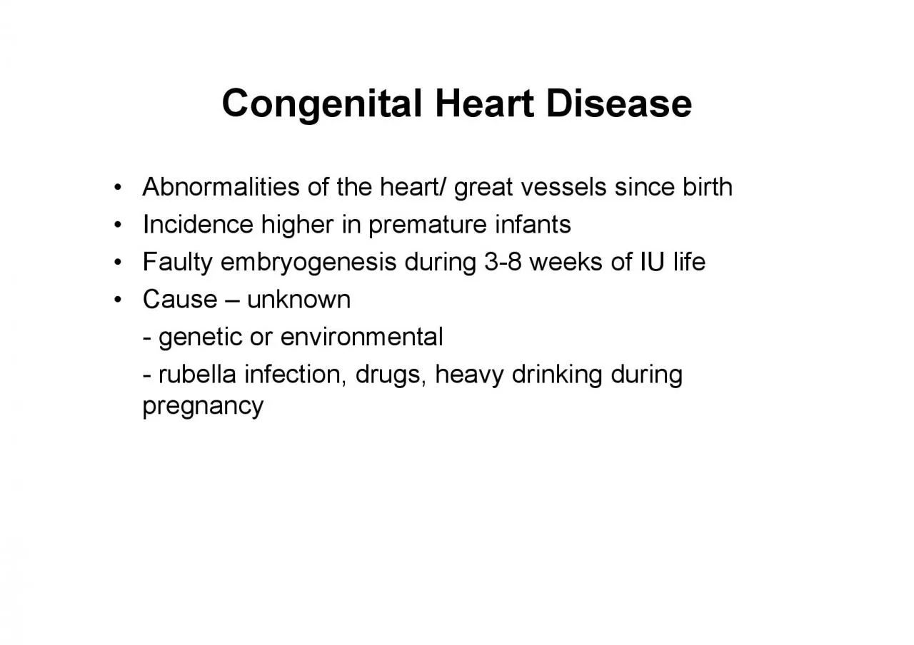
/
Abnormalities of the heart great vessels since birth Incidence higher Abnormalities of the heart great vessels since birth Incidence higher

Abnormalities of the heart great vessels since birth Incidence higher - PDF document

susan
susan . @susan
Follow
342 views
Uploaded On 2022-08-24

Abnormalities of the heart great vessels since birth Incidence higher - PPT Presentation

Classification 1Dextrocardia may be accompanied by situs inversus 2 Left to Right shunts Acyanotic or Late Cyanotic group Ventricular septal defect VSD 2530 Atrial septal defect ASD101 ID: 941187

stenosis pulmonary blood mci pulmonary stenosis mci blood atresia aorta arteriosus 150 heart ductus tricuspid pulm valve aortic hypertrophy

Share:

Link:

Embed:

Download Presentation from below link

Download Pdf The PPT/PDF document "Abnormalities of the heart great vessels..." is the property of its rightful owner. Permission is granted to download and print the materials on this web site for personal, non-commercial use only, and to display it on your personal computer provided you do not modify the materials and that you retain all copyright notices contained in the materials. By downloading content from our website, you accept the terms of this agreement.


Presentation Transcript

Abnormalities of the heart/ great vessels since birth Incidence higher in premature infants Faulty embryogenesis during 3-8 weeks of IU life Cause – unknown -genetic or environmental -rubella infection, d

rugs, heavy drinking during Classification 1.Dextrocardia may be accompanied by situs inversus 2. Left to Right shunts (Acyanotic or Late Cyanotic group) Ventricular septal defect (VSD) -25-30% Atrial sept

al defect (ASD)-10-15% Patent ductus arteriosus (PDA)-10-20% Right to Left shunts (Cyanotic group) Tetralogy of Fallot (TOF)-6-15% Transposition of great arteries -4-10% Persistent truncus arteriosus –

; 2% Tricuspid atresia and stenosis 1% Coarctation of Aorta 5-7% Pulmonary stenosis and atresia 5-7% Abnormal communication b/w 2 chambers or blood vs; blood flows according to pressure gradient dusky

blueness of mucus membranes and skin ( increased synthesis of Hb + RBC Emboli from peripheral veins donot undergo filteration enter Lt heart embolize to systemic infarction & abscess Clubbing (hypertrophic ost

eoarthropathy) of tips of �� &#x/MCI; 0 ;&#x/MCI; 0 ;Shunt &#x/MCI; 1 ;&#x/MCI; 1 ;L→R shunt: &#x/MCI; 2 ;&#x/MCI; 2 ;• ↑pulm blood flow →↑ medial hypertrophy + intim

al proliferation to prevent pulmonary edema. prolonged ↑ ( even systemic pressure) reversing the flow from R L: unoxygenated blood in late cyanosis or Eisenmenger syndrome Once significant pulmonary HT deve

lops, surgical Rx of Hole between the two ventricles, incomplete closure of Left to right shunt – majority Dilated right heart – too much blood to lungs – increase in pulmonary pressure -90% in

membranous septum -10% lie below pulm valve or within muscular septum -Mostly single. Multiple VSDs in muscular septum: Swiss Cl features: depend on size of lesion Small lesions recognized later or may spont

aneously Large VSDs recognized early in life, cause Lt-Rt shunts volume hypertrophy of RV pulmonary HT since Not corrected till 1 yr to wait for spontaneous closure Abnormal fixed opening in atrial septum c

aused by incomplete tissue formation Not to confuse with patent foramen ovale present in 30% of normal individuals Unnoticed in infancy and childhood Usually presents late in life (30), late cyanotic heart dis

. shunt at atrial level (pulm vascular resistance is less than systemic and compliance of Rt ventricle is Pulm blood flow increased to 2-4 times, hypertrophy of Pulmonary HT, RHF unusual Morphology: 3 types

according to location Secundum ASD (90%)-deficient or fenestrated oval Primum ASD-occur adjacent to AV valves Sinus venosus-near entrance of SVC AVSD – Atrio ventricular septal defect �� &#x/MCI;

0 ;&#x/MCI; 0 ;Patent Ductus Arteriosus &#x/MCI; 1 ;&#x/MCI; 1 ;• &#x/MCI; 2 ;&#x/MCI; 2 ;Ductus arteriosus is normal connection b/w aorta and bifurcation of pulmonary A Normally clos

es at 1persistence is abnormal Cause: possibly due to -seen in children with respiratory distress syndrome -pharmacologic closure with indomethacin (PGE2 inhibitor) Most often does not produce functional diffi

culties at birth A narrow ductus: no effect on growth and development Harsh machinery like murmur 1 Tetrology of Fallot (TOF)-6-15% 3 Persistent truncus arteriosus – 2% Tricuspid atresia and stenosis-1

% Combination of shunts with obstruction with functional shunting of blood • 4 features: Displacement of aorta to right side so that it overrides Sub-pulmonary stenosis (obstruction) Right ventricular

hypertrophy Clinical manifestations dependant on extent of pulmonary stenosis & VSD 2 types : Cyanotic and Acyanotic (-Pulm stenosis is greater resistance to flow of blood -Effects: -pressure hypertrophy of

RA and RV -small tricuspid valve -small lt atrium & ventricle -enlarged aortic orifice -VSD larger, pulmonary stenosis mild: L Boot shaped heart Transposition of Great arteries -Aorta arises from RV and Pulm

onary A from LV cyanosis from birth -Aorta arises from RV, Pulmonary A from LV + Pulm -Physiologically corrected circulation Persistent Truncus Arteriosus Arch that separates aorta from pulmonary A fails to d

evelop. A single large vessel receives blood from both Often associated VSD Early systemic cyanosis Poor prognosis Tricuspid Atresia and stenosis -Often associated with pulmonary stenosis or atresia -Atresia-a

bsence of tricuspid orifice, there is dimple in floor of rt atrium -Stenosis-tricuspid ring is small Obstructions (Obstructive CHD) Localised narrowing in any part of the aorta -Obstruction is just distal to

ductus arteriosus which is -Characterized by HT in upper extremities, weak pulses and low BP in the lower extremities, effects of arterial insufficiency such as coldness and claudication -With time, developmen

t of collaterals b/w pre-stenotic and post-stenotic segment with enlargement of -narrowing proximal to ductus arteriosus which remains -lower half of body cyanosed while upper part of body receives blood from

aorta Aortic stenosis and atresia Most common anomaly of aorta is congenital bicuspid valve. Not much functional significance except predisposes it to calcification Congenital aortic atresia rare & incompat

ible with life Aortic stenosis-congenital or acquired(RHD) 3 types of congenital AS: : thick fibrous ring under the aortic valve May be assoc with hypoplastic heart synd: fatal in Stenosis -commonest form of

obstructive CHD -occurs as component of TOF or isolated defect -fusion of cusps of pulmonary valve forming diaphragm •Atresia -no communication b/w rt ventricle & lungs -blood goes to left heart through i