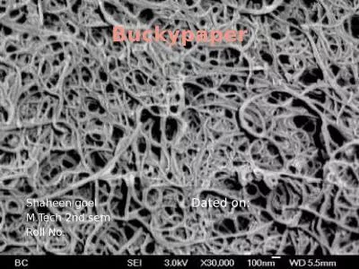
Prof and head of neurology department Faculty of medicine Fayoum university Obstructive sleep apnea in epilepsy a preliminary Egyptian study a preliminary Egyptian study A preliminary Egyptian study ID: 571185
Download Presentation The PPT/PDF document "Hala A. Shaheen" is the property of its rightful owner. Permission is granted to download and print the materials on this web site for personal, non-commercial use only, and to display it on your personal computer provided you do not modify the materials and that you retain all copyright notices contained in the materials. By downloading content from our website, you accept the terms of this agreement.
Slide1
Hala A. ShaheenProf and head of neurology department, Faculty of medicine, Fayoum university
Obstructive sleep apnea
in
epilepsy:
a
preliminary Egyptian studySlide2
a preliminary Egyptian study
A
preliminary Egyptian studySlide3
Hala A. Shaheen
,
Ann A.
Abd El-Kader , Amira
M. El Gohary , Neveen
M. El-
Fayoumy
, Lamia M.
Afifi
Faculty of medicine, EgyptSlide4
Introduction
Epilepsy
and obstructive sleep apnea are two
common disorders that coexistSlide5
Introduction
Epilepsy
and obstructive sleep apnea profoundly
exacerbate each other’s [Peppard etal 2000] Slide6
Introduction
Several
mechanisms may
contribute to the increased incidence of OSA in patients with epilepsy,Slide7
Introduction
The adverse effect of
antiepileptic drugs
CNS depression, Effect on upper airway tone,
Weight gain Slide8
Introduction
Reduced
physical activity
of patients with epilepsy Also seizures prolonged REM
where OSA became worse
[
Sharafkhaneh
A,
etal 2005)Slide9
IntroductionAppearance
of OSA symptoms coincided with clear increase in
seizure
frequency or an emergence of status in patients with epilepsy [Nieto F, etal 2011] Slide10
Introduction
A variety
of
seizure-provoking mechanisms in OSA patients are postulated Slide11
IntroductionObstructive sleep apnea (OSA) is
due
to intermittent
blockage of the upper airway with Consequent reduction or cessation of airflow during sleep [Frucht M, etal 2000]Slide12
Introduction
It
leads to
Cerebral hypoxemia,cortical arousal, Sleep fragmentation, and Slide13
Introduction
Decrease time spent in deep sleep
, and Sleep deprivation [Wyler
A, Weymuller E 2012].Slide14
Introduction
OSA with
subsequent significant morbidity
such as Slide15
IntroductionM
orbidity
increased risk of cardiac, respiratory, and metabolic consequences [Parra etal 2010
]Slide16
Introduction
Mortality
OSA
postulated as a cause of sudden unexpected death in patients with epilepsy [Malow B, etal 2008] Slide17
Introduction
To
make matters worse, obstructive sleep apnea is notoriously
underdiagnosed particularly in patients with epilepsy [Decary A, etal 2000]Slide18
Introduction
The
extent and clinical relevance of the association between epilepsy and sleep apnea are not previously studied in
Egypt Slide19
What we wanted to know was theSlide20
Aim of work
This
study aimed to look for the
frequency of sleep apnea in Egyptian patients with epilepsy in comparison to a control group. Slide21
Aim of work
Try
to find out sleep apnea relation
toClinical data, Sleep complaint Polysomnographic findingsSlide22
Introduction
The
identification and treatment of OSA may have far-reaching consequences in
improving patient’s quality of life [Trupp R, etal 2004] Slide23
Introduction
Continuous
positive airway pressure (
CPAP) treatment was found to improve seizure control in those patients [Gami A, etal 2005] Slide24
Patients and methods:Slide25
“
“
PatientsSlide26
Patients
and methods:
This
is a
case
control study of 26 patients with epilepsy
and
26
normal controls
PatientsSlide27
Patients
and methods:
Inclusion
criteria
Children
with
idiopathic epilepsySlide28
Patients
and methods:
Exclusion criteriaSlide29
Patients
with any
neurological
disease apart from epilepsy; patients with psychiatric illness; patients with history of hypnotics or sedatives
intake; or those with liver or kidney failure
were excluded from the study. Slide30
Patients and methods:
The
patients were divided into two subgroups according to apnea/hypopnea index
(AHI)
: Group
(1)
patients without
OSA
group
(2)
patients with
OSA Slide31
Patients and methods
:
control group
H
ealthy
children their
age and
sex matched
to our patients
were
also
studiedSlide32
Methods
Clinical assessment
Epilepsy
history
and detailed neurological
examination
Epilepsy history:
Neurologist interviewed patients and one of their close relative to inquire about age of onset of epilepsy and disease
duration Slide33
Methods
Clinical assessment
Seizures
characteristics
such
as
Seizure
frequency,
status
epilepticus
,
Circadian
rhythm, and
D
osage of medications
Slide34
MethodsClinical assessment
Type
of epilepsy was determined according to International League against Epilepsy classification Slide35
MethodsSleep
history
They
were asked about total nocturnal
sleep time
,
repeated
awakenings
during sleep, history of excessive
daytime
sleepiness, insomnia, and
nocturnal
snoring
Slide36
Methods
Electrophysiological assessment
Electroencephalogram(EEG)
was done for all patients using a Schwarzer GmbH medical diagnostic
equipmentSlide37
Methods
Electrophysiological assessment
Video
EEG monitoring was done for all patients using a
Schwarzer GmbH medical diagnostic equipment and a digital video-camera Panasonic AG6040) Slide38
Methods
Electrophysiological assessment
Overnight
polysomnography
was performed for both patients and controls using a Schwarzer. Epos 32 GmbH, medical diagnostic
polysomnogram
,
Germany
The
software used was
Somnologica
version 3.1Slide39
Methods
The
polysomnographic
parameters were :
Total
sleep time (TST),
Sleep efficiency,
Percentage
of each
stage
Number
of awakenings,
A
rousal
index
and
leg movementsSlide40
Methods
Polysomnographic
assessment
Apnea
events were counted according to the criteria established by the American Academy of Sleep Medicine
Slide41
Statistical analysis:
Statistical package for social science (SPSS) version 15 were used for data management.
Chi square
test was used for comparison between qualitative variables groupsSlide42
Statistical analysis:
Independent
sample T test
was used for normally distributed quantitative variables as comparing age mean, seizure duration, and sleep efficiency among patient subgroups.Slide43
Statistical analysis:
Mann–Whitney
test
were used for non normally distributed quantitative variables as age of onset, number of awakening, sleep latency, PLM, and apnea and hypopnea index. Slide44
Statistical analysis:
Pearson’s correlation
coefficient was calculated for the association between the apnea/hypopnea index and clinical and polysomnographic data.Slide45
Statistical analysis:
The logistic regression
analysis
was done to test for significant predictors of OSA among the patients.P value 0.05 was considered significant Slide46
ResultsSlide47
Results
The
patients’ age ranged from 4.5 to 18
years. Ten (38.5%) of them were
girls. Sixteen (61.5%) were
boys
. Slide48
Results
The
control
group were chosen to be age-and sex-matched
to the patients group.There were no obese childrenSlide49
Type of epilepsySlide50
ResultsClinical
Mean
age of onset of epilepsy was
7.49 ± 4.2 years. Mean
duration was
5
.09± 4.57 minutes.
Slide51
ResultsClinical
The
seizure frequency ranged from once per month to six per day with mean
of 47.6±53.2 seizures per month
Slide52
Results
Clinical
Seizures were uncontrolled (more than once per month) in 24 patients (
92.3%) and
three patients (11.5%) had history of
status
epilepticus. Slide53
ResultsClinical
Circadian
distribution of the attacks was
diurnal in two patients (7.7%),
nocturnal
in 12 (
46
.2%),
and
both
diurnal and nocturnal in 12 (
46.
2%). Slide54
Results
Clinical
Six patients (23.1%) diagnosed had not received medications yet,
Ten (38.5%) were on
monotherapy
,
and
10 (
38.5
%) were on
polytherapy
Slide55
Sleep
clinical
results
The
patients’ sleep
complaint
Repeated
awakenings
during sleep in 15 patients (57.7%)
Daytime
sleepiness
in 13 patients (50%).
Snoring
was encountered in only two patients (7.7%). Slide56
Polysomnographic
r
esults
The
patients had significantly Higher arousal index and
percent
of stage 2 from total sleep time but
lower
SWS percent from TST and
sleep
efficiency,
in
comparison to the control
group.Slide57
Polysomnographic data
Patients
group
Controls
group
P-value
Sleep onset in min.
18.73 ± 17.5
14.1 ± 12.16
.53
Sleep efficiency %
73.17 ± 7.33
83.12 ± 10.59
.006
No. of awakenings
12 ± 6.03
13.67 ± 5.1
.449
Sleep latency to S1
102.01 ± 114.58
102.43 ± 176.07
.530
Sleep latency to S2
51.7 ± 74.76
17.35 ± 13.21
.157
Sleep latency SWS
81.82± 78.9
74.6 ± 47.55
.489
Sleep latency REM
196.88 ± 108.38
224.08 ± 90.67
.572
% of S1 from TST
8.18 ± 7.18
7.22 ± 6.86
.530
% of S2 from TST
57.01 ± 17.12
44.05 ± 14.98
.022
% of SWS from TST
23.11 ± 10.27
36.08 ± 14.9
.012
% of REM from TST
12.57 ± 9.33
12.62 ± 6.28
.706
Arousal index
8.23 ± 10.88
.89 ± .62
.000
Periodic Leg Movement index
.82 ± 1.33
.38 ± .35
.545
Apnea index REM
.39 ± 1.10
.00 ± .00
.109
Apnea index in NREM
.31 ± .39
.00 ± .1
1
Hypopnea index in REM
1.75 ± 4.91
.00 ± .00
.255
Hypopnea index in NREM
.7 ± .97
.00 ± .45
.775
Apnea hypopnea index
1.21 ± 1.46
.00 ± .75
.704Slide58
Results
Eleven patients with
epilepsy
(42.3%) have Obstructive sleep apnea Slide59
Frequency of sleep apnea in patients Slide60
Comparison between clinical
variables in
epilepsy
patients’groupswith and without OSASlide61
Comparison
Results
Seizure
frequency was significantly
higher in the patients with OSA. All other clinical epilepsy, sleep, and EEG findings did not differ significantlySlide62
ResultsIf sleep
deprivation
is the assumed mechanism, one
might expect that seizures during both sleep and wakefulness would be facilitated as in our study. Slide63
Results
If
sleep
fragmentation and frequent stage shifts resulting from apneas are responsible for provoking seizures, then seizures during sleep may be facilitated preferentially
[Wyler A etal 1981
]Slide64
Clinical
variables
Group (1) Patients without OSA
No. (%)
Group (2) Patients
with OSA
No. (%)
P-value
Age of onset
7.97 ± 3.37
6.83 ± 5.27
.310
Duration of illness
5.23 ± 3.86
4.89 ± 5.6
.435
Sex Female
Male
7 (46.7%)
3 (27.3%)
.315
8 (53.3%)
8 (72.7%)
Type
Generalized
Focal
Focal with secondary generalization
3 (20%)
1 (9%)
.305
5 (33.3%)
7 (63.6%)
7 (46.7%)
3 (27.3%)
Seizure frequency
26.55 ± 36.18
63.07 ± 59.38
.046
Status epilepticus
2 (13.3%)
1(9%)
.738
Circadian rhythm
Diurnal
Nocturnal
Nocturnal and diurnal
Treatment
.550
2 (13.3%)
0 (0%)
7 (46.7%)
6 (40%)
5 (45.4%)
6 (54.6%)
No Medication
Monotherapy
Polytherapy
3 (20%)
3 (27.3%)
.910
6 (40%)
4 (36.3%)
6 (40%)
4 (36.3%)Slide65
ResultsComparison
Results
Comparison
of sleep parameters patients’ subgroup
Apart
from apnea and hypopnea indices, all other
polysomnographic sleep
parameters did not differ between patients’
subgroupsSlide66
Polysomnographic data
Group (1)
patients without OSA
Mean ± SD
Group (2)
patients with OSA
Mean ± SD
P value
Sleep onset in min.
21.84 ± 21.46
14.49 ± 9.3
.659
Sleep efficiency %
85.3 ± 9.39
80.15 ± 11.83
.227
No. of awakenings
10.8 ± 5.02
13.64 ± 7.12
.404
Sleep latency to S1
92.66 ± 128.12
114.76 ± 97.61
.421
Sleep latency to S2
48.83 ± 69.57
55.63 ± 84.66
.659
Sleep latency SWS
79.23 ± 70.47
85.35 ± 92.67
.697
Sleep latency REM
182.69 ± 114.59
216.23 ± 101.34
.516
% of S1 from TST
8.53 ± 7.71
7.71 ± 6.72
.795
% of S2 from TST
57.81 ± 17.8
55.93 ± 16.94
.697
% of SWS from TST
22.07 ± 9
24.54 ± 12.08
.406
% of REM from TST
11.62 ± 9.88
13.87 ± 8.81
.311
Arousal index
5.57 ± 6.45
11.85 ± 14.57
.233
Periodic Leg Movement index
.86 ± 1.28
.75 ± 1.45
.546
Apnea index REM
.00 ± .00
.93 ± 1.58
.005
Apnea index in NREM
.16 ± .21
.51 ± .48
.062
Hypopnea index in REM
.03 ± .13
4.1 ± 7.05
.000
Hypopnea index in NREM
.23 ± .24
1.34 ± 1.22
.006
Apnea hypopnea index
.36 ± .27
2.36 ± 1.63
.000Slide67
Correlation between
apnea/hypopnea
index,
Clinical and
polysomnographic dataSlide68
Correlation
Apnea
index
in REM positively correlates with latency to deep sleepSlide69
Correlation
between Apnea index
and
latency to deep sleep
CorrelationSlide70
ResultsCorrelation
Hypopnea
index in REM
positively
correlates with number of awaking (r=0.393, P=0.047). Slide71
Correlation
number of awakening and hypopnea index Slide72
Discussions
It
is well-known that sleep
apnea is not the same throughout the course of the night. It tends to be worse in (REM) sleepSlide73
DiscussionsP
atients
with epilepsy and OSA had significantly
longer sleep latency and higher arousal index. A significant tendency towards light sleep than slow wave deep sleepSlide74
Discussions
In this study, no epilepsy or sleep data have been
found to be significant
predictors of OSA in patients with epilepsySlide75
Discussions
Contradictory
to previous study that reported that older
, heavier male, and sleepier epileptics are more prone to haveOSA [Raffaele M, etal 2003].Slide76
left
centrotemporal
sharp wavesSlide77
Polysomnographic
recording
O
bstructive sleep
apnoea
Slide78
ConclusionsSlide79
ConclusionsObstructive sleep apnea is
frequent
in patients with epilepsy
. Obstructive sleep apnea contribute to increase seizures frequency.Slide80
Recommendations
Investigating
sleep apnea in all patients with
epilepsy even those without sleep complaintSlide81
Egypt
Fayoum
neurology
Conference 10-12
November 2016
has01@
fayoum.edu.eg
FinallySlide82
Thank You