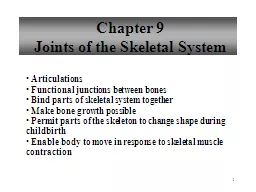
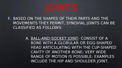
Joint Classification Synovial Joints Characteristics of synovial joint Types of synovial joints Naming joints Pectoral Girdle Upper Limb Pelvic Girdle Lower Limb Joints Of The human body Joint is a point of connection between two bones ID: 650752
Download Presentation The PPT/PDF document "Joints of the human body" is the property of its rightful owner. Permission is granted to download and print the materials on this web site for personal, non-commercial use only, and to display it on your personal computer provided you do not modify the materials and that you retain all copyright notices contained in the materials. By downloading content from our website, you accept the terms of this agreement.
Slide1
Joints of the human bodySlide2
Joint Classification
Synovial Joints
Characteristics of synovial jointTypes of synovial joints
Naming joints:Pectoral GirdleUpper LimbPelvic GirdleLower Limb
Joints Of The human bodySlide3
Joint is a point of connection between two bones
Strands of connective tissue, ligaments, hold the bones together and ensure the stability of jointsSlide4
JOINT CLASSIFICATION
Joints are classified according to their motion capabilities:
Three terms Synarthroses - Immovable
Amphiarthroses - Slightly movableDiarthroses - Allow the greatest amount of motionSlide5
Joint Classification
Joints are further classified by the material that joints them:
Fibrous joint -allow no movement -E.g. sutures of the skull Cartilaginous joints -allow limited movement -E.g. inverterbral discs Synovial joints
-allow large range or movements
-E.g. hip jointSlide6
Characteristics of synovial joints
Hyaline cartilage
A protective layer of dense white connective tissue that covers the ends of the articulating bonesJoint CavitySynovial Membrane
Covers joint cavity, except over the surfaces of the articular cartilagesSecretes the lubrication fluidSynovial FluidLubricates the jointCapsuleMay or may not have the thickenings called intrinsic ligamentsExtrinsic ligamentsSupport the joint and connect the articulating bones of the jointSlide7
Types of sYNovial Joints
There are thee basic types of synovial joints:
Unilateral (rotation only about one axis)Biaxial joints (movement about two perpendicular axes)Multiaxial Joints
(movement about all three perpendicular axesSlide8
Types of Synovial joints cont’d
Synovial Joints are further classified into:
1) Hinge joint2) Pivot joint3) Condyloid4) Saddle shaped joint5) Ball and socket joint
6) Plane jointSlide9
Hinge (ginglymus) Joint
Uniaxial
Has one articulating surface that is convex, and another that is concaveE.g. Humero
-ulnar elbow joint, interphalangeal jointSlide10
Pivot Joint
Uniaxial
E.g. head of radius rotating against ulnaSlide11
Condyloid (knuckle) joint
Biaxial (flexion-extension, abduction-adduction)
The joint surfaces are usually oval One joint surface is an ovular convex shape, and the other is a reciprocally shaped concave shape surfaceE.g. Metacarpophalangeal Joint Slide12
Saddle Joint
Biaxial (flexion-extension, abduction-adduction)
The bones set together as in sitting on a horseE.g. carpometacarpal joint of the thumbSlide13
Ball and socket joint
Multiaxial ( rotation in all planes )
A rounded bone is fitted into a cup = like receptacleE.g. shoulder and hip jointsSlide14
Plane (gliding) Joint
Uniaxial (permits gliding movements)
The bone surfaces involved are nearly flatE.g. intercarpal joints and acromioclavicular joint of the vertebraeSlide15
Joints of the pectoral girdleSlide16
Sternoclavicular joint
Connects sternum to the clavicle
The only joint connecting the pectoral girdle to the axial skeleton True Synovial joint strengthened by an intracapsular disc and extrinsic ligamentsSlide17
Acromioclavicular joint
Unites the lateral end of the clavicle with the acromion process of the scapula
Where shoulder separations often occur in sports such as hockey, baseball and footballSlide18
Glenohumeral joint
Connects the upper limb and the scapula
A Typical multiaxial joint Has a wide range of movement at this jointCompromise = relative lack of stabilitySlide19
Upper Limb JointsSlide20
Elbow Joint
There are three joints at the elbow
humero-ulnar joint Medial (with respect to anatomical position)
Between the trochlea of the jumerus and the olecranon process on the ulnaHumero-radial joint lateral between the capitulm of the humerus and the head of the radius
Radio-ulnar joint
between the radius and the ulnaSlide21
Elbow joint con’tSlide22
Joints of the wrist
Radio-carpal joint
-between distal end of the radius and the carpals-movements : Flexion – extension and abduction – adductionSlide23
Joints of the HAND
Intercarpal
joints -between the bones of the carpus
-gliding jointsCarpometacarpal joint -between carpals and metacarpals -the characteristics of the carpometacarpal joint of the thumb allows the range of movement necessary for oppositionSlide24
Joints of the hand cont’d
Metacarpophalangeal joints
-joint between metacarpals and carpals -the knuckles
-movements : flexion-extension, and adduction-abductionInterphalangeal joints -joints between the phalanges -permit flexion-extensionSlide25
Joints of the pelvic girdleSlide26
Hip joint
B
etween the head of the femur and the cup (acetabulum ) of the hip bone (os coxae)-Like shoulder joint, hip joint is:
-ball and socket joint -multiaxial joint that allows : flexion-extension, adduction-abduction and circumductionSlide27
Hip joint con’t
-
unlike shoulder joint, hip joint is very stable-in fact it is the body’s most stable synovial joint due to -deepened socket (via lip or fibrocartilaginous labrum)
-an intrinsic and very strong extrinsic ligamentsDislocations in sports is not common, but can occur in car collisionsDislocate the head posteriorly or drive it through the posterior lip of the acetabulumSlide28
Lower limb jointsSlide29
Knee joint
Tibiofemoral or knee joint
Incredible range of movement(flexion-extension)Slide30
Knee joint cont’d
However, the knee joint is relatively stable due to additional supports from:
MenisciShock absorbing fibrocartilaginous discsAnterior and posterior cruciate ligamentsIn the center of the jointLateral and medial collateral ligaments
Extending from the sides of the femur to the tibia and fibulaThe musculature that surrounds itSlide31
Knee joint cont’d
Movements:
Primary action is flexion – extension (e.g. squat or jump) When flexed, medial and lateral rotation can also occurSlide32
Ankle Joint
Talocrural or ankle joint
Involves several bones:Medial and lateral malleoli of the tibia and fibulaHead of the taluscalcaneus ( heel bone ) Slide33
Foot and toe joints
I
ntertarsal jointsBetween tarsalsTransverse tarsal jointBetween the proximal and distal row of tarsal bones
Movement – inversion- eversion of the sole of the foot This action enables you to adjust to uneven ground when walking or runningSlide34
Foot and toe joints cont’d
As in the hand, there are joints between the tarsal bones, metatarsals and phalanges.
Tarsometatarsal Metatarsophalangeal Interphalangeal They are strengthened by planter ligaments which aid in maintaining the arch of the footSlide35
Muscles of the human bodySlide36
Over 600 in the human body
Allow the skeleton to move
To cause movement, both ends of each muscle must be attached to boneOrigin or proximal attachment – the attachment closer to the center of the bodyInsertion or distal attachment – the attachment away from the center of the bodySlide37
Facial musclesSlide38
Various functions of the facial muscles
Enable you to change expression and display your emotions outwardly
Allow you to close your eyes and blinkEssential for opening and closing the mouth for:Keeping food in the mouthAllowing you to chewAllowing you to form words
TongueFunction:Allows you to move food around in your mouth and get it to a position to be swallowedAllows you to pronounce words and speak intelligentlySlide39
Neck musclesSlide40
Neck muscles
There are muscles posterior, lateral and anterior tot eh neck or cervical region
These muscles Maintain the position in which the sits on the 1st cervical vertebra (atlas) i.e. they hold up our head
Also permit a wide range of movementSlide41
Sternocleidomastoids
The most important anterior pair of muscles
Movements:Acting together, they flex the head toward the chestIndividually, each muscle tilts the face up and toward the opposite sidSlide42
Erector spinae muscles
Important posterior neck muscles
Attach to sacrum inferiorly and to the skull superiorlyMaintain your erect positionTherefore they are also called anti-gravity muscles
When someone faints, these muscles no longer function and the body falls face forward to the groundSlide43
Trapezius, upper fibres
Important lateral neck muscles
Movements:Acting alone tilt the head to the same sideActing together, they assist in neck extension