/
4.1 Joints and Muscles 4.1 Joints and Muscles

4.1 Joints and Muscles - PowerPoint Presentation

sherrill-nordquist
sherrill-nordquist . @sherrill-nordquist
Follow
457 views
Uploaded On 2016-04-03

4.1 Joints and Muscles - PPT Presentation

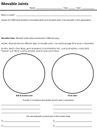
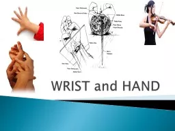
What would life be like without Joints Move a joint that you use often How do different joints move Essential Question 1 What role do joints play in the human body Joints are the places where two bones meet and allow movement amp flexibility and provides support to ID: 273094

muscles muscle joint joints muscle muscles joints joint origin motion fibers tissue insertion attachment bones move movement contraction question

Share:

Link:

Embed:

Download Presentation from below link

Download Presentation The PPT/PDF document "4.1 Joints and Muscles" is the property of its rightful owner. Permission is granted to download and print the materials on this web site for personal, non-commercial use only, and to display it on your personal computer provided you do not modify the materials and that you retain all copyright notices contained in the materials. By downloading content from our website, you accept the terms of this agreement.


Presentation Transcript

Slide1

4.1 Joints and MusclesSlide2

What would life be like without

Joints

Move a joint that you use often

How do different joints moveSlide3

Essential

Question

1. What role do joints play in the human body?

Joints are the places where two bones meet and allow movement & flexibility and provides support to the human skeleton.

2. How are joints classified by both structure and function?

Functionally, joints are classified by how much motion they allow. Structurally, joints are classified as fibrous, cartilaginous, or synovial.Slide4

Joint Classification

Immovable/Fibrous

Do not move—EX:

joints in dome of skull and between teeth and jawbone

Partially Moveable/Cartilaginous

Move little—linked by cartilage—EX: vertebrae in spine

Immovable joints and slightly movable joints are restricted mainly to the axial skeleton where protection and stability are key Slide5

Joint ClassificationFreely Moveable/Synovial

Move in many directions— found at the hip, shoulders, elbows, knees, wrists, and ankles —filled with synovial fluid (acts as lubricant)—these joints have synovial cavities

Freely movable joints are found on the appendicular skeleton and permit flexibility in the limbs.Slide6

Activity 4.1.1 Bones, Joints, Action!

Obtain a body system graphic organizer (skeletal view)

Research the six main types of synovial joints

Complete activity through question 6Slide7

Essential Question

3. What are the different types of synovial joints?

Pivot joint

Ball-and-Socket joint

Saddle joint

Condyloid

(Ellipsoidal) joint

Hinge joint

Plane (Planar or Gliding) jointSlide8

Types of Synovial Joints Slide9

Types of Synovial Joints Slide10

Activity 4.1.1 Bones, Joints, Action!http://www.youtube.com/watch?v=9fZkne0GE9g-

cow elbow dissection

Create groups of 3 or 4

Obtain Gloves, Goggles and Cow Elbow

Look for the movement of the joint, cartilage, tendons and ligaments

Complete the rest of Activity 4.1.1 and Conclusion QuestionsSlide11
Slide12

Dissected Cow ElbowSlide13

Activity 4.1.1 Bones, Joints, Action!How are cow elbows and human elbows similar and different?

I

n

the cow joint, the ulna and the radius are fused; whereas in the human, they are two separate

bones

Human elbow

joint allows for rotation and overall dexterity (not needed by the cow

)

What type of synovial joint was modeled by cow elbow?Slide14

Connective Tissue

Connective tissue protects, supports, and binds together other body tissues.

Connective tissue is made up of different types of cells in varying amounts of a nonliving substance around the cells, called the

matrix

.

Fibrous

connective tissue which is found in tendons and ligaments. Fibrous connective tissue is composed of large amounts of closely packed collagenous fibers

.

Cartilage is a form of fibrous connective tissue that is composed of closely packed collagenous

fibers

in a rubbery gelatinous substance

called

chondrin

.

 Slide15

Essential Question

4. What role do cartilage, tendons, and ligaments play at a joint?

Cartilage

-

Cushions/protects bones where they meet and rub against each other. The cartilage found in joints is

hyaline cartilage—the same kind found in a fetal skeleton & it’s referred to as articular cartilage where it attaches to articular bone surfaces.

Tendons

-

Fibrous tissue that connects muscles to bones

Ligaments

-

Fibrous straps that fasten bones to other bones

Slide16

Motion with the Cow ElbowHow did your cow elbow move?

Think about the type of synovial joint

How do human elbows move?Slide17

Flexibility

What makes our bodies flexible?

JointsSlide18

Joints

Joints

with a large range of motion has limited

stability

Joint

with limited mobility, such as the sutures in the skull, have great

stability

M

ove your hip

and

shoulder

and describe the range of motion of each

joint

J

oints

make up for a lack of stability by the addition of

muscle

Joints

that are very stable and produce little movement are assisted by limited amounts of

muscle

Joints

that are very flexible, but offer little stability are surrounded by large amounts of

muscleSlide19

Describing MotionHow do we describe the motions of a joint ?

Bending

Flexing

Scientists

and medical professionals use precise terms to describe the direction of motion as well as the relationship of one body part to

another

Depression and Elevation

(Make this Motion)Slide20

Elevation and Depression Movement

Elevation

DepressionSlide21

Activity 4.1.2: Range of Motion

In groups of 3 research the following Terms and Document on your Body Organizers

(Complete Steps 1 & 2)

Depression

and elevation

Rotation and circumduction

Flexion and extension (and hyperextension)

Abduction and adduction

Plantar flexion and dorsiflexion

When you know all the Motion as a group demonstrate them to your teacherSlide22

Essential Question

5. What terms describe the path of movement at a joint

?Slide23

Essential Question6. What is range of motion

?

Range of motion

is the range through which a joint can be moved & can be measured using a goniometer to determine angles. Slide24

Essential Question

7. How do you measure the range of motion of a particular joint movement?

Each specific joint has a normal range of motion that is expressed in degrees.

Devices to measure range of motion in the joints of the body include the goniometer and inclinometer which use a stationary arm, protractor, fulcrum, and movement arm to measure angle from axis of the joint.

As measurement results will vary by the degree

of

resistance, two levels of range of

motion

results are

recorded in most cases.Slide25

Normal ROM for Joints in Adults Slide26

Finish Activity 4.1.2: Range of Motion

You will need

Activity 4.1.2 Range of Motion

Activity 4.1.2 Student Resource Sheet - ROM

ROM

Schematics

Goniometer

http://

www.youtube.com/watch?feature=player_embedded&v=ZUF7tpkVAIY-

or

http://

www.youtube.com/watch?v=J_R-igYFj98&feature=player_embedded

Instructions on how to use a goniometerSlide27

Key TermsSlide28
Slide29

Essential Question

8. How

do bones, muscles and joints work together to enable movement and locomotion for the human body?

Our bones provide support and give our bodies shape, but cannot move on their own. The muscles provide the movement. The joints help attach bones to one another to provide flexibility & allow the muscles to help give the bones a way to move. Slide30

Lesson 4.2 MusclesSlide31

Essential Question

1. How do muscles assist with movement of the body and of substances around the body

?

Our muscles are what allow all movement of our bodies (and within our bodies). They help us involuntarily by helping food move down the esophagus and into the stomach (peristalsis) and helping blood move through our bodies (the heart is a muscle). They also help us move our bodies voluntarily from place to place (the muscles in our limbs). Our bodies each have about 650 muscles & are ~ 50% muscle by weight!Slide32

Activity 4.2.1 Muscle Rules Part 1

With a partner research the following 3 muscle tissues

skeletal muscle

smooth muscle

cardiac muscle

Create this table in your Journal

View prepared slides

Complete Part 1 onlySlide33

Essential Question

2. How do the structure and function of the three types of muscle tissue compare?

Cardiac

- They are striated muscle fibers form the wall of the heart & function involuntarily.

Skeletal

-They are attached to bone, mostly in the legs, arms, abdomen, chest, neck and face. They are striated muscle fibers (lined under microscope) & attach to bone by a tendon. They hold the skeleton together and give the body shape. They are voluntary (we control them) and contract quickly and powerfully), but they tire easily.

Smooth

-They are smooth (not striated) & are controlled automatically by our nervous system. They are also called ―involuntary‖ muscles. They make up the walls of the stomach and intestine to help break down and move food. They also line the walls of blood vessels. They take longer to

c

ontract than skeletal muscles, but also don’t tire as easily.

Slide34

Skeletal Muscle

Voluntary – we control the movement

Striated – looks like long fibers

Linked to bones by tendons

Function – to help us move / move our bonesSlide35

Smooth Muscle

Involuntary Action –

controlled by our CNS

Non-striated

Found in arteries, veins, intestines, etc.

Function : Maintain organ dimensions – stretch and recoilSlide36

Cardiac Muscle

Involuntary

Striated – but, may be branched which is unlike skeletal muscle.

Found in walls of the heart

Function : To pump the heart!

Highly resistant to fatigue

w/lots of mitochondriaSlide37

Let’s Start By Building a Muscle from Spaghetti

Pick up one piece of spaghetti.

Each piece of spaghetti will represent one skeletal muscle cell or fiber

Each muscle fiber is enclosed by a delicate membrane called the

endomysium

.

(For the purposes of this activity, the yellow outer coating of the spaghetti represents this membrane.)

Activity 4.2.1 Muscle Rules

Part 2Slide38

Spaghetti Muscle Cont’d

Pick up a handful of spaghetti

. This bundle of fibers

represents a

fascicle

.

Each fascicle, however, is covered by a membrane called the

perimysium

.

Place the bunch of spaghetti on the end of a piece of plastic wrap.

Roll the spaghetti up in the plastic used to represent the

perimysium

.

Hold up the completed

fascicle.

Pull the ends taut, and notice that this tissue has little to no bumps.

These ends represent

dense regular connective

tissue. Slide39

Spaghetti Muscle Cont’d

Fascicles group together to form a skeletal muscle.

Combine your fascicle w/ three other pairs’ to form a whole muscle.

These fascicles are bound together by an even tougher outer membrane called the

epimysium

.

Wrap the combined fascicles in another piece of plastic wrap.

This layer of wrap will represent the

epimysium

.

Twist the plastic wrap on each end of the completed muscle.

At the ends of the muscle, the

epimysia

blend together to form tendons, cordlike structures that attach muscle to bone, cartilage or other connective tissue. Slide40

Essential Question

3. How are muscle fibers and membranes organized to form a whole skeletal muscle?

The

epimysium

(“upon muscle”) is the outermost layer of connective tissue.

The

perimysium

(“around muscle”) is made of connective tissue and forms casings for bundles of muscle fibers.

The

endomysium

(“within muscle‖) is connective tissue surrounding each individual muscle fiber.

Each fascicle is a small cluster of muscle fibers, with

endomysium

between the individual fibers. Blood vessels run between the fascicles, bringing the tissue nutrients & removing waste. Nerves also run throughout, controlling the movement of the muscles. Together, the network of nerves and blood vessels are referred to as the plexusSlide41

Activity 4.2.1 Muscle Rules Part 3

Will need your

Manikins

Clay

Lab Journals

We will create a muscle togetherSlide42

Step 1Locate the ventral side and use a pencil to place a dot on the lateral and medial side of the radial groove (about halfway up the humerus). Slide43

Step 2Locate the ulna just below the fold of the elbow. Help the students see the hollowed out area in the

antecubital

region. Place a pencil dot above this area.Slide44

Step 2 Cont’d - Rule 1

These

dots each represent an attachment point for a muscle.

Note

that there are at least two attachments (in this case three) and the muscle will cross a joint at the elbow.

This leads us to Muscle

Rule #1:

Muscles must have at least two attachments and must

cross at least one joint. Slide45

Step 3 – Brachialis Muscle

Using terra cotta clay, form two balls about the diameter of a nickel

.

Rolling the clay between the tabletop and a palm, roll each ball into a long carrot. The total length of the carrots should stretch from the humeral attachment to the

ulnar

attachment.

Bring the fat part of the carrots together, leaving the tops free

(

rabbit ears

).Slide46

Step 4 – Rule 2

Using your left

thumb to represent the humeral attachments and

your

left middle finger to represent the

ulnar

attachment, place the left hand on the right arm where the attachments would

be.

Make

sure to cross the joint.

Pull your

forearm towards

your heart

and

watch

the position of

your

fingers.

You should

notice that

your

index finger and thumb are closer together than when

you

started

.

This lead to

rule 2:

Muscles always “pull” and get shorter. Slide47

Step 5 – Rule 3

Repeat

the motion and identify which attachment is “pulling” or moving closer to the other

attachment

.

The

attachment that moves is known as the

insertion

of the muscle.

The

insertion is usually the distal attachment.

The

attachment that does not move and pulls the other attachment toward it is referred to as the

origin

.

The

origin is usually the proximal attachment.

This leads to Rule 3:

The

attachment that moves is known as the insertion and the attachment that remains stationary is known as the origin. Slide48

Step 6

Extend your

arms out in front of their bodies.

Notice

this angle

is

180°.

Show

the movement

again of

the muscle

you

have

just built

.

This

time

pay

attention to what happens to this angle when the muscle shortens.

Notice that

the angle decreases.

Do you remember

what we call motion at a joint that decreases the angle between articulating

bones?

Flexion

and thus a muscle such as this is referred to as a

flexor

.

Slide49

Step 7 – Rule 4

Flex your arms one more time, but stop at the end of the movement.

If muscles only pull, then how can the arm be straightened?

What do we call motion at a joint that increases the angle between articulating bones?

Extension

and thus a muscle that controls this movement is referred to as an

extensor

.

Muscles

that decrease the angle between ventral surfaces of the body are known as flexors. Muscles that increase the angle between ventral surfaces of the body are known as extensorsSlide50

Step 8Place a pencil dot halfway up the dorsal side of the humerus.

Place

another dot just distal of the elbow onto the ulnaSlide51

Step 9 –triceps medial head

Using

terra cotta clay, form a ball the diameter of a nickel. Roll the ball into an

even tube

.

Attach the ends of the clay tube to dots on the humerus and on the ulna.

Since

the back of the humerus is flat, the muscle shapes to the bone and is also flat.

Use your

thumbs to flatten

the clay

.

Remove

any clay that

makes its way

to the

ventral

side. Slide52

Step 9 Cont’d

Act

out the action of this muscle. With the right arm in the flexed position, place the left thumb on the back of the humerus and the left index finger on the back of the elbow.

“Pull

” with

your

index fingers and the angle should increase to 180°.

Repeat

the motion and

think of Rules

2, 3 and 4.

Since

the angle in this motion increases, the muscle is an extensor

. Slide53

The TricepsOrigin = proximal half of dorsal humerusInsertion = distal of elbow on the ulna

Action = extends elbowSlide54

Flexors and Extensors

Flexors

are on the ventral side of the body and extensors are located dorsally

.

For smooth movements to occur, can both extensors and flexors be contracting at the same time?”

When

the flexors are pulling, the extensors are relaxing.

This brings us to

Rule #5:

Muscles

work in opposing pairs

.Slide55

Rule # 6

Muscle striations point to the attachments and show the direction of pull.Slide56

Naming Muscles

Each muscle is given a Latin name based on one or more of its

features

Take a look at the following muscle names and brainstorm what you can tell about these muscles simply by their names

Trapezius

and Rhomboid minor

Gluteus

maximus

and Gluteus

minimus

Frontalis

and

Temporalis

Orbicularis

Oculi

and Transverse

abdominis

Flexor

Carpi

Ulnaris

and Extensor

digitorum

longus

SternoCleidomastoid

and

Brachioradialis

Biceps

Brachii

and Triceps

BrachiiSlide57

Essential Question

Muscles

each have an insertion, where they attach to the moveable bone and an origin, where they attach to the stationary bone.

4. What do skeletal muscle structure and attachment to bones tell you about function?Slide58

Essential Question

5. How are muscles named?

Several factors are considered when naming a muscle, including

1) Location (EX:

tibialis

anterior is on the front of the tibia)

2) Shape (EX: deltoid ―resembles‖ (-

oid

) a ―triangle‖ (

delt

))

3) Points of attachment (EX:

sternocleidomastoid

—the muscle attaches to the sternum and the tendons attach to the mastoid process of the skull.)

4) Relative size (EX:

gluteal

or ―rump‖ region – the gluteus

maximus

is bigger and the gluteus

minimus

smaller).

5) Number of muscle ―heads‖ or divisions (EX: Biceps means ―two-headed‖ and has two divisions)

6) Direction of muscle fibers (EX: the rectus

abdominis

muscle is located in the front of the abdomen and its fibers are oriented in a ―straight‖ (

rect

), vertical direction).

7): Association with characters (EX:

sartorius

means ―presence of‖ (-us) a ―tailor‖ (

sartori

)! Tailors used to sit cross-legged upon the ground. The

sartorius

is actually located along the inner aspect of each thigh. Thus, when it contracts, it flexes (bends) the lower leg like an ancient tailor. Slide59

Activity 4.2.2: Building a Better Body Slide60

HBS - A.4.2.2

How to Build a Better Body

Please grab your

maniken

and a tool kit and sit with your partner!

Use the wrench to take the arm off your

Maniken

.

DO NOT LOSE THE SCREWS!Slide61

Muscle #1: Intercostals

We will

build the external intercostals of

the

chest

. These muscles are found in

between

the

ribs and extend from the front of the ribs,

around

back and past the bend in the bones.

Describe

the function of the muscles that are found between the ribs.

These

muscles help move air in and out of the chest.

When you eat

ribs,

you are

actually eating the

intercostal

muscles between the bones, not the ribs themselves.  

Place a strand of spaghetti between each rib, starting at the back of the rib where it attaches to the vertebral column, all the way around to the rib’s attachment at the sternum.  

Use your thumb or one of the clay tools to flatten down these strands. The

intercostal

muscles do not stick out of the chest. Slide62

Muscle #2 – Serratus Anterior 

Attach the stand-off to the torso. The indentation in the stand-off should face the midline of the model. Do not yet tighten the screws completely.

The Maniken

®

displays vertical dashes midway around the ribs to indicate where the bone becomes cartilage.

Origin = lateral surface of ribs 1-8 (bone only)

Insertion = medial border of the scapulaSlide63

Muscle #2 – Serratus Anterior 

Take small pieces of spaghetti and attach these strands from the medial side of each rib (where the dashes are shown) to the stand-off on the arm

.

Attach

one strand from each of ribs 1-8 to form a saw-like structure – a “serrated” edge.

This

muscle helps move the scapula forward and is often used at the end of big

movements

such as a bench press, a

baseball

pitch, or a swimming stroke.

Attach the

arm of

your

Maniken

®

.

The

screws should thread in as easily as

they

unthreaded on removal

. Slide64

Muscle #3- Pectoralis Minor

Where are the origin and insertion of the

pectoralis

minor?

Origin = anterior surface of ribs 3 – 5 (just past the origins of the

serratus

anterior)

Insertion = coracoid process of the scapula (piece of the scapula visible on the front)

Use spaghetti strands to form the

pectoralis

minor. Place one small strand at the origin of each rib and run these three strands together as they attach at the scapula. The

muscle is built in a manner similar to the

serratus

anterior.

Act out the movement of this muscle.

This muscle works to rotate the shoulder

forward

. Slide65

Muscle #4 – Pectoralis Major

Even though this muscle only has one name, there are actually three different “heads” or pieces to this muscle.

Each part will be built separately and will be formed from a carrot-shaped tube that has been rolled flat.

Keep these muscles thick and striate each muscle as it is built.

Have students first construct the abdominal head of the

pectoralis

major.

Given the name only, ask students where they think this muscle might attach.

What are the origin and insertion of the abdominal head of the

pectoralis

major?

Origin = ribs 5-7 (actually attaches to fascia of abdominal muscles)

Insertion = lateral edge of the most proximal part of the

humerus

Make a long carrot out of terra cotta clay. Flatten the carrot slightly to make a tongue.Slide66

Muscle #4 – Pectoralis Major

Gently lay the muscle across the chest of the

Maniken

®

from the origin to the insertion.

The long end of the carrot should point towards the

shoulder

and the wide end should run down towards

the 5

th

through 7

th

rib. The muscle will have a teardrop shape. Keep

the insertion very narrow and the origin much wider. Do not worry

about perfect shape at this point. You will trim the muscle to fit the

Maniken

®

.

Use the wire tool or a pencil to carefully outline the shape of the muscle and trim off any jagged edges.

Take the muscle off the model and use the knife to trim the edges you have marked with your tool or pencil. Gently roll out the muscle if you need to stretch it a bit to fit from the origin to the attachment.

Attach the muscle to the model. Ask students to striate the muscle. Remember that the striations of the muscle indicate the direction the muscle moves. Slide67

Pectoralis MajorAct out the motion of this portion of the

pectoralis

major.

Which sports or exercises utilize this muscle?

Tennis serve or a volleyball spike.

We will now create the largest portion of the muscle – the sternal or

sternocostalis

head.

Where do you think this muscle might attach.

Origin = ribs 1-5 on the lateral edge of the sternum (no clay should be on the sternum)

Insertion = lateral edge of the

humerus

, inferior to the insertion of the abdominal head.

Make a short, fat carrot out of terra cotta clay. Flatten the carrot slightly to make a thick triangle. Do not worry about perfect shape at this point. You will trim the muscle to fit the

Maniken

®

. Slide68

Pectoralis Major

Gently lay the muscle across the chest of the model from the origin to the insertion.

The long end of the carrot should point towards the

humerus

and the wide end should run along the lateral edge of the sternum.

The origin of this muscle will overlap the origin of the abdominal head.

Use the wire tool or a pencil to carefully outline the shape of the muscle.

Take the muscle off of the model and use the knife to trim the edges you have marked with your tool or pencil. Gently roll out the muscle if you need to stretch it a bit to fit from the origin to the attachment. Make sure no clay extends over the sternum.

Attach the muscle to the

Maniken

®

. Ask students to striate the muscle. Remember that the striations of the muscle indicate the direction it moves. Slide69

Pectoralis Major

Ask students to act out the motion of this portion of the

pectoralis

major.

Which sports or exercises utilize this muscle?

This muscle adducts the arm across the chest and is at the route of a tennis forehand shot.

The butterfly machine in the gym allows a person to isolate and train this portion of the muscle.

Create the smallest portion of the muscle – the

clavicular

head.

What are the origin and insertion of the

clavicular

head of the

pectoralis

major?

Origin = medial half of inferior edge of the clavicle

Insertion = lateral edge of the proximal

humerus

, inferior to the insertion of the sternal headSlide70

Pectoralis Major

Make a small carrot out of terra cotta clay.

Flatten the carrot slightly to make a shape

similar to an isosceles triangle. Do not worry

about perfect shape at this point. You will trim the muscle to fit the

Maniken

®

.

Gently lay the muscle across the chest of the model from the origin to the insertion. The long end of the carrot should point towards the

humerus

and the slightly wider end should run up against the bottom of the clavicle. The insertion of this muscle will cross over the insertion of the other two muscles on its way to the humeral attachment. Slide71

Pectoralis Major

Use the wire tool or a pencil to carefully outline the shape of the muscle.  

Take the muscle off the model and use the knife to trim the edges you have marked with your tool or pencil. Gently roll out the muscle if you need to stretch it a bit to fit from the origin to the attachment.  

Attach the muscle to the

Maniken

®

. Striate the muscle. Remember that the striations of the muscle indicate the direction it moves. Slide72

Building MusclesThinking about the muscles we built, why would one exercise not tone all of the muscles.Slide73

Essential Question

6. What

are the requirements for muscle contraction

?

Calcium and ATP are cofactors required for the contraction of muscle cells.

ATP supplies the energy

Calcium is required by two proteins that regulate muscle contraction by blocking the binding of myosin to filamentous

actin

Troponin

Tropomyosin

In a resting

sarcomere

,

tropomyosin

blocks the binding of myosin to

actinSlide74

Essential Question

7. What role do calcium and ATP play in muscle contraction?

1) Calcium ions cause

troponin

and

tropomyosin

to shift, exposing myosin binding sites

2) Myosin heads connect with

actin

binding sites & move the thin filament, contracting the muscle

3) The ADP & P that caused the myosin heads to cock back are left behind during the power stroke

4) Introduction of ATP causes myosin heads to release the

actin

5) ATP is broken down into ADP & P, causing myosin heads to cock back and prepare for another power strokeSlide75

Muscle Contraction

http://www.youtube.com/watch?v=hqynCsign8E-

Video

discussing muscle contractionSlide76

Activity 4.2.4 Laws of Contraction

Pair up

You will need:

5 Test tubes

5

Micorscopic

slides

Salt solution, no ATP

0.25% ATP in distilled water

0.25% ATP in salt solution

0.10% ATP in salt solution

0.05% ATP in salt solution

Disposable transfer pipettes (1ml)

Laboratory journal

Teasing needles (from dissection kits) or straight pins

Forceps or tweezers

Millimeter ruler

Microscope

Frog MuscleSlide77

Essential Question8. What is a

sarcomere

?

The contractile unit of a myofibril;

sarcomeres

are repeating structural units of striated muscle fibrils, delimited by the Z bands along the length of the myofibril.

9. How does a

sarcomere

contract and lengthen to cause muscle contraction?Slide78

Muscle Contraction-SacomereSlide79

Molecular Muscle MovementMuscle filaments are called myofibrils. Myofibrils are made up of two kinds of filament:

Thin filaments made of

actin

protein

Thick filaments made of

myosin

protein.

Actin

and myosin filaments work together to make muscles contract. These fibers are located between protein sheets called Z-discsSlide80

Muscle Contraction

Actin

and myosin are layered. Myosin filaments have hooked parts that will stretch and pull themselves along the

actin

filaments when ATP attaches to them.Slide81

Muscle Filament ContractionSlide82

Muscle Filament Relaxation

Calcium (Ca

2+

)

must be removed for the muscle fibers to relax

Moving (Ca

2+

) back into the

sarcoplasmic

reticulum is ACTIVE TRANSPORT (requires ATP)

If the muscle cannot remove the (Ca

2+

)

the muscle cannot relax and will stay contracted.Slide83

Rigor MortisSlide84

Rigor MortisHow is the condition rigor mortis related to muscle contraction?

After death

actin

and myosin shorten muscle fibers.

ATP is needed to release the myosin heads from the

actin

fibers and allow muscles to relax, but ATP reserves are quickly depleted, causing muscles to remain contracted.

It can take 10 minutes to hours to occur, with maximum stiffness 12-24 hours after death.

Eventually tissue decays and

lysosomal

enzymes leak and cause muscles to relax.Slide85

Essential Question

11. How do nerves interact with muscles?

In order for muscles to contract (shorten and thicken), they must receive a message from the CNS to do so. The messages come through efferent neurons (nerves that move away from the CNS).

Afferent neurons send messages back from muscles to the CNS.

If there are problems with nerves, it can lead to issues with muscle function (i.e. Carpal Tunnel Syndrome)Slide86

Activity 4.2.6: You’ve Got Nerve

Building Nerves

Part 1 Brachial Plexus and Radial NerveSlide87

Activity 4.2.6: You’ve Got NervePart 2 Carpal Tunnel

Carpal tunnel syndrome is related to pinching of the medial nerve.

How can repetitive movement cause damage.

Complete Part 2Slide88

Essential Question12. How can we assess muscle function?

Heart rate can help assess cardiac muscle function. Strength tests can help assess function of voluntary muscles.

What equipment and testing have we used that could be used on muscles.

Labview

EMG data Slide89

Key Terms

Actin

-A contractile protein that is part of the thin filaments in muscle fibers

Afferent neurons

-Nerve cells that carry impulses towards the central nervous system

Cardiac muscle-

Striated muscle fibers (cells) that form the wall of the heart; stimulated by the intrinsic conduction system and autonomic motor neurons

Carpal tunnel syndrome-

A condition caused by compression of the median nerve in the carpal tunnel and characterized especially by weakness, pain, and disturbances of sensation in the hand and fingers

Contract

-To shorten and thickenSlide90

Key Terms

Efferent neurons

- Nerve cells that conduct impulses away from the central nervous system

Endomysium

- The delicate connective tissue surrounding the individual muscular fibers within the smallest bundles

Epimysium

- The external connective-tissue sheath of a muscle

Fascicle

- A small bundle or cluster, especially of nerve or muscle fibers

Insertion

- The attachment of a muscle tendon to a moveable bone or the end opposite the origin

Muscle

- An organ composed of one of the three types of muscular tissue (skeletal, cardiac, and smooth), specialized for contraction to produce voluntary and involuntary movements of parts of the bodySlide91

Key Terms

Myofibril

- A threadlike structure, extending longitudinally through a muscle fiber (cell) consisting mainly of think filaments (myosin) and thin filaments (

actin

,

troponin

, and

tropomyosin

)

Myosin-

The contractile protein that makes up the thick filaments of muscle fibers

Nerve-

A cordlike bundle of neuronal axons and/or dendrites and associated connective tissue coursing together outside the central nervous system

Origin -

The attachment of a muscle tendon to a stationary bone or the end opposite the insertion

Perimysium

-

The connective-tissue sheath that surrounds a muscle and forms sheaths for the bundles of muscle fibersSlide92

Key Terms

Plexus-

Network of interlacing blood vessels or nerves

Rigor mortis-

Temporary rigidity of muscles occurring after death

Sarcomere

-

Any of the repeating structural units of striated muscle fibrils

Skeletal muscle-

An organ specialized for contraction, composed of striated muscle fibers (cells), supported by connective tissue, attached to bone by a tendon or

aponeurosis

, and stimulated by somatic motor neurons

Sliding filament mechanism-

The explanation of how thick and thin filaments slide relative to one another during striated muscle contraction to decrease

sarcomere

length

Smooth muscle-

A tissue specialized for contraction, composed of smooth muscle fibers (cells), located in the walls of hollow internal organs, and innervated by the autonomic motor neuronsSlide93

Key Terms

Striation-

Any of the alternate dark and light cross bands of a myofibril of striated muscle

Tropomyosin

- A protein of muscle that forms a complex with

troponin

regulating the interaction of

actin

and myosin in muscular contraction

Troponin

- A protein of muscle that together with

tropomyosin

forms a regulatory protein complex controlling the interaction of

actin

and myosin and that when combined with calcium ions permits muscular contraction