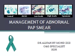
PAP SMEAR DR ALIFAH BT MOHD ZIZI OampG SPECIALIST SGH BETHESDA SYSTEM 2001 It was designed to provide uniform diagnostic language to facilitate communication between cytologists ID: 359755
Download Presentation The PPT/PDF document "MANAGEMENT OF ABNORMAL" is the property of its rightful owner. Permission is granted to download and print the materials on this web site for personal, non-commercial use only, and to display it on your personal computer provided you do not modify the materials and that you retain all copyright notices contained in the materials. By downloading content from our website, you accept the terms of this agreement.
Slide1
MANAGEMENT OF ABNORMAL PAP SMEAR
DR ALIFAH BT MOHD ZIZIO&G SPECIALISTSGHSlide2
BETHESDA SYSTEM 2001
It was designed to provide uniform diagnostic language to facilitate
communication between cytologists
and clinician
3 general categories
Within Normal LimitsBenign Cellular ChangesEpithelial Cell Abnormality Slide3
BETHESDA SYSTEM 2001
Adequacy of the sample is paramount 8000 – 12,000 squamous cells for conventional PS/10 HPF5000 cells/10 HFP for liquid-based sample
Presence of endocervical cells (at least 10) is recommended (not required for women < 40 y.o
) Slide4
WHAT IS ABNORMAL PAP SMEAR?
Abnormal due to inadequacy
Abnormal due to inflammation
Abnormal due to
infection
Abnormal due to dysplastic changesSlide5
1. INADEQUATE OR UNSATISFACTORY SMEARSlide6
SATISFACTORY SPECIMEN..
Appropriate labeling and identifying information Relevant clinical information Adequate numbers of well preserved and well visualized squamous epithelial
cells. An adequate endocervical / transformation zone component (from a patient with a cervix).
Quality of the Pap smear will still be noted when:
1. More than
10 well preserved endocervical or metaplatic cells are seen2. No blood or inflammation obscuring the Pap smearSlide7
INADEQUATE/UNSATISFACTORY SMEAR
A smear that is
unreliable for the detection of cervical epithelial cell abnormalitiesSlide8
INADEQUATE/ UNSATISFACTORY SMEAR
1. Sampling
Scanty cells
Blood
, mucous,
pus2.PreparationToo thick due to poor spreadingAir drying artifact
Broken slide
3
.Mainly
endocervical
cellSlide9
HOW TO DEAL WITH INADEQUATE/
UNSATISFACTORY SMEAR ??
Correct timing of smear
Do not use cream or gel
Cleaning of excessive mucusChoice of sampling devicesCorrect spreadingRapid fixation (< 10 second)Correct timing of smearDo use cream or gelSlide10
PAP SMEAR
UNSATISFACTORY
TX ANY INFECTION
GIVE A COURSE OF ESTROGEN IF POST MENOPAUSE WITH ATROPHY
REPEAT 6/12
2ND SMEAR UNSATISFACTORY
REPEAT 6/12
3
RD
SMEAR UNSATISFACTORY
NEGATIVE FOR INTRAEPITHELIAL
LESSION COLPOSCOPYROUTINE SCREENINGSlide11
2. INFLAMMATORY SMEARSlide12
Inflammation on Pap smear results,
does not indicate any particular pathology Therefore, does not necessitate routine treatment. Slide13
POSSIBLE CAUSES……
InfectionChronic cervicitis
Atrophic
cervicitis
Chemical or mechanical irritation to cervix-
tampoon, douchingSlide14
PAP SMEAR
NEGATIVE FOR MALIGNANT CELL
INFLAMMATORY
TX ANY INFECTION OR ATROPHY
REPEAT 6/12
2
ND
SMEAR INFLAMMATORY
REPEAT 6/12
3
RD
SMEAR INFLAMMATORYNORMALCOLPOSCOPYROUTINE SCREENINGSlide15
3. ABNORMAL SMEAR DUE TO INFECTION Slide16
COMMON INFECTIONS….
Tricomonas vaginalis
Fungal
ie
candidiasisBacterial VaginosisActinomycesHerpes Simplex
ORGANISM
TREATMENT
TRICHOMONAS
VAGINALIS
T. METRONIDAZOLE 400MG TDSFUNGAL INFECTION (CANDIDA)CANNESTAN PESSARY 200MG ONBACTERIA VAGINOSIST. METRONIDAZOLE 400MG TDSSlide17
PAP SMEAR
NEGATIVE FOR MALIGNANT CELL
SPECIFIC MICROORGANISM
TREAT ANY INFECTION
NORMAL
ROUTINE SCREENING
REPEAT PAP SMEAR 6/12 Slide18
4. ABNORMAL SMEAR DUE TO DYSPLASTIC CHANGESSlide19
DYSPLASTIC CHANGES
SQUAMOUS
CELL ABNORMALITY
GLANDULAR ABNORMALITY
ASCUS ASC-HLGSILHGSIL
INVASIVE
SQUAMOUS
CELL CARCINOMA
AGS
AISINVASIVE ADENOCARCINOMASlide20
Spectrum of Changes in Cervical Squamous Epithelium Caused by
HPV Infection*CIN = cervical intraepithelial neoplasia
Adapted from Goodman A, Wilbur DC. N Engl J Med
. 2003;349:1555
–15
64. Normal Cervix
HPV
Infection/
CIN
* 1
CIN
2 / CIN 3 /Cervical CancerSlide21
%
Regress
Persist
Progress to CIS
Progress to Invasion
CIN 1
60
30
10
1
CIN 2
40
35
20
5CIN 3
30<56-18 (5y), 36(10y
)NATURAL HISTORY……..Slide22
SQUAMOUS CELL ABNORMALITY…Slide23
ABNORMAL PAP SMEAR DUE TO DYSPLASTIC CHANGES –
SQUAMOUS CELL ABNORMALITIES
1. Atypical
Squamous
Cells
(ASC) Atypical Squamous Cells-Undetermined Significance (ASC-US) Atypical
Squamous
Cells, Cannot Exclude High Grade
Lesion
(
ASC
-H)2. Low-grade Squamous Intraepithelial Lesion (LSIL) (Mild Dyskaryosis / HPV/CIN 1)
3. High-grade Squamous Intraepithelial Lesion (HSIL)(Mod or Severe Dyskaryosis / CIN 2,3) 4. Invasive Squamous Cell CarcinomaSlide24
1. Undetermined Significance (
ASC-US)Cytologic changes suggestive of a low grade
squamous lesion but
lack criteria
for definitive interpretation
.2. Cannot Exclude High Grade Lesion (ASC-H)Cytologic changes suggestive of a high grade
squamous
lesion but
lack criteria
for definitive interpretation.
1.ATYPICAL
SQUAMOUS CELL (ACS)Slide25
PAP SMEAR
ATYPICAL
SQUAMOUS CELL (ASC
)
ASCUS
REPEAT 6/12NEGATIVE FOR INTRAEPITHELIAL
LESSION
RESUME NORMAL SCREENING
HPV
DNA TESTING
POSITIVE
NEGATIVECOLPOSCOPYSlide26
PAP SMEAR
ASC
-H
COLPOSCOPYSlide27
2. LOW GRADE INTRAEPITHELIAL LESSION
(LGSIL) / CIN 1CIN
I being the morphologic manifestation of a self-limited sexually transmitted HPV infection
60% of
CIN
I regress spontaneously30% of CIN I persists. 10% of CIN I lesions progress to CIN III,1% may ultimately progress to invasive cancer. Slide28
Assessment of client
yes
No
Presence of at least 1 criteria:
-Age > 30 yrs
Poor complianceImmunocompromised
Sx
Hx
of
pre-invasive
lesion +ve for high risk HPV (16,18,31,33,45,52,58)Immediate colposcopyRepeat smear in 6/12
NILMLSILResume routine screening scheduleColposcopy=60%Slide29
MANAGEMENT APPROACH
A
lesion that persist after 1-2 years or any progression during follow up suggest need of treatment
If
HPV
testing is available, +ve HPV: indication for treatment- Treatment- local ablative/
excission
-
Follow up after treatment for
CIN1
-repeat smear in 6/12
-repeat smear and colposcopy in 12/12-If normal, yearly pap smear x 2 years then back to normal routineSlide30
3.HIGH GRADE INTRAEPITHELIAL
LESSION (HGSIL)/ CIN 2-3CIN 2-3 is a cervical cancer precursor1.CIN
240% of CIN II regress
30
% of
CIN II persist20% of CIN II progress to CIN III5% of CIN II progress to CIN III2. CIN 333% of CIN III regress18% of CIN III
progress to invasive disease over a 10 years
36% of
CIN
III
progress to invasive disease over a 20 yearsSlide31
PAP SMEAR
HGSIL
COLPOSCOPY
AND BIOPSY
Subsequent management depends on:
Whether lesion identifiedWhether colposcopy satisfactory
Annual smear following treatmentSlide32
MANAGEMENT APPROACH
EXCISION METHOD
LLETZ
Cold
knife cone biopsy
HysterectomySlide33
ABLATIVE METHODS
Cryocautery
Electrodiathermy
Cold
coagulationSlide34
PAP SMEAR
INVASIVE
SQUAMOUS CANCER
COLPOSCOPY
AND BIOPSY
Subsequent management depends on: Stage of the disease
4. INVASIVE SQUAMOUS CELL CANCERSlide35
GLANDULAR ABNORMALITYSlide36
ABNORMAL PAP SMEAR DUE TO DYSPLASTIC CHANGES-
GLANDULAR CELL ABNORMALITIES
1.Atypical
Glandular Cells
(AGS)
(undetermined or favour neoplastic)2.Adenocarcinoma in Situ
(
AIS
)
3. Invasive
AdenocarcinomaSlide37
GLANDULAR ABNORMALITIES
The most common significant lesions associated
with AGC (Atypical Glandular Cells) are
actually
squamous
Management should include colposcopy and endocervical samplingSlide38
ATYPICAL ENDOMETRIAL CELLS
Always
perform endometrial sampling
If endometrial sampling is negative
:
colposcopy with endocervical samplingSlide39
GLANDULAR ABNORMALITIESSlide40
OTHERS…Slide41
PAP SMEAR
ATROPHY
LOCAL ESTROGEN CREAM
1G
ON FOR 2 WEEKS THEN TWICE WEEKLY FOR 6 WEEKS
ATROPHY SMEAR
REPEAT IN 6 MONTHS Slide42
PAP SMEAR
REACTIVE CELLULAR CHANGES DUE TO RADIATION, REPAIR OR
IUCD
REACTIVE CELLULAR CHANGES
REPEAT IN 1 YEAR Slide43
ABNORMAL
PAP SMEAR IN PREGNANCY
Reported abnormal smear during pregnancy
1%- 8%
Follow-up should be similar to non
pregnant state-every trimester Regardless of gestation, suspicious lesion shouldbe biopsied. Cervical biopsy does not
increase the
risk of miscarriage
If evidence of invasive
cancer- require
excissionSlide44
THANK YOU…….