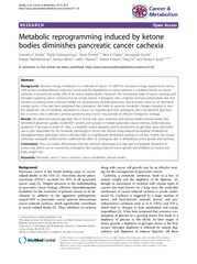
/
Metabolic reprogramming induced by ketone bodies diminishes pancreatic cancer cachexia Metabolic reprogramming induced by ketone bodies diminishes pancreatic cancer cachexia

Metabolic reprogramming induced by ketone bodies diminishes pancreatic cancer cachexia - PDF document

tatiana-dople
tatiana-dople . @tatiana-dople
Follow
465 views
Uploaded On 2017-08-15

Metabolic reprogramming induced by ketone bodies diminishes pancreatic cancer cachexia - PPT Presentation

cancerandmetabolismcontent2122 RESEARCH Open Access Metabolic reprogramming induced by ketone bodies diminishes pancreatic cancer cachexia Surendra K Shukla Teklab Gebregiworgis Vinee Purohit 13 Nina V Chaika Venugopal Gunda Prakash Radhakrishnan ID: 77184

cancerandmetabolismcontent2122 RESEARCH Open Access Metabolic

Share:

Link:

Embed:

Download Presentation from below link

Download Pdf The PPT/PDF document "Metabolic reprogramming induced by keton..." is the property of its rightful owner. Permission is granted to download and print the materials on this web site for personal, non-commercial use only, and to display it on your personal computer provided you do not modify the materials and that you retain all copyright notices contained in the materials. By downloading content from our website, you accept the terms of this agreement.


Presentation Transcript

Acorrection to thisarticlehas beenpublished:http://www.cancerandmetabolism/content/2/1/22 RESEARCHOpenAccessMetabolicreprogramminginducedbyketonebodiesdiminishespancreaticcancercachexiaSurendraKShukla,TeklabGebregiworgis,VineePurohit,NinaVChaika,VenugopalGundaPrakashRadhakrishnan,KamiyaMehla,IraklisIPipinos,RobertPowers,FangYuandPankajKSinghAberrantenergymetabolismisahallmarkofcancer.Tofulfilltheincreasedenergyrequirements,tumorcellssecretecytokines/factorsinducingmuscleandfatdegradationincancerpatients,aconditionknownascancercachexia.Itaccountsfornearly20%ofallcancer-relateddeaths.However,themechanisticbasisofcancercachexiaandtherapiestargetingcancercachexiathusfarremainelusive.Aketogenicdiet,ahigh-fatandlow-carbohydratedietthatelevatescirculatinglevelsofketonebodies(i.e. *Correspondence:pankaj.singh@unmc.eduTheEppleyInstituteforResearchinCancerandAlliedDiseases,UniversityofNebraskaMedicalCenter,Omaha,NE68198,USADepartmentofPathologyandMicrobiology,UniversityofNebraskaMedicalCenter,Omaha,NE68198,USAFulllistofauthorinformationisavailableattheendofthearticle Cancer & Metabolism ©2014Shuklaetal.;licenseeBioMedCentralLtd.ThisisanOpenAccessarticledistributedunderthetermsoftheCreativeCommonsAttributionLicense(http://creativecommons.org/licenses/by/2.0),whichpermitsunrestricteduse,distribution,andreproductioninanymedium,providedtheoriginalworkisproperlycredited.TheCreativeCommonsPublicDomainDedicationwaiver(http://creativecommons.org/publicdomain/zero/1.0/)appliestothedatamadeavailableinthisarticle,unlessotherwisestated.etal.Cancer&Metabolismhttp://www.cancerandmetabolism.com/content/2/1/18 accountforthecachexiasyndromeandresultinapoorresponsetochemotherapy,fatigue,andareducedqualityoflifeforcancerpatients[5].Cancercellsexhibitreprogrammingofseveralmetabolicpathwaysalongwithmultiplegenetic,epigenetic,andgrowthsignalingalterations[6,7].Mostcancercellsdem-onstrateanincreaseinglucoseuptake,ahigherrateofglycolysis,andanincreaseinlactatesecretiondespitethepresenceofoxygen,aphenomenonknownastheWar-burgeffect[8].Aerobicglycolysisplaysanimportantroleinrapidcellulargrowthasitprovidesseveralintermedi-atesrequiredforbiomasssynthesisbyroutingthecarbonfluxthroughthepentosephosphatepathway[9].Thein-creasedconversionofpyruvateintolactatebyaerobicgly-colysisleadstoacidosisintumormicroenvironmentsthatfacilitatesinvasionandmetastasisofcancercells[10].Aer-obicglycolysisisalsoanenergy-inefficientprocessrequir-inglargeamountsofglucose.Correspondingly,tumorcellsserveasaglucosesink[11].Additionally,lactatepro-ducedfromtumorcellspassestotheliverandgetscon-vertedtoglucosebymeansoftheCoricycle,anotherenergy-inefficientprocess[9].Alongwithglucoseuptakeandenhancedaerobicglycolysis,cancerpatientsalsopresentglucoseintoleranceandincreasedhepaticglucoseproduction[12].Anincreasedrequirementforglucosemightbethecriticalstimulusneededforenhancedhepaticglucoseproduction.Tumorcellsalsohavealterationsinthemetabolismofglutamine,anitrogensourceandargu-ablythemostsignificantmetaboliteprecursorfortumorcellsafterglucose[13].Aketogenicdietisahigh-fatandlow-carbohydratedietthatleadstoelevatedcirculatinglevelsofketonebodies(i.e.,acetoacetate,-hydroxybutyrate,andacet-one)andanalternativeenergysource[14].Ketogenicdietspossessanticonvulsantandantiinflammatoryac-tivities[15,16].Ithasalsobeenproposedthataketo-genicdiettreatmentresultsinsystemicmetabolicchangeslikeincreasedglucosetolerance,reducedfattyacidsynthesis,andweightloss[17].Keepinginviewthesignificantroleofinflammationandmetabolicalter-ationsincancer,aketogenicdietmayprovideaneffi-cienttherapeuticstrategy.Furthermore,mostcancercellslackkeymitochondrialenzymestometabolizeke-tonebodiesandgenerateATP,whilemyocytesandothertissues,includingthebrain,stillretainthisability[18].Hence,aketogenicdietmayactagainstthecancer-inducedcachexiawhilecausingminimalsideef-fectsaspreviouslyithasbeenshownthata27-mMke-tonebodyconcentrationcanbeachievedsafelywithoutgivingrisetoclinicalacidosis[19,20].Inthepresentstudy,wehaveevaluatedanticancerousandanticachec-ticpropertiesofketonebodiesincellcultureconditions,aswellastheeffectofaketogenicdietontumorburdenandcachexiainanimalmodelsFurthermore,ourstudiesestablishaketonebody-inducedmetabolomicreprogrammingasthemechanismofactionofaketo-genicdietagainstcancerandcancer-inducedcachexia.CellsandreagentsThehumanpancreaticcancercelllineCapan1,mousemyoblastC2C12,andmouseembryofibroblast(preadi-pocyte)3T3L1wereobtainedfromAmericanTypeCul-tureCollection(Manassas,VA,USA).S2-013isaclonedsublineofahumanpancreatictumorcellline(SUIT-2)derivedfromalivermetastasis[21].AllthecelllineswereculturedinDulbeccosmodifiedEaglesmedium(DMEM)supplementedwith10%fetalbovineserum,penicillin(100mg/mL),andstreptomycin(100mg/mL)andincubatedat37°Cinahumidifiedchamberwith5%.Sodium-3-hydroxybutyrate,lithiumacetoacetate,dihydroethidium(DHE),3-[4,5-dimethylthiazol-2-yl]-2,5-diphenyltetrazoliumbromide(MTT),BCPCF,andhydroxybutyricacidwerepurchasedfromSigmaChe-micals(Sigma-Aldrich,St.Louis,MO,USA).Cellviabilityandcaspase3/7activityassayCellviabilitywasdeterminedbyperformingMTTassay.Capan1andS2-013cells(5×10cellsperwell)wereseededin96-wellplatesfor12handthentreatedwithdifferentconcentrationsofsodium-3-hydroxybutyrateorlithiumacetoacetatefor72h.Aftertreatment,cellswereincubatedwithMTTreagentfor2h;theresultantfor-mazancrystalsweredissolvedindimethylsulfoxideandtheabsorbancewasrecordedat590nm.Untreatedcellswereutilizedasacontrolfortheviabilityassays.Caspase3/7activitywasdeterminedbyutilizingaPromegaCaspase-Glokit(Madison,WI,USA).Capan1andS2-013cells(0.6×10cellsperwell)wereseededin6-wellplatesfor12handthentreatedwithdifferentconcen-trationsofsodium-3-hydroxybutyrateandlithiumacet-oacetatefor48h.Caspase3/7activitywasthendeterminedasperthemanufacturersprotocol.GlucoseandglutamineuptakeassayTodetermineglucoseuptake,Capan1andS2-013cells(5×10cellsperwell)wereseededin24-wellplates.After12h,cellsweretreatedwithmultipleconcentrationsofsodium-3-hydroxybutyrateandlithiumacetoacetatefor24h.Aftertreatment,cellswerestarvedforglucosefor2handthenincubatedfor20minwith1Ci[H]-2-deoxyglucose(DG)foraglucoseuptakeassay.Cellswerewashedwithphosphate-bufferedsaline(PBS)andlysedwith1%sodiumdodecylsulfate(SDS).Thelysateswerethensubjectedto[H]countingbyutilizingascintillationcounter.Scintillationcountsfromcellstreatedwithlabeledandexcessunlabeled2-DGwereutilizedascon-trolsforbaselinecorrection.Theresultswerenormalizedetal.Cancer&MetabolismPage2of19http://www.cancerandmetabolism.com/content/2/1/18 tothecellcounts.Fordeterminingglutamineuptake,Capan1andS2-013cells(5×10cellsperwell)wereseededin24-wellplates.After12h,cellsweretreatedwithsolventcontrol,multipleconcentrationsofsodium-3-hydroxybutyrate,orlithiumacetoacetatefor24h.Posttreatment,cellswerestarvedforglutaminefor2handthenincubatedfor3minwith1CitritiatedGlutamine,L-[3,4-H(N)].CellswerewashedwithPBSandlysedin1%SDS.Thelysateswereusedfor[H]countingbyutilizingascin-tillationcounter.Scintillationcountsfromcellstreatedwithlabeledandexcessunlabeledglutaminewereutilizedascontrolsforbaselinecorrection.Theresultswerenormal-izedtothecellcounts.LactatereleaseassayCapan1andS2-013cells(5×10cellsperwell)wereseededin24-wellplates.After12h,cellsweretreatedwithindicatedconcentrationsofsodium-3-hydroxybutyrateorlithiumacetoacetatefor24h.Theculturesupernatantswerethenutilizedfordetermininglactaterelease.TheassaywasperformedbyutilizingaLactateAssayKit(EtonBioscienceInc.,SanDiego,CA,USA),asperthemanufac-turersprotocol.ATPassayTotalATPlevelincellswasdeterminedbyusinganATPassaykit(Roche,Indianapolis,IN,USA).After12h,cellsweretreatedwithdifferentconcentrationsofsodium-3-hydroxybutyrateandlithiumacetoacetatefor24handATPlevelwasdeterminedasperthemanufacturerprotocol.ATPlevelwasnormalizedwithtotalproteinconcentration.ReactiveoxygenspeciesassayReactiveoxygenspecieslevelwasdeterminedbyusingoxidation-sensitivefluorescentdyeDHE.Capan1andS2-013cells(0.1×10cellsperwell)wereseededin12-wellplatesonglasscoverslips.After12h,cellsweretreatedwithsolventcontrolorindicateddosesofsodium-3-hydroxybutyrateandlithiumacetoacetatefor24h.Controlandtreatedcellswereincubatedat37°Cin2.5mMDHEcontainingDMEM.Afterincubation,cellswerewashedwithcoldPBSandfixedwithHistoChoice®fixative(Sigma-Aldrich)for15minatroomtemperature.CellswerewashedwithPBSandthecoverslipsweremountedontoglassslidesusingreal-mount.FluorescenceintensitypercellwasdeterminedbyscanningwithZeissAxiovert200Mmicroscope(Oberkochen,Germany)andanalyzingtheimageswithSlideBook5.5software(Intelli-gentImagingInnovations,Inc.,Denver,CO,USA).GeneexpressionanalysisbyqRT-PCRTotalRNAwasisolatedbyutilizingRNeasycolumns(Qiagen,Venlo,TheNetherlands)asperthemanufacturerprotocol.TotalRNA(5g)wasreversetranscribedbyutilizingVerso-cDNAsynthesiskit(ThermoScientific,Pittsburgh,PA,USA)accordingtothemanufacturerguidelines.Quantitativereversetranscriptionpolymer-asechainreaction(qRT-PCR)wasperformedwithgene-specificprimersat95°Cfor10sand60°Cfor60s(40cycles)in10Lreactionmixcontaining3cDNA,2Lprimers,and5LSYBRGreenMasterMix(AppliedBiosystems,GrandIsland,NY,USA)usinganABI7500thermocycler.Beta-actinwasutilizedasanin-ternalcontrol.ThesequenceofdifferentsetsofprimersusedinthestudyisgiveninAdditionalfile1.Quantifi-cationwasperformedwiththeCtmethod[22].ImmunoblottingForimmunoblotting,cellswerewashedtwicewithcoldPBSandlysedinRIPAlysisbufferbyincubatingat4°Crotatoryshakerfor30min.Celldebriswasremovedbycentrifugationat13,000rpmfor10minandthesuper-natantwascollected.ProteincontentwasmeasuredbyperformingBradfordassay.Westernblottingwasper-formedasdescribedpreviously[23].ThemembraneswereprobedwithprimaryantibodyagainstGLUT1(Abcam,Cambridge,UK),c-Myc-9E10(SantaCruzBio-technology,Dallas,TX,USA),HKII(CellSignalingTechnology,Beverly,MA,USA),andHSP90(SantaCruzBiotechnology).Luciferaseassayc-Myc-promoter(del1)-luciferasereporterconstructwasobtainedfromAddgene(Cambridge,MA,USA)[24].Cellsweretransfectedwith1gofplasmid,and16hposttransfection,cellsweretreatedwithdifferentketonebodiesfor24h.AsyntheticRenillaluciferasereporterpRL-TKwasutilizedasatransfectioncontrol.LuciferaseactivitywasdeterminedbyutilizingDual-LuciferaseRe-porterAssaySystem(Promega).ChromatinimmunoprecipitationForchromatinimmunoprecipitation,cellsweretreatedwith20mMsodium-3-hydroxybutyrateandlithiumacetoacetatefor24halongwithsolventcontrol.Chro-matinimmunoprecipitationwasperformedbyutilizingc-Mycantibody(9E10)asdescribedpreviously[25].MouseIgGwasutilizedasacontrol.qPCRdatawerenormalizedtoagenomicregionlocatedwithinGUSBgeneandrepresentedasfoldenrichmentrelativetotheIgGcontrol.PrimersequencesusedforqPCRamplifica-tionaredescribedinAdditionalfile1.TumorgrowthmeasurementCongenitallyathymicfemalenudemice(NCr-nu/nu)werepurchasedfromtheNationalCancerInstitute.Miceweretreatedaspertheguidelinesofourinstitutionalanimalcareetal.Cancer&MetabolismPage3of19http://www.cancerandmetabolism.com/content/2/1/18 andusecommittee(IACUC).S2-013cells(5×10)wereusedfororthotopicinjectionsintothepancreasofnudemice.After7daysofimplantation,miceweredividedingroupsofnineanimalseachandfedadlibitumwithanor-maldietoraketogenicdiet(compositiongiveninTableS2inAdditionalfile1).After3weeksoftreatment,miceweresacrificedandtumorweight,tumorvolume,muscleweight,carcassweight,etc.wererecorded.Tumortissueandotherorganswereflashfrozeninliquidnitrogenforfurtherana-lysis.AnimalprotocolswereinaccordancewiththeNIHGuidefortheCareandUseofLaboratoryAnimalsandwereapprovedbytheUniversityofNebraskaMedicalCen-terAnimalCareandUseCommittee.ImmunohistochemistryImmunohistochemistrywasperformedasdescribedprevi-ously[26].Ki67(ThermoFisherScientific,Waltham,MA,USA),c-Myc(Epitomics,Burlingame,CA,USA),andCleavedCaspase3(CellSignalingTechnology)primaryantibodieswereutilized.Thestainedsectionswereimagedat×20underanuprightmicroscopeandrepresentativeim-ageswerecapturedandpresented.MetaboliteextractionandNMRsamplepreparationAfterconfirmingtheconfluenceofthecells,themediawasaspiratedandthecellswerewashedtwicewith1×phosphatebuffertoremoveremnantsofthemediabeforelysingthecells.Thecellswerethencoldshockedwith1mLofcryogenicallycold80%methanol/watermixture.Theplateswiththe80%methanol/waterwereincubatedina80°Cfreezerforatleast15min.ThecellsfromthecoldplateswerescrapedwithacellscraperandpipettedintoanEppendorftubeandcentrifugedat13,000rpmfor5min.Thesupernatantwascollectedand250LofMilli-Qwater(Millipore,Billerica,MA,USA)wasaddedtotheremainingcelldebrisforre-extraction.Aftermixingthecelldebriswiththewaterbypipetting,thesamplewasagaincentrifugedat13,000rpmfor5min.Thenewsuper-natantwascombinedwiththepreviouslycollectedsuper-natant.Finally,thesamplewasdriedusingspeedvacuumevaporator(SpeedVac®Plus,Savant,ThermoScientific,Waltham,MA)toevaporatethemethanolandsubjectedtofreezedrying(Labconco,KansasCity,MO)tolyophilizethewaterconsecutively.ThedriedsamplewasmadereadyforanNMRexperimentbydissolvingin600Lof50mMphosphatebufferin99.8%DO(Isotec,St.Louis,MO)atpH7.2(uncorrected)with50M3-(tetramethysilane)propionicacid-2,2,3,3-d(TMSP)(500Mfor2DHSQC)forspectralreferencing.NMRexperimentanddataanalysisTheNMRspectrawereacquiredonaBrukerAVANCEDRX500MHzspectrometerequippedwith5mmtriple-resonancecryogenicprobe(C,andN)withaZ-axisgradient.TheNMRdatacollectionwasautomatedusingaBACS-120samplechanger,ATM(automatictun-ingandmatching),andBrukerIconNMRsoftware.Theone-dimensional(1D)protonnuclearmagneticresonanceHNMR)datawasacquiredusinganexcitationsculptingpulsesequencetoremovethesolventpeakandmaintainaflatbaseline[27].Thespectrawerecollectedat300Kwith32Kdatapoints,128scans,16dummyscans,andaspec-tralwidthof5,483Hz.OurMVAPACKsoftware(http://bionmr.unl.edu/mvapack.php)[28]wasusedtoprocessthe1DHNMRspectra.TherawNMRdatawasonlyFouriertransformedandautomaticallyphased.Theresult-ingNMRspectrumwasbinnedusinganadaptiveintelli-gentbinningalgorithmthatautomaticallyadjustsbinsizestoavoidsplittingNMRresonancesbetweenmultiplebins[29].ThespectralregionbeforetheTMSPwasusedasatrainingsettoremovethenoisefromthedatausingthemethodstatedbyHalouskaetal.[30].Thespectrawerethennormalizedusingstandardnormalvariate(SNV)andscaledusingParetoscaling.Theprocesseddatawasuti-lizedtogenerateplotsofprincipalcomponentanalysis(PCA)andorthogonalprojectionstolatentstructuresdiscriminantanalysis(OPLS-DA)scoresandbackscaledloadings(Additionalfile2)usingourMVAPACKsoft-ware[28].Metaboliteidentificationfromthe1DNMRspectrawasaccomplishedusingtheChenomxNMRSuite7.6(http://www.chenomx.com/)andthebackscaledloadings.The2DChetero-nuclearsinglequantumcoher-ence(HSQC)NMRspectrawerecollectedat300Kwith64scans,16dummyscans,anda1.5-srelaxationdelay.Thespectrawerecollectedwith2Kdatapointsandaspectrumwidthof4,735Hzinthedirectdimensionand64datapointsandaspectrumwidthof17,607Hzintheindirectdimension.The2DCHSQCNMRspectrawereprocessedusingNMRPipe(NIH,Bethesda,Mary-land)[31]andanalyzedusingNMRViewJVersion8.0.3.Peakintensitieswerenormalizedbytheaveragepeakin-tensityforagivenspectrumandthenassignedtoame-taboliteusingchemicalshiftreferencesfromtheHumanMetabolomicsDatabase[32],MadisonMetabolomicsConsortiumDatabase[33],andPlatformforRIKENMetabolomics[34].Chemicalshifterrorsof0.08and0.25ppmfortheHandCchemicalshifts,respect-ively,wereusedtomatchtheexperimentalchemicalshiftswiththedatabases.Inadditiontochemicalshifts,peaksplittingpatternsandpeakshapeswerealsousedtoverifymetaboliteassignments.The2DCHSQCNMRexperimentisamorereli-ableapproachformetaboliteidentificationbecauseofthesignificantlyhighersignaldispersion,andthecorrelationbetweenHandCchemicalshiftsforeachC-Hpairinamolecule[35].Moreimportantly,the2DCHSQCexperimentsimplifiestheanalysisofthemetabolomeetal.Cancer&MetabolismPage4of19http://www.cancerandmetabolism.com/content/2/1/18 becauseonlycompoundscontainingaC-carbonderivedfromthe-glucoseaddedtothemediawillbedetected.Thus,using-glucosewillhighlightmetabolitechangesassociatedwiththeglycolyticfluxintumorcells.Thisavoidsthechallengewiththe1DHNMRexperimentswherethespectraweredominatedbycatabolicproductsofketonebodies.Thus,theidentificationofmetabolitesfromthe2DCHSQCexperimentsismorereliableandpertinenttotheanalysisofmetabolicchangesresult-ingfromketonebodyeffectsonpancreaticcancercachexia.Eachmetabolitepeakfromthetwosets(controlandketonebody-treated)oftriplicate2DCHSQCNMRspectrawerefurthernormalizedbyusingthemaximumpeakintensityforthemetaboliteandthenscaledfrom0to100.ThepeakintensitiesforeachmetabolitewerethenaveragedandcomparedbetweenthecontrolandtreatedgroupsusingaStudenttest.Metaboliteswithvalue0.1wereusedtogenerateaheatmapusingtheRstatisticalpackage[36].Arelativechangeinpeakintensityimpliesacorrespondingmetaboliteconcentra-tionchange.Absoluteconcentrationsarenotmeasur-ablefromthe2DCHSQCbecauseotherfactors,suchascouplingconstants,relaxation,anddynamics,alsocontributetopeakintensities.Measurementofbloodglucoseand-hydroxybutyrateconcentrationBloodglucoselevelofmicewasmeasuredafter16hofstarvationbyutilizingContourUSBbloodglucosemeter(BayerHealthCare,Mishawaka,Japan),asperthemanu-facturersprotocol.Bloodketonelevelwasmeasuredbyutilizingabloodglucoseandketonemonitor(NovaBiomedical,Waltham,MA,USA)asperthemanufac-turersprotocol.Theconcentrationof-hydroxybutyratewasmeasuredbycomparingtheTMSP-normalizedme-thylpeaksofHNMRcollectedforsixanimals.C2C12and3T3L1differentiationandconditionedmediumpreparationC2C12mousemyoblastsweregrowninDMEMwith10%FBS.Toinducedifferentiation,cellswereswitchedto2%horseserumand10g/mLinsulin-containingDMEMandgrownfor72h.3T3L1mouseembryofibroblastswereculturedinDMEMwith10%FBS.Fordifferentiationof3T3L1preadipocytes,after2daysatconfluency,cellsweretreatedwithdifferentiationmedium:DMEMcon-taining10%FBS,1Mdexamethasone,0.5mMmethyli-sobutylxanthine(IBMX),and1g/mLinsulinfor2days.After2days,differentiationmediumwasreplacedwithDMEMcontaining10%FBS.Mediumwaschangedregu-larlyafter48h.Forconditionedmediumpreparation,cellswereplatedatadensityof50,000cells/cm,andafter12hofseeding,cellswerewashedtwicewithPBSandculturedinserum-freeDMEMforthenext24h.Conditionedmediumwascentrifugedat1,200for10minandfilteredwitha0.2-msyringefilterandusedimmediatelyorstored80°C.MeasurementofintracellularpHCytosolicpHwasmeasuredbyusingfluorescencespec-troscopyusingBCPCF-AMasdescribedbyMarinoetal.al.StatisticalanalysisComparisonsbetweendifferentgroupswereperformedbyusingANOVA(one-way;GraphPadPrismversion4.03)withDunnettsposthoctest.Studenttestwasusedforinvivostudies.Avalueof.05wasconsideredtobesignificant.Ketonebodiesdiminishpancreaticcancercellgrowthandinduceapoptosisinadose-dependentmannerWeinvestigatedtheeffectofketonebodies(sodiumhydro-xybutyrateandlithiumacetoacetate)oncellsurvivalinmultiplepancreaticcancercelllines.Initially,weevaluatedtheeffectofmultipledosesofsodiumhydroxybutyrateandlithiumacetoacetate(1to20mM)onthesurvivalofCapan1andS2-013pancreaticcancercellsbyperformingMTTassays.Ketonebodieswereobservedtoinhibitcellsurvivalinadose-dependentmanner.Ketonebodiessignifi-cantlyinhibitedcellgrowthatconcentrationsof10and20mMaftera72-htreatment(Figure1A,B).Toevaluateiftheketonebody-inducedsurvivaldiminutionisspecifictocancercells,wesubjectedimmortalized,non-transformedpancreaticepithelialcelllinesHPNEandRAPANtoincu-bationwithsodiumhydroxybutyrateandlithiumacetoace-tate.Weobservednosignificanteffectonsurvivalofthesecellsundertreatmentwithketonebodies(Additionalfile3).Similarly,toruleoutthepossibilityoforganicacidandlith-iumionbeingresponsibleforreductionincancercellsur-vival,ratherthanketonebodiesthemselves,weevaluatedthesurvivalofS2-013andCapan1undertreatmentwithhydroxybutyricacidandlithiumchloride.WeobservednosignificanteffectonS2-013andCapan1cellsurvivalafter72hoftreatment(Additionalfile4).Furthermore,weinvestigatedtheeffectofsodiumhydroxybutyrate,lithiumacetoacetate,and-hydroxybutyrateonintracellularpH.After6hoftreatmentwithdifferentdosesofsodiumhydroxybutyrate,lithiumacetoacetate,and-hydroxybuty-rateonS2-013,Capan1,HPNE,andRAPANcells,weobservedsignificantdecreaseinintracellularpH(0.2to0.4units)inallthecelllines(Additionalfile5).Althoughintra-cellularacidificationupontreatmentwithketonebodiesmightcontributetotheantiproliferativeeffectsofketonebodies,itisnottheprimarycauseofcellsdeathaswedidnotobservesignificantcelldeathupontreatmentwithetal.Cancer&MetabolismPage5of19http://www.cancerandmetabolism.com/content/2/1/18 isomerofhydroxybutyratethat causedsimilarintracellular pHchangeasdidtheotherketonebodies.Furthermore,we observedasimilareffectonintracellularpHinnon- transformedpancreaticepithelialcelllinesHPNEand RAPANaftertreatmentwithsodiumhydroxybutyrate,lith- iumacetoacetate,and S -isomerofhydroxybutyratebutno significantcelldeath,asmentionedearlier.Bright-fieldmi- croscopyimagesofthecellstreatedwith10and20mMof NaHBandLiAcAcarepresentedinFigure1C,D.Further- more,weinvestigatedtheeffectofketonebodiesonfive pancreaticcancercelllinesandobservedthatcellgrowth issignificantlyinhibitedinadose-dependentfashion (Figure1E).Wealsoinvestigatedtheeffectofketonebody treatmentoncaspaseactivityinCapan1andS2-013cell lines.Caspase3/7activityincreasedupontreatmentofthe pancreaticcancercellswithketonebodiesinadose- dependentmanner(Figure1F). Ketonebodiescausemetabolicalterationsinpancreatic cancercells Previousstudiesindicatethataketogenicdietinduces metabolicalterationsinmice[17].Hence,wenextinvesti- gatedtheeffectofketonebodiesonpancreaticcancercell metabolism.Becausecancercellsdemonstrateanincrease inglycolysis[38],weexaminedtheeffectofketonebodies onglucoseuptakeandlactatereleaseinCapan1andS2- 013cells.TreatmentofCapan1andS2-013cellswith ketonebodiesresultedinadecreaseinglucoseuptake (Figure2A,B)andreleaseoflactate(Figure2C,D)ina dose-dependentmanner.Sinceglutaminealsosupports pancreaticcancercellgrowth[7],wealsoevaluatedtheef- fectofketonebodiesonglutamineuptake.Ourresults indicateareduceduptakeofglutaminebyCapan1and S2-013pancreaticcancercellsundertreatmentwith ketonebodies(Figure2E,F).Furthermore,weobserveda Figure1 Ketonebodiesinhibitgrowthandinduceapoptosisinpancreaticcancercelllines. Capan1 (A) andS2-013 (B) cellsweretreated withdifferentconcentrationsofsodium-3-hydroxybutyrate(NaHB)andlithiumacetoacetate(LiAcAc)for72h,andcellviabilitywasdetermined byMTTassay. Bar representspercentviabilityunderindicatedtreatmentsrelativetotreatmentwithsolventcontrol.Representativebright-field imagesofCapan1 (C) andS2-013 (D) cellsundertreatmentwith10-and20-mMconcentrationsofNaHBandLiAcAcfor72h. (E) Multiple pancreaticcancercelllinesweretreatedwith10-and20-mMconcentrationsofNaHBandLiAcAcfor72h,andrelativecellviabilitydetermined byMTTassayisplottedinthe barcharts . (F) Capan1andS2-013cellstreatedwith10-and20-mMconcentrationsofsodium-3-hydroxybutyrate andlithiumacetoacetatefor48handtherelativecaspase3/7activityareplotted.Valuesrepresentedaremean±SEM.* P 0.05;** P 0.01. Shukla etal.Cancer&Metabolism 2014, 2 :18 Page6of19 http://www.cancerandmetabolism.com/content/2/1/18 Figure2(Seelegendonnextpage.)etal.Cancer&MetabolismPage7of19http://www.cancerandmetabolism.com/content/2/1/18 reductioninintracellularATPlevelsupontreatmentwithketonebodies(Figure2G,H).Reactiveoxygenspecies(ROS;anaturalby-productofoxidativemetabolism)levelswerealsodownregulatedupontreatmentwithketonebodies(Figure2I,J).Overall,ourresultsindicatethatke-tonebodiesdiminishtheoverallenergetichealthofpan-creaticcancercellsbyreducingglucoseuptake,lactaterelease,glutamineuptake,cellularATPcontent,andROSlevels.KetonebodiesdiminishtheexpressionofglycolyticTodeterminethemechanismofreducedglucoseuptakeandlactatereleaseupontreatmentofpancreaticcancercellswithketonebodies,wenextexaminedifketonebody-mediatedchangeswereduetoalterationsingeneexpressionlevels.Weevaluatedtheexpressionofgenesencodingglucosetransporter-1(GLUT1)andglycolyticenzymeshexokinaseII(HKII)andlactatedehydrogen-aseA(LDHA)inketonebody-treatedandcontrolcells.InbothCapan1andS2-013cells,weobservedareducedexpressionofGLUT1andLDHAbutnosignificantchangeinexpression(Figure3A,B)upontreat-mentwithketonebodies.Furthermore,weanalyzedtheproteinexpressionlevelsofGLUT1andHKIIinS2-013andCapan1cellsundertreatmentwithketonebodiesorcontrol.OurresultsindicateareducedexpressionofGLUT1butalmostnochangeinHKIIlevelsincellstreatedwithketonebodies(Figure3C,D).Toeliminatethepossibilityofnon-specificeffectsoforganicacidandlithiumionongeneexpressionandmetabolism,wede-terminedGLUT1,HKII,andLDHAgeneexpressionandglucoseuptakeinS2-013cellsaftertreatmentwith-hydroxybutyricacidandlithiumchloride.Wefoundanegligibleeffectoftheseagentsonmetabolicgeneex-pressionandglucoseuptake(Additionalfile6).Ketonebodiesdiminishc-MycexpressionandactivitySinceweobservedadownregulationofglucoseandglu-tamineuptakeupontreatmentwithketonebodiesandacorrespondingdecreaseinglycolyticgeneexpression,wenextinvestigatedthemolecularmechanismbehindthisde-crease.Specifically,weevaluatedtheeffectofketonebodiesontheactivityandexpressionofc-Myc,acommonregula-torofbothglucoseandglutaminemetabolism[39].UsingtheTFsearchprogram,thepromotersequencesofGLUT1LDHAwerebothfoundtocontainc-Mycbindingsites.Chromatinimmunoprecipitationassayswerethenper-formedtoevaluatethec-MycoccupancyattheGLUT1LDHApromoters.Ourresultsindicateareducedoccu-pancyofc-MyconGLUT1LDHApromotersforS2-013cellstreatedwithketonebodies(Figure4A,B).Fur-thermore,weperformedreal-timePCRtodeterminethec-MyctranscriptlevelsinS2-013andCapan1cellsaftertreatmentwithketonebodiesorcontrol.Weobservedsig-nificantreductioninc-Mycexpressionaftertreatmentwithketonebodies(Figure4C,D).Wealsoevaluatedc-Mycpro-teinexpressionaftertreatmentwithketonebodiesandob-servedareductioninc-Myclevelsinadose-dependentmanner(Figure4E,F).Toconfirmifthealterationsinc-Mycexpressionwereduetoalteredc-Myctranscription,weevaluatedthetranscriptionalactivityofc-Mycpromoterbyutilizingc-Mycpromoter-luciferasereporterconstructs[24].Weobservedsignificantlyreducedc-Mycpromoteractivityundertreatmentwithketonebodies(Figure4G).Altogether,ourresultsdemonstratethatthetreatmentofpancreaticcellswithketonebodiesleadstoareducedMycexpressionatmRNAlevelandareducedc-Mycoccu-pancyontheGLUT1LDHApromoters.Ketonebodiesinhibittumorcell-inducedmusclefiberandadipocytedegradationincell-basedassaysMalignantcellshavebeenshowntolackkeymitochon-drialenzymesrequiredformetabolizingketonebodiestoproduceATP,whilemusclecellsretainthiscapacity[18].Ofnote,-hydroxybutyrateimprovesbodyweight,whilereducingproteolysisinmusclecells[40].Hence,wedevelopedacellculture-basedsystemtoevaluatetheeffectofketonebodiesonmusclefibersandadipocyteadiposedepositsundercoculturewithcancercells.ThiswasachievedbydifferentiatingC2C12premyocytesintomyotubesand3T3L1preadipocytesintodifferentiatedadipocytes.TreatmentwithCapan1orS2-013cancercell-conditionedmedium(CCCM)induceddegradationofmyotubesanddepletionofadiposedepositsin3T3L1adipocytes.Treatmentwithketonebodiesdemonstrated (Seefigureonpreviouspage.)Figure2Ketonebodiesinducemetabolicalterationsinpancreaticcancercelllines.andCapan1cellsweretreatedwithdifferentdosesofketonebodiesfor24h,andglucoseuptakewasdeterminedbyperformingH-2DGuptakeassay.representcountsnormalizedwithcellnumberandplottedrelativetocontrol.LactatereleasewasdeterminedbycolorimetricassayusingculturemediumofandCapan1cellstreatedwithdifferentconcentrationsofNaHBandLiAcAcfor24h.Valueswerenormalizedwithtotalcellnumberandrepresentedrelativetocontrols.S2-013andCapan1cellsweretreatedwithindicatedconcentrationsofketonebodiesfor24h,andglutamineuptakewasdeterminedbyperformingtritiatedGlutamine,L-[3,4-H(N)]uptakeassays.Countswerenormalizedwithcellnumberandplottedrelativetocontrol.ATPlevelsinS2-013andCapan1cellspost24-htreatmentwithketonebodiesweredeterminedbyperformingATPbioluminescenceassays.Valueswerenormalizedtototalproteinconcentrationandrepresentedrelativetocontrol.ReactiveoxygenspecieslevelofS2-013andCapan1cellsundertreatmentwithketonebodieswasdeterminedbyutilizingafluorescenceprobe,dihydroethidium(DHE),andfluorescenceintensitynormalizedtocellcountwasplotted.Valuesrepresentedaremean±SEM.*0.05;**0.01.etal.Cancer&MetabolismPage8of19http://www.cancerandmetabolism.com/content/2/1/18 asignificantprotectionofmyotubes(Figure5A,B)and adipocytes(Figure5D,E)againstCCCM.Furthermore, weanalyzedthegeneexpressionlevelsof muscle-specific ringfingerprotein1 ( MuRF1 or Trim63 )and Atrogin ( Fbxo32 ;alsoknownas muscleatrophyF-boxprotein or MAFbx )inmyotubes,and zincalpha-2-glycoprotein1 ( Zag or Azgp1 )andhormone-sensitivelipase( HSL or Lipe )inadipocytestreatedwithCCCMundertreatment withketonebodiesorcontrol.Theseproteinsareupreg- ulatedduringcancer-inducedcachexia[41].Ketone bodieswereshowntosignificantlylower MuRF1 and Atrogin mRNAlevelseveninthepresenceofCCCM (Figure5C).Wealsoobservedasignificantdecreasein Zag and HSL expressionupontreatmentwithketone bodies(Figure5F).Hence,ourresultsindicatetheutility ofketonebodiesininhibitingcancer-inducedcachexia. Ketonebodiesaltercentralcarbonmetabolismin pancreaticcancercells Toevaluatetheeffectofketonebodiesonmetabolite levelsinpancreaticadenocarcinomacells,weperformeda seriesofNMR-basedmetabolomicsstudies.S2-013cells weretreatedwith20mMsodium-3-hydroxybutyratefor 24h,andthenasetof1D 1 HNMRspectrawereacquired forcellextractsofsodium-3-hydroxybutyrate-treatedor controlS2-013cells.OPLS-DAoftheNMRspectraindi- catesthatthemetabolomesfromtheketonebody-treated cellsandthecontrolcellsclusteredintotwoseparate groupsinaOPLS-DAscoreplot,indicatingthatthe treatedcancercellsaremetabolicallydifferentiatedfrom thecontrols(Figure6A).TheOPLS-DAmodelwasvali- datedusingCV-ANOVAyieldinga p valueof8.74×10  6 (Additionalfile2).Thebackscaledloadings(Additional file7)ofthe1D 1 HNMRdataindicateareductionin cellularglutamineandglutamatelevel.Manycancersare dependentonglutaminemetabolismandhaveenhanced glutamineuptake.Asignificantreductionofcholine- containingcompoundsisalsoobservedinketone body-treatedcells.Totalcholine-containingcompound concentrationistypically increasedinmultiplecancer phenotypesandreferredasametabolichallmarkofcan- cer.Itisparticularlynot eworthythatketonebody Figure3 Ketonebodiesrepresstheexpressionofkeyglycolyticenzymes. RelativemRNAexpressionlevelsof GLUT1 , HKII ,and LDHA inCapan1 (A) andS2-013 (B) cellstreatedwith10-and20-mMconcentrationsofNaHBandLiAcAcfor24h.TotalRNAwasisolatedfromNaHB-and LiAcAc-treatedaswellascontrolcells,andrelativemRNAlevelsofdifferentgenesweredeterminedbyperformingqRT-PCR.  -Actinwasutilizedasan internalcontrol.ProteinexpressionofGLUT1andHKIIwasdeterminedbyimmunoblottingthetotalcelllysatesfromS2-013 (C) andCapan1 (D) cells treatedwith10and20mMNaHBandLiAcAcfor48h.  -Tubulinwasutilizedasaninternalcontrol.Valuesshownaremean±SEM.* P .05; ** P .01. Shukla etal.Cancer&Metabolism 2014, 2 :18 Page9of19 http://www.cancerandmetabolism.com/content/2/1/18 treatmentappearstoreversethesetrends[42].Ketone bodytreatmentalsoelevatesmyo-inositol,taurine,and alanine.Myo-inositolanditsderivativeareknownto suppresspancreaticcancer[43]. Tofurtherexploretheimpactofketonebodiesonother metabolicprocess,wethenlabeledtheS2-013metabolome with 13 C 6 -glucoseandcomparedthecellextractsfrom sodium-3-hydroxybutyrate-tr eatedS2-013cellswithcontrol cellsusing2D 1 H- 13 CHSQCNMRexperiments.The2D 1 H- 13 CHSQCspectraidentifiedthe 13 C-labeledmetabolites inthecellextractsderivedfrom 13 C 6 -glucose.Importantly, 13 C 6 -glucosehighlightedmetabol itechangesassociatedwith Figure4 Ketonebodiesreducec-Mycexpressionanditsrecruitmenttoglycolyticgenepromoters. Recruitmentofc-Myconto GLUT1 (A) and LDHA (B) promotersinS2-013cellsundertreatmentwith20mMNaHB,LiAcAc,orcontrolwasconfirmedbyperformingChIPusing anti-c-MycAbandIgGcontrol,followedbyqRT-PCRanalysis.Relativec-MycmRNAlevelsinCapan1 (C) andS2-013 (D) cellstreatedwith10and 20mMNaHB,LiAcAc,orcontrolfor24h.TotalRNAwasisolatedandrelativemRNAlevelof c-Myc wasdeterminedbyqRT-PCR.  -Actinwas utilizedasaninternalcontrol.Capan1 (E) andS2-013 (F) cellsweretreatedwithindicateddosesofketonebodiesfor48h,andc-Mycprotein levelwasdeterminedbyimmunoblottingthewholecelllysates.HSP90wasusedasaninternalcontrol. (G) c-Myc-promoter-fireflyluciferase reporterand Renilla luciferasereporterplasmidsweretransientlytransfectedintoS2-013cells.After16hoftransfection,cellsweretreatedwith solventcontrolorketonebodiesfor24h.Normalizedfireflyto Renilla luciferaseactivityratioisplottedinthe barchart .Valuesrepresentedare mean±SEM.* P 0.05;** P 0.01. Shukla etal.Cancer&Metabolism 2014, 2 :18 Page10of19 http://www.cancerandmetabolism.com/content/2/1/18 glycolyticfluxintumorcells.TheNMRmetabolomics studiesidentifiedmultiplemet abolitesexhibitingstatisti- callysignificantconcentrationchangesduetoketonebody treatment(Figure6B).Theseidentifiedmetaboliteswere thenincorporatedintoanetwo rkusingCytoscape[44]and Metscape[45]bylinkingnearest-neighbormetabolites. Overall,glucose-derivedmetabolitesinvolvedinglycolysis, aminoacidmetabolism,andTCAcyclewerealteredupon treatmentwithketonebodies(Figure6C).The1D 1 Hload- ingsarealsoconsistentwiththisgeneralobservation.Spe- cifically,bothexperimentsidentifychangesinaminoacid metabolism.Again,theketonebodiesarebeingusedasa Figure5 Ketonebodiesinhibittumorcell-conditionedmedium-induceddegradationofmyofibersandadipolysis. DifferentiatedC2C12 cellsweretreatedwithS2-013 (A) andCapan1 (B) cell-conditionedmediumwithorwithoutsolventcontroland10and20mMNaHBandLiAcAc for72h,andbright-fieldimageswererepresentedforindividualtreatments. (C) DifferentiatedC2C12cellswereculturedinCapan1andS2-013 cell-conditionedmediumwithorwithoutketonebodytreatmentfor24h.TotalRNAwasisolatedandrelativemRNAlevelsof MuRF1 and Atrogin weredeterminedbyperformingqRT-PCR.  -Actinwasutilizedasaninternalcontrol.Differentiated3T3L1cellswereculturedinS2-013 (D) and Capan1 (E) cell-conditionedmediumwithorwithoutketonebodytreatmentfor72handstainedwithnilered.Fluorescentandbright-field imagesforindividualtreatmentsarepresented. (F) Differentiated3T3L1cellswereculturedinCapan1andS2-013cell-conditionedmediumwith orwithoutketonebodytreatmentfor24h.TotalRNAwasisolatedandrelativemRNAlevelsof Zag and HSL weredeterminedbyqRT-PCR.  -Actinwasutilizedasaninternalcontrol.Valuesrepresentedaremean±SEM.Allstatisticalanalyseswereconductedwithone-wayANOVAwith Dunnett ’ sposthoctestandCMasthereferencegroup.* P 0.05;** P 0.01. Shukla etal.Cancer&Metabolism 2014, 2 :18 Page11of19 http://www.cancerandmetabolism.com/content/2/1/18 metabolicsubstrateandarereplacingthecellularneed forglucoseandglycolysis.Theimportanceofadetailed analysisofmetabolicchangesishighlightedbyakeyex- ample.The2D 1 H- 13 CHSQCexperimentsindicatethat theconcentrationsofglutamateandglutaminederived fromglucosehavedecreasedsignificantly.Similarly,the 1D 1 Hloadingsindicatethatthe overall concentrations ofglutamateandglutaminehavealsodecreasedupon ketonebodytreatment.Themetabolitesderivedfrom ketonebodiesdonotcontaina 13 C-carbonandarenot detectedinthe2D 1 H- 13 CHSQCexperiment.Corres- pondingly,the1D 1 Hloadingsidentifymajorchanges forunlabeledmetabolitesthatarenotvisibleina 2D 1 H- 13 CHSQCspectrum.Thus,the2D 1 H- 13 C HSQCexperimentsarecomplimentarytothe1D 1 Hex- perimentsandprovideamoredetailedanalysisofaspe- cificsubsetofthemetabolome. Tostudyifketonebodiesaregettingmetabolizedby cancercells,wetreatedS2-013cellswith 13 C 4 -labeledhy- droxylbutyrateandidentifieditscatabolicproductsby usinga2D 1 H- 13 CHSQCNMRexperiment(Additional file8).Becauseofthelownaturalabundanceof 13 C (1.1%),theonlypeaksobservableina2D 1 H- 13 CHSQC spectrummustoriginatefromthe 13 C 4 -labeledhydroxyl butyrate.Asareferencepoint,thecellularextractwas spikedwith500  MofTMSP.Thesinglepeakoriginating fromthenaturalabundantTMSP 13 C-methylgroupsis barelydetectableinthe2D 1 H- 13 CHSQCspectrum.A negativecontrol( datanotshown ),whereS2-013cells arenottreatedwitha 13 C-labeledmetabolite,yieldeda nullspectrum.Thus,thefactthatmultipleintense peaksareobservableinthe2D 1 H- 13 CHSQCspectrum isaclearevidencethattheS2-013cellsuptakehydroxyl butyrate.The 1 Hand 13 Cchemicalshiftsmeasured fromthe2D 1 H- 13 CHSQCspectrumwerethencom- paredagainstreferenceNMRspectraavailablefromthe HumanMetabolomicsDatabase[32],MadisonMetabo- lomicsConsortiumDatabase[33],andPlatformfor RIKENMetabolomics[34]toidentifyother 13 C-labeled metabolitespresentinthecellextract.Weobserved chemicalshiftsconsistentwith3-hydroxybutyrate,the original 13 C-carbonsource,andbetaine, N -acetylgluco- samine,homocarnosine,andsuccinate,whichallmust becatabolicproductsof3-hydroxybutyrate. Inhibitingglycolyticfluxintumorcellspreventscachexia phenotypeincelllinemodels Metabolicalterationsincancercells[46]maycontribute tothesecretionofcachecticcytokinesandmetabolites involvedincancer-inducedcachexia.Basedonourfind- ingsthatketonebodiesmediatedadecreaseintheex- pressionof GLUT1 resultinginametabolicflux,we evaluatedifinhibitingtheglycolyticfluxaltersthecach- ecticpotentialincancercells.WepretreatedS2-013 Figure6 Ketonebodiesmodulatemetabolitelevelsinpancreaticcancercells.(A) OPLS-DAscoreplotgeneratedfrom1D 1 HNMRspectra collectedfromcelllysatesofS2-013cells( redsquare )andS2-013cellstreatedwith20mMNaHB( greendiamond );each point intheOPLS-DAscoreplot representsasingle1D 1 HNMRspectrum. Ellipses enclosethe95%confidenceintervalsestimatedbythesamplemeansandcovariancesofeachclass. Theleave-n-outcrossvalidationyieldedaqualityassessment( Q 2 )valueof0.959and R 2 valueof0.997.TheOPLS-DAmodelwasvalidatedusing CV-ANOVAyieldinga p valueof8.74×10  6 . (B) Heatmapgeneratedfrom2D 1 H- 13 CHSQCNMRspectraldataforS2-013cells.The heatmap representstriplicatemeasurementsofmetaboliteintensitiesrecorded(* P .1;** P 0.05;*** P .001). (C) Metabolicpathwaydepicts 13 Ccarbonflow fromglucosetotheintermediatesofglycolyticpathway,citricacidcycle,aminoacidmetabolism,andnucleotideanalogues.The arrows represent relativeincrease( greenarrowup )ordecrease( redarrowdown )inmetaboliteconcentrationsduetoketonebodytreatment. Shukla etal.Cancer&Metabolism 2014, 2 :18 Page12of19 http://www.cancerandmetabolism.com/content/2/1/18 cellswith3-sodiumhydroxyb utyrate,lithiumacetoace- tate,orglycolyticinhibitor3-bromopyruvicacid(BPA) andthencollectedtheconditionedmediumandevalu- atedthecachecticpotentialbyutilizingC2C12myotube and3T3L1adipocytesystems.Wealsoassayedcondi- tionedmediumfrom GLUT1 knockdownS2-013cells (S2-013-sh GLUT1 )forcachecticpotential.Treatment withconditionedmediumgeneratedfromketonebodies orglycolyticinhibitor-pretreatedS2-013cells,orGLUT1 knockdownS2-013cells,demonstratedasignificantpro- tectionagainstmyotubedegradation(Figure7A,B)and adipocytefatdepletion(Figure7C,D)incomparisonto controls.Furthermore,weanalyzedthegeneexpression levelsof MuRF1 and Atrogin inmyotubesand Zag and HSL inadipocytestreatedwiththeabove-described CCCM.Weobservedasignificantreductionof MuRF1 and Atrogin geneexpressioninC2C12myotubesunder treatmentwithCCCMfromketonebodyorinhibitor- pretreatedcells,orGLUT1knockdowncell-derived conditionedmedia,incomparisontocontrolCCCM- treatedmyotubes(Figure7E).Similarly,weobserved areducedexpressionof Zag and HSL in3T3L1adi- pocytesinthepresenceofpretreatedCCCMincom- parisontothecontrols(Figure7F).Toensurethe inhibitionofglycolysisbyBPAandGLUT1knock- down,weperformedglucoseuptakeassayinS2-013 cellstreatedwithBPAorsolventcontrolaswell asS2-013-sh GLUT1 andS2-013-shScrcells.Weob- servedasignificantreductioninglucoseuptakeunder theseconditions(Additionalfile9).Asweobserved thatketonebodiesactlikemetabolicinhibitors,we furtherexploredifglycolyticproductlactateand alanine,whichcouldbesecretedintothemedium,in CCCMcouldabrogatetheeffectofketonebodytreat- mentoftumorcellsontheexpressionofcachectic markersinadipocytesandmyotubes.Weobservedno significantalterationincachecticmarkerexpression afteraddinglactateoralanine(Additionalfile9), Figure7 Pretreatmentoftumorcellswithketonebodiesorglycolyticinhibitiondiminishestheircachecticpotential. S2-013cellswere treatedwithsolventcontrol,20mMNaHB(NaHB-S2-013),20mMLiAcAc(LiAcAc-S2-013),and10  M3-bromopyruvicacid(BPA-S2-013)for24h.The cellswerethenwashedtwicewithphosphate-bufferedsalineandculturedinserum-freeDMEM.After24h,theconditionedmediumwascollected. TheconditionedmediumwasalsopreparedfromGLUT1knockdownS2-013(S2-013-sh GLUT1 )andcontrolcells(S2-013-shScr).Differentiated myotubesfromC2C12cellswereculturedin (A) control,S2-013-CM,NaHB-S2-013-CM,LiAcAc-S2-013-CM,andBPA-S2-013-CMor (B) control, S2-013-shScr-CM,andS2-013-sh GLUT1 -CMfor72h,andbright-fieldimageswererepresentedforindividualtreatments.Differentiated3T3L1cellswere culturedin (C) control,S2-013-CM,NaHB-S2-013-CM,LiAcAc-S2-013-CM,andBPA-S2-013-CMor (D) control,S2-013-shScr-CM,andS2-013-sh GLUT1 -CM for72handstainedwithnilered,andimagesforindividualtreatmentsarerepresented. (E) DifferentiatedmyotubeformC2C12cellswereculturedin similarconditionsfor24h.TotalRNAwasisolatedandrelativemRNAlevelsof MuRF1 and Atrogin weredeterminedbyqRT-PCR.  -Actinwasutilized asaninternalcontrol. (F) Differentiated3T3L1cellswereculturedintheabove-mentionedconditionsfor24h.TotalRNAwasisolatedandrelative mRNAlevelsof Zag and HSL weredeterminedbyqRT-PCR.  -Actinwasutilizedasaninternalcontrol.Valuesrepresentedaremean±SEM.Allstatistical analyseswereconductedwithone-wayANOVAwithDunnett ’ sposthoctestandS2-013-CMasthereferencegroup.* P .05;** P .01. Shukla etal.Cancer&Metabolism 2014, 2 :18 Page13of19 http://www.cancerandmetabolism.com/content/2/1/18 whichindicatesthatthesemetabolicby-productsare notsolelyresponsibleforthecachecticphenotype. Aketogenicdietreducestumorgrowthandcachectic phenotypeinanimalmodels Todeterminetheeffectofaketogenicdietontumor growth,weimplantedS2-013pa ncreaticcancercellsortho- topicallyintothepancreasofathymicnudemice.Oneweek later,S2-013cell-implantedmicewerefed adlibitum ona ketogenicdietornormalchowfor3weeks.After3weeks onthediets,miceweresacrificedandtumorweight,tumor volume,muscleweight,andcarcassweightwererecorded. S2-013tumor-bearingmicefedontheketogenicdiet demonstratedreducedtumorweightandtumorvolume (Figure8A,B).Theketogenicdietalsoreduceddesmoplasia asobservedbyMasson ’ strichromestain(Figure8C).Fur- thermore,weinvestigatedtheeffectoftheketogenicdiet ontumorcellproliferationandapoptosisbyimmunohisto- chemicallystainingthetumor sectionsforKi67andcleaved caspase3,respectively.Weobservedthattheketogenicdiet reducedtumorcellproliferati onandincreasedapoptosisas indicatedbythedecreasedpercentageofcellswithKi67- positivestainingandincreasedstainingforcleavedcaspase 3,incomparisontothetumorsfrommicefedonacontrol Figure8 Aketogenicdietreducestumorgrowthandproliferationandrevertsthecachecticphenotype. Intothepancreasofathymic nudemice,0.5×10 6 S2-013cellswereorthotopicallyimplanted.After1weekofimplantation,miceweredividedintwogroupsandfedwith eitheracontroldietoraketogenicdiet.Threeweeksposttreatment,miceweresacrificedandtumorweight (A) andtumorvolume (B) were measured. (C) Masson ’ strichromestaining(bluestainindicatesdesmoplasticregion)andimmunohistochemistryimagesfromthecontroldiet andtheketogenicdiet-fedmicetumorsections. (D) Bloodglucoseandketonelevelsinthecontrolandtheketogenicdiet-fedmicebefore necropsy. (E) Muscleweightandcarcassweightofthecontrolandtheketogenicdiet-fedmice. (F) Immunohistochemistryofc-Mycinthe controlandtheketogenicdiet-fedmicetumorsections.Valuesshownaremean±SEM.* P 0.05;** P 0.01. Shukla etal.Cancer&Metabolism 2014, 2 :18 Page14of19 http://www.cancerandmetabolism.com/content/2/1/18