/
PILA GLOBOSA ( APPLE SNAIL) PILA GLOBOSA ( APPLE SNAIL)

PILA GLOBOSA ( APPLE SNAIL) - PowerPoint Presentation

tatiana-dople
tatiana-dople . @tatiana-dople
Follow
541 views
Uploaded On 2019-12-21

PILA GLOBOSA ( APPLE SNAIL) - PPT Presentation

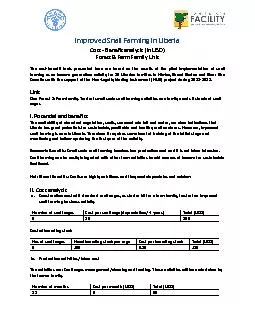
PILA GLOBOSA APPLE SNAIL Systematic Position Phylum Mollusca Class Gastropoda Order Prosobranchiata Suborder Pectinibranchiata Family Pilidae Genus Pila Species ID: 771096

renal blood auricle aorta blood renal aorta auricle visceral sinus chamber vein pulmonary posterior ventricle cephalic sac side sinuses

Share:

Link:

Embed:

Download Presentation from below link

Download Presentation The PPT/PDF document "PILA GLOBOSA ( APPLE SNAIL)" is the property of its rightful owner. Permission is granted to download and print the materials on this web site for personal, non-commercial use only, and to display it on your personal computer provided you do not modify the materials and that you retain all copyright notices contained in the materials. By downloading content from our website, you accept the terms of this agreement.


Presentation Transcript

PILA GLOBOSA ( APPLE SNAIL)

Systematic Position Phylum - Mollusca Class - Gastropoda Order - Prosobranchiata Suborder - Pectinibranchiata Family - Pilidae Genus - Pila Species - globosa

CIRCULATORY SYSTEM

INTRODUCTION Circulatory system of Pila is open type . It is quite complicated due to dual mode of respiration. It comprises the pericardium, heart, arteries, sinuses, veins and blood.Pericardium: It is more or less ovoid, thin-walled sac situated vertically but somewhat obliquely on the left side of the body whorl, just behind the pulmonary sac between the two renal chambers (dorsally) and junction of oesophagus and stomach (ventrally). The cavity of pericardium is true coelom as it communicates with the posterior renal chamber. It encloses heart. (2) Heart: It comprises of two chambers: an auricle and a ventricle, connected by auriculoventricular aperture guarded by semilunar valves (which allows the blood to flow in one direction i.e. towards ventricle). The auricle is thin walled and receives oxygenated and purified blood from two veins, viz. efferent ctenidial vein from gills and efferent pulmonary vein from lungs while it gets deoxygenated blood from post. renal chamber via. efferent renal vein. The ventricle has a thick, muscular wall and receive mixed type of blood from auricle and sends it to whole body through large artery (Aortic trunk). A pair of semilunar valves is also present in aortic trunk also. Heart of Pila

(3) Arteries: The aortic trunk immediately divides into two branches, viz. the anterior Cephalic Aorta and the posterior Visceral Aorta . The cephalic aorta is dilated near its base into a sac, the aortic ampulla which helps in proper distribution of blood in the head region of pila. The cephalic aorta supplies blood to pericardium, skin, oesophagus, left and right side of mantle, left & right nuchal lobes, radular sac, eyes, tentacles, foot and to copulatory organs in males. The visceral aorta extends backward into the visceral mass and gives off arteries to peircardium of its side, skin, digestive glands, stomach, intestine, kidney, hepatopancreas and gonads.(4) Sinuses & Veins: The dexoygenated blood from the various parts of the body collects in small spaces called the lacunae which later on joins to form larger spaces termed the sinuses. Both of them lacks a definite wall and constitutes the Haemocoel. Sinuses are of 4 main types, viz. perivisceral, periintestinal, branchiorenal and pulmonary sinus. The cephalic aorta sends its blood to perivisceral and pulmonary sinus while visceral aorta circulates its blood via. periintestinal and branchiorenal sinus (Chart).z(5) Blood: The blood consists of plasma and corpuscles. The plasma is light-blue in colour due to respiratory pigment, haemocyanin dissolved in it. Corpuscles are colourless , amoeboid and of several types. They are called leucocytes . Function : Blood transports food , O 2 , CO 2 and nitrogenous wastes to desired places. It also keeps the tissues moist and protect the animal from microorganisms

CIRCULATORY SYSTEM The circulatory system is open and consists of heart, arteries, veins and the sinuses and has attained great complexity because of its amphibious nature. The heart is situated on the left side of the visceral mass very close to the posterior end of the ctenidium . The heart is enclosed in a thin-walled coelomic cavity called pericardium. The pericardium is situated between the pulmonary chamber and the posterior renal sac on the left side of the body whorl . It extends anteriorly upto the stomach and digestive gland and communicates with the posterior renal chamber by a reno-pericardial aperture. Heart consists of two chambers: an auricle and a ventricle . The auricle is thin walled, highly contractile and lies in the dorsal part of the pericardium. Ventricle is thick walled, spongy and muscular situated just below the auricle in the same vertical axis. Ventricle has a reduced cavity due to a coarse meshwork of muscular strands. The auricle communicates with the ventricle by a auriculo-ventricular aperture which is guarded by two semilunar valves to prevent the backflow of blood from the ventricle to the auricle.

The auricle receives oxygenated blood from the ctenidium and the pulmonary sac through the efferent ctenidial vein and the pulmonary vein. It also receives blood from the posterior renal chamber by efferent renal vein. The lower end of the ventricle gives rise to an aorta which immediately divides into two branches: 1. An anterior cephalic aorta 2. Posterior visceral aorta

The opening between the ventricle and aorta is guarded by two semilunar valves which prevent the backflow of blood into the ventricle. CEPHALIC AORTA: The cephalic aorta supplies blood to the head region. It is swollen into a thick walled contractile aortic ampulla at its base which helps in the propulsion of the blood. The cephalic aorta gives off three branches on its outer side: 1. A fine cutaneous artery to the skin. 2. A thick oesophageal artery to the oesophagus. 3. A thick pallial artery to the left side of the mantle, left nuchal lobe and the osphradium. The inner side of the cephalic aorta gives off a single pericardial artery to the pericardium which finally enters the posterior renal chamber and gives off branches to both the renal chambers and part of reproductive system. The cephalic aorta enters the perivisceral sinus (space surrounding the buccal mass and oesophagus) along the left side of the oesophagus and then crosses beneath the oesophagus and gives off many arteries to the buccal mass, oesophageal wall, right side of the mantle, right nuchal lobe, copulatory organ, eyes, tentacles

Heart and blood vascular system of pila

The visceral aorta passes through the visceral mass and supplies blood to the visceral organs. It gives out: 1. A small pericardial artery to the pericardium. 2. A large and stout gastric artery to the stomach. 3. Intestinal arteries to the intestine. 4. A hepatic artery to the digestive gland and the gonads. 5. Renal arteries to the anterior and posterior renal chamber. Finally the visceral aorta terminates into branches that supply the rectum and the anus

The blood that is supplied to the various organs of the body by the arteries and its branches is finally collected in small spaces called the lacunae. These lacunae unite to form large sinuses (Fig. 13). Sinuses are spaces between the body wall and visceral organs and together with the lacunae form the haemocoel . They connect arteries to veins and serve as capillaries. There are four blood sinuses in Pila : 1. A peri -visceral sinus surrounding the anterior part of the alimentary canal. It contains deoxygenated blood. Three channels carry blood from this sinus to the renal chamber and the pulmonary sac. 2. Peri-intestinal sinus surrounding the terminal part of the intestine and the the genital duct. It collects blood from the digestive gland and the visceral organs. 3. Branchio-renal sinus lying outside the anterior renal chamber and leading into the afferent ctenidial vein. It receives blood from the roof of both the renal chambers. 4. Pulmonary sinus present in the wall of the pulmonary sac. It receives blood from the peri-visceral sinus.

The blood from different organs of the body is collected by three main veins and poured into the auricle either directly or through the respiratory and excretory organs. 1. Afferent ctenidial vein: It collects deoxygenated blood from the peri - visceral sinus, the rectum and the terminal part of the genital ducts and carries it to the ctenidium where the blood is oxygenated. The efferent ctenidial vein collects the oxygenated blood from the ctenidium, mantle, and the copulatory organs and pours it into the auricle. 2. Afferent renal vein: It receives blood from the peri-intestinal sinus and carries it to the posterior renal chamber. The efferent renal vein then collects blood from the posterior renal chamber and conveys it to the auricle. 3. Pulmonary vein collects blood from the pulmonary sinus and sends it to the auricle.

BLOOD Blood is colorless because of the absence of haemoglobin. It contains the respiratory pigment haemocyanin which is dissolved in blood plasma. Haemocyanin contains copper and thus it becomes blue when it combines with oxygen and colorless when it combines with carbondioxide. Blood also contains some colorless stellate amoeboid cells which are phagocytic in nature and help in the removal of waste substances. Some intracellular digestion also takes place in these amoeboid cells.

COURSE OF CIRCULATION The cephalic and visceral aorta supplies blood to different parts of the body. The blood is then collected from various parts of the body in two main sinuses, the peri -visceral and periintestinal . From these sinuses, the blood passes either into the ctenidium , pulmonary sac or the kidney. During aerial respiration, the blood flows from the peri-visceral sinus into the pulmonary sac and after aeration comes to the auricle by the pulmonary vein. During aquatic respiration, the blood flows from the perivisceral sinus to the ctenidium and after aeration comes to the auricle by the efferent ctenidial vein. The blood from the peri- intestinal sinus takes two courses: it either goes to the anterior renal chamber from where it goes into the ctenidium where it is purified and finally through the efferent ctenidial vein enters the auricle, or from the peri-intestinal sinus, the blood goes to the anterior renal chamber and then into the posterior renal chamber. However, the blood can enter the posterior renal chamber directly from the peri-intestinal sinus. Whatever may be the case, the blood is finally carried from the posterior renal chamber by the efferent renal vein to the auricle. This blood is not aerated. Thus, the aerated and non-aerated blood gets mixed up in the auricle. This mixed blood then enters the ventricle and is distributed to the arteries. The renal chambers remove the excretory products from the blood.

THANK YOU