/
The Canadian Journal of CME / May 2001  approach each patient in an or The Canadian Journal of CME / May 2001  approach each patient in an or

The Canadian Journal of CME / May 2001 approach each patient in an or - PDF document

tatiana-dople
tatiana-dople . @tatiana-dople
Follow
444 views
Uploaded On 2015-08-26

The Canadian Journal of CME / May 2001 approach each patient in an or - PPT Presentation

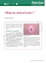
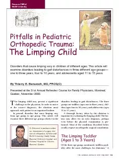
Disorders that cause limping vary in children of different agesThis article willxamine disorders leading to gait disturbances in three different age groups ID: 115779

Disorders that cause limping vary

Share:

Link:

Embed:

Download Presentation from below link

Download Pdf The PPT/PDF document "The Canadian Journal of CME / May 2001 ..." is the property of its rightful owner. Permission is granted to download and print the materials on this web site for personal, non-commercial use only, and to display it on your personal computer provided you do not modify the materials and that you retain all copyright notices contained in the materials. By downloading content from our website, you accept the terms of this agreement.


Presentation Transcript

The Canadian Journal of CME / May 2001 approach each patient in an organized fashion.In general, disorders that cause limping varyfrom age group to age group. This article willexamine three different age groups relative to thedisorders leading to gait disturbances. The threegroups are toddlers (ages one to three years), chil-dren (ages four to 10 years), and adolescents (agesto 15 years).important in evaluating the limping child. The his-tory may allow for an early diagnosis, perhapsformed. Most of the conditions described belowusually require an orthopedic surgical consultation.Of the three age groups mentioned, toddlers prob-ably offer the most challenges for clinicians. Disorders that cause limping vary in children of different ages.This article willxamine disorders leading to gait disturbances in three different age groupsÑone to three years, four to 10 years, and adolescents aged 11 to 15 years. By Thierry E.Benaroch,MD,FRCS(C)Presented at the 51st Annual Refresher Course for Family Physicians, Montreal,Quebec, November 2000. McGill University Dr.Benaroch is assistant profes-sor, department of surgery, divi-sion of orthopedics, full-time staff,McGill University Health Centre,Montreal ChildrenÕs Hospital, andShrinersÕHospital for Children, Pitfalls in Pediatric Orthopedic Trauma:The Limping Child reliable history is difficult to obtain, even whentaken from the childs parents.freely with his/her parents. Lack of spine motionquickly evident. Tenderness to palpation, warmth,redness and swelling of extremity are all helpful innarrowing the differential diagnosis.I. Infection versus non-infection.This has to bedifferentiated in every age group. Transient syn-ovitis and septic arthritis often must be differenti-ated from one another. Although both conditionspatients who have septic arthritis are usually moreirritable and frequently refuse to walk. Transientsynovitis„probably the most common cause ofcome, whereas, septic arthritis, if untreated, hastions usually present with a rapid onset of joint orbone pain, usually progress to a febrile systemic The Canadian Journal of CME / May 2001imited, but not painful, range of motion of the knee and ankle, hyperreflexia andclonus provide confirmation of a neurologic disorder, such as cerebral palsy. illness and lead to the toddlers refusal to use theextremity. There may be a history of mild traumaor concurrent illness or infection. On examination,Range of motion of the affected joint causes obvi-ous pain to the child. X-rays are usually negative,except for soft tissue swelling in the acute phaseand radiographic bone changes, which are seened infection. The white blood cell count (WBC),C-reactive protein (CRP) and erythrocyte sedi-mentation rate(ESR) are usually elevated. Bloodcultures always should be drawn, as they willidentify the offending organism in up to 50% ofpatients with septic arthritis or osteomyelitis.Occasionally, bone scans are helpful to localizethe infection. Aspiration of the joint is necessary toorganism. An orthopedic surgeon and an infec-(b) Transient (toxic) synovitis.the older toddler. Transient synovitis is most com-age. In contrast to septic arthritis, children withsystemic illness. The clinical symptoms generallyshow a gradual and complete resolution over sev-eral days to weeks, usually averaging 10 days. Figure 1.a) Initial radiograph reveals no evidence of afracture.b) Two weeks later, some new periosteal bonemation (callus) is present confirming diagnosis of aspiral tibial fracture. It is during the acute phase, however, that theclinician must differentiate between septic arthritisand transient synovitis. The finding during physicalexamination may be similar, but children with sep-tic arthritis are usually more irritable. Temperatureis never greater than 38º C. ESR, WBC, CRPareusually within the normal ranges. The goals of. The toddlermay have difficulty walking or may have pro-gressed to the point where he/she refuses to walk.pick up an object from the floor, the child willeither refuse or will bend only at the hips whilethe spine. The toddler may not appear ill, but inover 80% of cases, the ESR will be elevated.Blood cultures may be positive and the organismaureus. Early radiographs will be normal. Abonescan is helpful in confirming the preliminary diag-nosis and assists in localizing the infection. Thetreatment of choice is systemic antibiotics, as thisleads to a more rapid resolution of symptoms thanII. ToddlerÕs fracture.the foot may produce a spiral fracture of the tibiawithout a fibular fracture. There may be no histo-ry of recognized trauma, yet the child presentswith a limp, or refuses to bear weight.Radiographs may demonstrate a spiral fracture orographs one to two weeks later will reveal subpe-riosteal new bone formation. If a fracture is sus-od of three weeks. The Canadian Journal of CME / May 2001 Figure 3.Knee x-rays reveal white thick metaphysealbands on both distal femurs and proximal tibias sugges-tive of leukemia.neoplasm in children under 16 years ofcomplaints are a presenting feature in20% of children with this disorder. Figure 2.An antero-posterior (AP) pelvic x-ray reveals anobvious left dislocated hip. III. Neurologic disorder (Cerebral Palsy). Verythe toddler. Athorough prenatal, perinatal, andpost-natal history is needed. Athorough examina-tion will help to differentiate the problem.confirmation. Areferral to a pediatric orthopedistand neurologist is in order.IV. Developmental dislocation of the hip.Examination of the toddlers gait will demonstratea limp, a short leg, one-sided toe-walking, or, ifa waddle. On supine examination, the toddlerspared to the normal side. After the age of sixmonths, a plain anteroposterior (AP) radiograph ofthe pelvis easily confirms the diagnosis (Figure 2).uvenile chronic arthritis (Monoarticular -This is the most common sub-group of juvenile arthritis. It usually is present Figure 4.AP and frog-leg pelvis reveals a smaller anddenser left femoral head compatible with early Legg-CalvŽ Perthes disease. mildly painful limp. Girls are four times morelikely to be affected than boys. Symptoms developinvolved in the lower extremity. Laboratory eval-uation, including ESR, WBC and rheumatoid fac-tent, a referral to a childrens rheumatologistand, therefore, are rarely responsible for a tod-dlers limp. If present, plain radiographs mayoften identify the abnormality. Two neoplasms,however, may be unremarkable on initial radi-ma have been shown to be responsible for painfullimps in toddlers.(a) Leukemia.five. Musculoskeletal complaints are a presentingfeature in 20% of children with this disorder.described as discomfort in an adjacent joint.which include lethargy, pallor, bruising, fever andbleeding. Furthermore, appreciation of skin bruis- The Canadian Journal of CME / May 2001 Figure 6.X-rays of the tibia and fibula demonstratesperiosteal reaction (callus) over the lateral aspect of thefibula.This most likely represents a healing stress fracture. Figure 5.a) AP pelvic x-ray reveals subtle right slippedemoral capital epiphysis (SCFE).b) Frog-leg pelvic x-rayon the same patient demonstrates a more obvious slip ofthe femoral epiphysis. the diagnosis. With the exception of bruising,bleeding and hepatosplenomegaly, the clinical pic-ture may be similar to that of septic arthritis,therefore, should always be included in the differ-ential diagnosis of these other disorders.and peripheral leukocyte counts. The earliest radi-bands (Figure 3). Bone scans may be normal.younger than five years of age. This diagnosis isextremely difficult to make in toddlers who are justlearning to walk. Although pain is the most fre-radiographs are negative, bone scans provide con- Figure 7.a) Oblique radiograph of a normal foot reveals a space between the calcaneus and navicular.b) In a calca-neonavicular coalition, the oblique radiograph reveals an osseus connection between these two bones. dlers and usually are more co-operative during anage group should be taken seriously, because thesethey are in secondary gains. Periodically, parentsor night. The pain responds to a rubdown andinfrequently requires medication. Prior to reassur-ing the parents that this represents benign grow-to avoid missing an underlying disorder. All of thedisorders mentioned for toddlers must be kept inI. Transient synovitis.seen most commonly in the three- to eight-years-of-age group and probably is responsible for themajority of limping due to an irritable joint. Themost important aspect is to differentiate this con-dition from a septic process.II.) Legg-CalvŽ Perthes disease (LCPD) idiopathic avascular necrosis of the childs hip.LCPD is most common in children aged four toaffected. Boys are involved four to five timesmore frequently than girls. These children presentfollowing activity.child. The earliest radiographic sign is anincreased density of the femoral head (Figure 4),physis is seen later in the course of the disease.This condition is not an emergency, but referral toa pediatric orthopedist within three to four weeksis necessary.III. ServerÕs disease/Calcaneus apophysitis.Severs disease or calcaneal apophysitis is aneight- to 10-year-old age group in girls, and inboys aged 10 to 12 years. This presents as a chron-ic, intermittent pain related to sports, whichinvolves jumping or running. It rarely hurts whilethe child is skating or skiing, where the heel isimmobile. The pain is located along the medialaspect of the posterior part of the heel. The rangeof motion of the ankle is usually normal. Treatmentconsists of ice, rest, limitation of activities andcushion heel inserts. This process is self-limiting.accurate history of the problem, however, thesports quickly. Likewise, the symptoms may bemany of the disorders already mentioned must being adolescent, however, several other disordersshould not be overlooked. These include slippedcapital femoral epiphysis, overuse syndromes,osteochondritis dissecans and tarsal coalition.I. Slipped capital femoral epiphysis (SCFE)disorder in which the epiphysis becomes posteriorly The Canadian Journal of CME / May 2001 lescence. Clinically, boys present around the age of14 years and girls around 12 years of age. Mostoften, adolescents who generally are overweight andpain in the hip, groin, thigh, or knee. The duration ofsymptoms is usually several months, but occasional-ly, the adolescent may present with acute excessivepain and actually is unable to walk at all. Always besuspicious of a hip problem that presents as kneeQuite often, hip pain is referred to the knee.for his/her age. Range of motion of the hip is lim-ited in internal rotation and abduction. As theassumes an externally rotated appearance. APradiographic views of the pelvis may miss the sub-tle slip, so a view of a frog leg pelvis or a true lat-chance to make the diagnosis (Figure 5). Arefer-ral to an orthopedic surgeon is mandatory andII. Overuse syndromes.As adolescents becomemore active in organized sports, overuse injuriesoccur with increasing frequency. These syndromestypically present with pain, but on rare occasions,also present as a limp. The knee is the most com-mon site for this. Patellar tendonitis or apophysitisof the tibial tubercle (Osgood-Schlatter disease)cause persistent pain. Point tenderness to palpa-tion is helpful in confirming these disorders. Rest,extremities. The tibia and fibula are most suscep-sclerotic line or periosteal reaction (Figure 6), orthey may be normal. If suspicion of a stress frac-ing the diagnosis. Treatment consists of rest in theIII. Osteochondritis dissecans becomes avascular. The etiology is unclear. Thiscan, on rare occasions, present with a limp. Theknee is affected most, but the hip and ankle canalso be involved. Radiographically, a tunnelŽclearly. Classically, it is located on the lateral sideof the medial femoral condyle. Patients with thiscondition should be referred to an orthopedic sur-geon within three weeks.IV. Tarsal coalitions.tion in which certain tarsal bones become fusedwith each other, most commonly the calcaneusThe adolescent presents with a rigid flatfoot, andthe subtalar joint motion (inversion, eversion) ismarkedly restricted and painful. X-rays (obliqueand Harris views of feet) are necessary to show thecoalition (Figure 7). Referral to a pediatricorthopaedic surgeon is necessary.1.Choban S, Killian JT: Evaluation of acute gait abnormali-2.Blatt SD, Rosenthal BM, Barnhart DC: Diagnostic utility3.Stahl JA, Schoenecker PL, Gilula LA: A2 1/2-year-oldmale with limping on the left lower extremity: Acute lym-1.MacEwen GD, Dehne R: The limping child. Pediatr Rev2.Phillips WA: The child with a limp. Orthop Clin North Am The Canadian Journal of CME / May 2001