/
Bone Morphogenetic Proteins (BMPs)rm a unique group of proteins within Bone Morphogenetic Proteins (BMPs)rm a unique group of proteins within

Bone Morphogenetic Proteins (BMPs)rm a unique group of proteins within - PDF document

tatyana-admore
tatyana-admore . @tatyana-admore
Follow
442 views
Uploaded On 2015-09-09

Bone Morphogenetic Proteins (BMPs)rm a unique group of proteins within - PPT Presentation

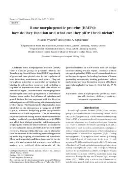
57 Journal of Oral Science Vol 45 No 2 5773 2003 of the requirements for the Degree of Doctor of Philosophy NS Bone morphogenetic proteins BMPshow do they function and what can they offe ID: 125109

57 Journal Oral Science Vol.

Share:

Link:

Embed:

Download Presentation from below link

Download Pdf The PPT/PDF document "Bone Morphogenetic Proteins (BMPs)rm a u..." is the property of its rightful owner. Permission is granted to download and print the materials on this web site for personal, non-commercial use only, and to display it on your personal computer provided you do not modify the materials and that you retain all copyright notices contained in the materials. By downloading content from our website, you accept the terms of this agreement.


Presentation Transcript

57 Bone Morphogenetic Proteins (BMPs)rm a unique group of proteins within theransforming Growth Factor beta (TGF-§) superfamilyof genes and have pivotal roles in the regulation ofbone induction, maintenance and repair. They actthrough an autocrine or paracrine mechanism by Journal of Oral Science, Vol. 45, No. 2, 57-73 2003 of the requirements for the Degree of Doctor of Philosophy. (N.S.) Bone morphogenetic proteins (BMPs):how do they function and what can they offer the clinician? 58 of a wide variety of cells, depending on the cellular micro-environment and the interactions with other regulatoryLigand-receptor interactions anddownstream eventsregion containing seven cysteine residues that is conservedsuperfamily(12). BMPs are synthesized inside the cell in a precursorpeptide sequences joined to the mature region. Proteolyticcleavage frees the mature region, which can then dimerizewith other BMPs. Mature BMP-2 is a homodimer of two114-peptides subunits (13). Dimeric molecules can beeither homodimers, when both subunits are the same, orheterodimers, consisting of two different subunits.Structural and chemical differences between thefor variations of their biologic potential and bindingBMPs initiate signaling from the cell surface when theythreonine kinase receptors (14,15), both of which have sub-categories (17). BMP receptors are composed of three parts:a short extracellular domain, a single membrane-spanningdomain, and an intracellular domain with the activeserine/threonine region (18). The 75kDa type II receptoris the primary binding site of the ligand and upon itsactivation, phosphorylation of the type I receptor (50-55kDa) occurs (14,17). It is only after the type I receptorpropagated to downstream substrates (9). It is believedrious signaling pathways have been proposed to beactivated by the ligand binding to the receptors. Recently,a novel family of proteins, the Smad family, have beenidentified as the downstream effectors of the phosphorylatedtype I receptor (21-23). More specifically, Smads 1 and5 become phosphorylated at the carboxy-terminal serineresidues and then translocate to the nucleus (24) where theyinteract with DNA binding proteins (25), or exhibit directtranscriptional activity (26), either as monomers or inassociation with Smad 4 (21). Specific Smads are expressedat different phases of the cell cycle and exhibit either aninhibitory or stimulatory function (27). Smads 6/7 havebeen shown to antagonize and inhibit phosphorylation ofSmads 1/5 by BMP-2 (28,29). Phosphorylation of Smad1leads to the transcriptional activation of the DPC4 genethat expresses Smad4 and the subsequent Smad1/4 complextranslocates to the nucleus (21). The lack of DNA bindingmotifs on Smads does not allow their direct associationwith genomic sequences. It has been shown that smallDrosophilarevealed that the geneschnurribecomes up-regulated after BMP binds to theirreceptors, leading to the formation of an active transcriptionactor similar to zinc finger proteins in mammals (30). Inaddition, the protein kinase TAK1 has been isolated andshown to transduce signals downstream following BMPbinding and function as a mitogen-activated protein kinase(31). Overall, BMPs regulate cell function at thetranscriptional level or higher (32), by increasing the rateof transcription and/or stabilizing the mRNA (33).Moreover, the large number (2,500/cell) of BMP receptors(6) and their variability (16,29) allows heteromericcomplexes to be formed with different signaling potential(16) capable of inducing various responsive cascadesAnother line of evidence indicates the involvement ofthe Ras/Raf pathway in the downstream response to BMPs(35). According to this model, the signal is transmittedthrough Ras to Raf and, subsequently, activates othertranscription factors such as AP-1 and GATA-2. BMP-2up-regulates Id (inhibitor of differentiation) gene expressionin osteoblastic cells and promotes their specific phenotypicxpression (32). BMP-2 has an effect on cell-matrixinteractions by suppressing the expression of the (3 integrinsubunit at the mRNA level (36), suggesting that BMPaction, in part, may be exerted through altered cell adhesionto the extracellular matrix (ECM), modified cell migrationand differentiation (37,38). In another report, cadherinxpression in osteoblasts was unaffected by BMP-2The family of Hedgehog genes (Sonic hedgehog, (Shh),Desert hedgehog, (Dhh) and Indian hedgehog, (Ihh)present a striking correlation with expression of BMPmodulators of BMP expression (41). In addition, growthactors such as TGF-1 exert a negative regulation onBMP-2 at the transcription level (42) and retinoic acidreceptors also affect BMP expression (43). Noggin,molecules that have recently been found to regulate BMPxpression and modulate their role in eliciting variousbiologic responses (44). A number of BMP-bindingproteins (lipovitellin 1, Ep45) (45) and antagonist molecules(noggin) (46), affect and control the presence of growth 59 actors thus defining various concentration gradients,which has been shown to play a role in cytodiferentiation.There are several reports on the associated action of BMPsand noggin (47,48) suggesting that transcription factorsamily are downstream targets of noggin-related calcium regulation (47). These transcriptionosteoblasts and thus act as autocrine regulators (49). In summary, the variety of BMP receptors and thenumerous pathways they regulate, together with the factthat BMPs exist in the form of homodimers, suggest thatthey may evoke synergistic, negative or additive net effects.This draws attention to the complexity of the signalingcascade and the variability of downstream responses toBMPs in bone formationBone formation can take place Intramembranous ossification occurs during embryonicdevelopment of the cranial vault bones by the directtransformation of mesenchymal cells into osteoblasts.Endochondral ossification, which is the process by whichlong bones develop, involves the formation of anintermediate cartilaginous anlagen that eventually becomesossified and contains all the cellular components of maturebone (50). In both mechanisms, the induction of bone andcartilage occurs through an epithelial-mesenchymalinteraction (51) that initiates specific cell differentiationThe development of cartilage and bone frommesenchyme, is characterized initially by a condensationof mesenchymal cells (52). This condensation can occurin two ways: either by cells moving toward a central focalpoint or by a localized region of increased proliferation.Direct cell-to-cell contact, diffusible molecules producedby the signaling cells, or matrix mediated interactions canresult in a cellular mass of increased proliferative activity(53-55). An early step in the endochondral bone formationdiscrete pre-cartilaginous nodules. Chondrogenic cellsactive state that involves mineral deposition in thecartilaginous matrix. The hypertrophic chondrocytes willthe invasion of the sites by blood vessels. The chondrocytesentually die and their matrix is partially destroyed duringascular invasion, which is when osteoblasts appear.Initially, osteoid will be deposited and remodeling willthrough BMPs (57) and retinoic acid, a derivative ofvitamin A, is a possible modulator of BMP expression (58).Cell adhesion molecules such as laminins (59), neuralcell adhesion molecules (N-CAM) (60), and integrins (36)are known to interact with BMPs and other growth factorsand have been localized at the areas of initial mesenchymalcondensation (60,61). Related to this intra-molecularattract various types of cells (19,62) and act as chemotactic,mitogenic or differentiating agents (63,64). BMPs mayaffect proliferation of cartilage-forming and bone-formingcells and can induce differentiation of mesenchymalprogenitor cells into various cell types, includingchondroblasts and osteoblasts (19,65). The latter propertyendochondral bone induction pathway and direct boneformation. In ectopic bone formation, associated withimplantation of BMPs, the sequence of events recapitulatesthe process of bone formation that is observed duringembryonic long bone development and many of the BMPproperties can be extrapolated from there. One of the most difficult subjects to study with regardthe responding cell population. The wide spectrum of cellsthat are sensitive to BMP action includes fibroblasts (61),mesenchymal connective tissue cells (66), muscle derivedconnective tissue cells (67), the astroglial lineage (68)and many more (61). Bone marrow stromal cells form anthat are capable of differentiating into various cell lineagesunder the appropriate conditions. Demineralized bonematrix, dexamethazone, beta glycerophosphate, vitaminD and BMP-1 have been shown to stimulate bone marrowstromal cells to take on an osteoblastic phenotype (69, 70).Bone marrow mesenchymal cells have the potential todifferentiate along the osteoblastic and adipocytic lineages.Studies have demonstrated a concentration specific responsewith lower doses of BMPs inducing the adipocyte lineageresponse (71). Treatment with rhBMP-2 protects andenhances cell commitment towards the osteoblasticphenotype (72,73). Osteoblasts and chondroblasts originate(74) or skeletoblast (75-78). Osteoprogenitors can beclassified as either determined or inducible, based on theirneed for additional signals in order to differentiate (58,79).This difference is important as it reflects the variationbetween cell commitment, when the fate of cells is 60 programmed, and cell differentiation, when the fate ofcells is expressed due to the permissive signals of themicro-environment. The answer to this debate is not clearyet but it is obvious that some form of control overphenotypic expression must occur to ensure that the tissuesdevelop in a coordinated fashion in the appropriate placesand amounts. (80). Evidence supports the hypothesis thatBMPs act on the skeletal progenitor cells and induce thedifferentiation of both the osteoblast (65,71,81-83) andactors affecting BMPs bone inductive ability areamounts, qualitative composition, possible presence ofinhibitors, correct processing and storage (86). In addition,important parameters of the inductive outcome (64,87).Low concentrations of BMP-2 (50ng/ml) up-regulatedthe expression of the collagen II gene whereas higherxpression in chondrocyte cell lines and increasedosteocalcin (OC) expression (72). These results clearlyshow that chondrocytes are able to express osteoblasticfeatures. It is more realistic to assume that BMPs induceytodifferentiation along those lineages when permissiveconditions for each cell type exist. Stability or structuralintegrity that allows blood vessels to grow (88), micro-environmental conditions that affect oxygen tension (89),affecting the cytoskeleton (90) through membrane receptorsare critical factors for cytodiferentiation. Differences inthe partial pressure oxygen and in the amount ofmesenchymal cells present in intramuscular andsubcutaneous sites are responsible for the lower dose ofeatment with cytochalasin D disrupts the cytoskeleton,concentrations of endogenous soluble and matrix factors,thus promoting the chondrogenic phenotype (92). Reducedserum, high cell density, and type I collagen have beendifferentiation (93). Cells at high density become attachedpromotes the chondrogenic phenotype. Furtherdifferentiation into hypertrophic chondrocytes andmineralization does not require BMP-2 but it is dependenton the presence of ascorbic acid and serum factors (94,95).ytokines (100,101), BMP-2 plays a regulatory role for thesequential progression of chondrocytes through theirmaturation (96,97), with development of hemopoieticbone marrow (98,99), and inhibition of myogenicdifferentiation (83). Differentiation of mesenchymal cellsinto pre-chondroblasts is induced by BMPs but thesubsequent osteoblastic lineage is regulated by othergrowth factors that work in an autocrine or paracrinemanner (102). BMPs acting through an autocrinemechanism reduce the expression of collagenase-3 andAlthough BMPs exert their action on both osteoblasts andchondroblasts, they do not change the fate of the respectiveprogenitors (104). Early exposure of undifferentiatedpathway, whereas later exposure accelerates osteoblasticdifferentiation (105). BMPs can stimulate osteoblastdifferentiation independently of cartilage formation (106).osteoblasts do not form from a transdifferentiation ofchondrocytes but rather as a result of a separate induction(1,70,93,107). Endothelial cells invading the cartilagemay serve as a homing target of the stem cells that laterdevelop into pre-osteoblasts (98). Numerous reports showan up-regulation of the osteoblastic phenotype by BMPs.Up-regulation of osteocalcin (OC), osteopontin (OP),a promotive effect of BMPs on mesenchyme-derived cells(64,82,86,106,108,109). BMPs can act on various cell typesand elicit a response that is specific to that stage of celldifferentiation (106). There is evidence that BMPs triggerin osteoblastic cells osteocalcin is upregulated and bonesialoprotein is expressed in differentiated osteoblasts priormodeling and remodeling. The morphogenetic activity ofInsulin-like growth factors (IGF-I, IGF-II), TGF2, PDGF, basic and acidic fibroblast growth factors, BMPsinto the forming bone matrix that serves as a reservoir (111).under these conditions are inactive. A heparin-binding sitehas been identified at the N-terminal segments of theBMP-2 that may function to localize the growth factor andrestrict its diffusion (13). Acid treatment associated withsubstrate rendering them biologically active (114) andable to affect cell proliferation and differentiation (70). 61 Guided streaming of specific cell types into the appropriatepathways makes BMPs important regulators of boneformation (113), with a pivotal role in bone remodeling(110). ÒBasic Multicellular UnitsÓ (BMUs) refer to theact in coordination (115). This is called coupling. Duringremodeling, which is a Òself-maintenanceÓ process, existingbone is resorbed and new bone is deposited. Resorptionduring each remodeling cycle is balanced by an equalother growth factors released from bone are proportionalto the extent of resorption (116,117). There is evidencethat BMP-2 promotes expression of cyclooxygenase-2and the osteoclast differentiation factor in osteoblast-likecells, thus regulating osteoclastogenesis (118). Based onthe above, the mitogenic, chemotactic and differentiatingeffect of BMPs may help to mediate coupling of boneformation to resorption during the adaptive response ofremodeling. It supports the cellular components andamplifies the molecular signals needed for the coordinatedinteraction of various cell types. BMPs in fracture healingFracture healing involves complex interactions amongmany local and systemic regulatory factors as well as celltypes that cluster at the fracture site. Fracture repairrepresents a situation in which cell differentiation is re-initiated in an otherwise mature organism (2).Mesenchymal stem cells congregate at the area and forma gap-spanning, highly cellular Òrepair blastemaÓ (119,120).cells, proliferation, differentiation into cartilage or bonecell lineages, mineralization and remodeling, and marrowThe first demarcation of osteoprogenitor cells duringfracture repair, referred to as the Òstacked-cell layerÓ (120),is derived from mesenchymal stem cells (119). They aredifferentiate into pre-osteoblasts. In the case of boneoriginally continuous bone, a sequence of cellular andmolecular events is initiated in response to the trauma,of platelets, releasing numerous growth factors. The bloodclot then begins to organize and the formation of aprovisional callus that bridges the fracture site becomesapparent. Increased vascular permeability allows fluidand plasma proteins to leave the blood vessels. Variouscell types then emerge from the vessels in significantnumbers. In acute inflammation, neutrophils (PMNs) arethe first leukocytes to provide an effective defense inhas a strong and dose-dependent promotive effecton the osteogenic activity of rhBMP (123), andglucocorticoids exert similar effects (69) whereas bindingof BMP to free heparin at the fracture site could help tolocalize the growth factor by restricting its diffusion (13).acidic conditions that develop and, together with theactivate latent forms of growth factors such as TGF-regulate a positive feed-back system that amplifiesactivation of TGF-1 from platelets, thus stimulatingcartilage and bone formation (113). BMPs can also inducechemotaxis of monocytes and stimulate their expression1 mRNA (62). Finally, blood-clot formationfollowing tissue injury results in the lysis of platelets thatrelease numerous growth factors that are involved inound healing and contribute to bone repair (121). mesenchymal stem cells can differentiate into chondroblastsor osteoblasts. If the fracture is mechanically unstable,cartilage will form and the bone fragments will bemechanically joined. If the fracture is mechanically stable,the chondrocytes within the blastema become hypertrophic,and along with their extracellular matrix, become erodedand replaced by osteoblasts and osteoid deposits (119,120).This process is similar to the one observed in embryoniclong bone development. If the original break ismechanically stable, the repair blastema can be spannedby vasculature and the mesenchymal cells differentiatedirectly into secretory osteoblasts (119, 120). Mesenchymalstem cells may originate from the periosteum, the marrowsystem. Although the exact origin is not determined, theimportant fact is that mesenchymal cells will be attractedto the fracture site and play an important role in the repairmechanisms that will be described later, and as such mustbe considered a necessary event for the healing of boneounds. TGF-autocrine regulator of bone formation (125). It has been1 downregulates alkalineBMP-2 mRNA expression. This provides evidence that1 acts as a powerful bone growth stimulant at thelevel of pre-osteoblasts (126), which is needed for the 62 differentiation pathways (127). TGF-1 stimulates DNAchemotactic for mesenchymal cells and osteoblast-likeformation and remodeling (113). EGF and FGFs are othermolecules with demonstrated involvement in the complexmolecular cascades involved in cellular change (128,129). formation that was later replaced by bone tissue with bonemarrow elements. Invasion of blood vessels into themarrow development (98). When larger doses of rhBMP-2 were used, bone formation was observed concurrentlywith cartilage formation, suggesting bone induction throughboth endochondral and intramembranous pathways. BMPsare believed to act through chemotactic, mitogenic ordifferentiating mechanisms. It is important to understandthat BMPs are not the only determinants of cell fates alongthe above-mentioned lineages. Specific nutrients, growthactors and cytokines at specific concentrations and in aspecific sequence of exposures are fundamental for the boneDuring fracture healing, BMP-2/4 affect precursor cellsto become chondroblasts and express proteins needed forproduction of woven bone (121). When lamellar bonereplaces woven bone, BMP expression is significantlyreduced (130). rhBMP-2 can induce bony trabeculae andbone marrow (99) with concomitant shortening of thebone formation (85). BMP-4 is also expressed in lessdifferentiated cells at fracture healing during distractionosteogenesis (131). rhBMP-2 does not increase the mitoticactivity of osteoblasts (132) and does not affect DNAsynthesis, but rather initiates sequences of gene expressionin these cells (133). rhBMP-2 up-regulates the expressionof BMP3/4 mRNA (109) with a mechanism that probablystabilizing the mRNA (33). Evidence of BMP-4 activatingthe transcriptional factors Msx-1/2 and Egr-1 in epithelial-mesenchymal interactions during tooth development makethis mechanism a valid working hypothesis (8). BMPs1/2/4/6 are expressed by osteoblasts before they formmineralized bone nodules and during expression of ALP,OC, OP (134), thereby becoming guiding factors inosteoprogenitor cells (106). Unlike the BMPs, the TGFchondrogenesis (63,110) but promote bone healing andfracture repair (125,135). However, acting at the level ofcell adhesion molecules (36), they may stimulatemesenchymal cell attraction and proliferation (136). Beingeffect (137) especially at the early stages of the bonerepair process (61, 138). BMP-2 up-regulates thephenotypic expression of osteoblasts (82,83,133,139) andreceptors (132). Additionalstudies have demonstrated a promotive effect of rhFGFBMPs, reducing the amount of growth factor needed toelicit a specific biologic response (141). The majority of studies investigating the role and actionof exogenous BMPs use a matrix to deliver the growth factorto the implantation site. Although the matrix may notcontribute any additional factors necessary for boneinduction (107), it is a fundamental and very importantcomponent of the growth process. Collagenous or syntheticmatrices have been used as delivery vehicles and theirmicroenvironment they create, play a role in the inductiveoutcome. Carriers can be solid xenogenic (HA) (89,142),of the above (149).One of the carrier functions is to maintain the factor atconcentration. However, BMPs also help to stabilize thecarrier by accelerating bone growth in its mass (150) dueto the stabilization brough about by the BMPs absorptionto the surface of the carrier matrix particles. As a result,rhBMP-2 was required in the absence of matrix (11). Theut the pharmacokinetics of the growth factor are notaffected by carrier properties (151). Collagen matrix retains~65% of the BMPs during initial impregnation and releasesit in two phases: an initial phase within hours ofimplantation and a second phase that depends on thenature of the carrier and its geometrical characteristics (152). It is believed that BMPs do not bind to the carrier (152),which makes certain designs more favorable for boneinduction over some others (153). In the case of collagensponge carriers, the mass, collagen cross-linking andsterilization methods affect BMP precipitation andsubsequent resistance of sponge degradation by collagenase(154). Properties of the best carrier may vary depending 63 on the specific implantation site and the intended therapeuticoutcome. Considerations include biodegradability,structural integrity, absence of immunogenicity, absorptionand rate of release of BMP (155). The latter characteristicof the carrier serves its second function, which is controlledrelease of the BMP. This allows for a more constant andprolonged application. This renders BMPs more efficientthe cells to respond (11). BMP-2 is retained in a hydrogelof the regenerate when compared to polymeric matrixproperties of the carrier. Recently, a novel approach has been suggested. Thisinvolves implanting matrices that actively concentratenative BMPs at the implantation site instead of passivelystoring and delivering rhBMPs which are a thousand timesless potent that the native BMP complex (158). The matrixalso serves as an environment in which bone can form andtherefore helps to define the region in which new bone canbe formed (159). Delivery vehicles with adequate structuralconsistency can function as primary scaffolds on whichcan be deposited (152,160). If the delivery matrix can actas a scaffold, then the cartilaginous intermediate may notbe necessary. Many investigators agree that it has not beenproved definitively that the chondrogenic process is essentialfor bone formation by BMP (89,161). The type of matrix(146). The material of the matrix and its geometricalparameters (pore size, and %volume) are factors thatdirectly (size of cells able to attach) or indirectly (effectenvironment and influence the mechanism of boneor fibrous collagen membrane lead to intramembranousossification (89,142,161), whereas fibrous glass membraneIn examining the action of BMPs, it is also importantto consider dose-related effects. It is evident that variousdoses elicit different responses on specific cell types atdifferent time intervals (162). The dose of the growth factordetermines its chemotactic, proliferative or mitogenicsignal and should therefore be well regulated. IncreasedBMP concentrations result in faster bone growth (11),with cartilage being more rapidly replaced by mineralizedosteoid (163). rhBMPs in the form of monomers,homodimers or heterodimers need to be evaluated andstandardized because they exhibit different biologicalThe carrier may also act synergistically by serving as areservoir of the inducible cell population. Bone marrowcan be combined with BMPs (164) and, when providingsuperior performance. Recently, investigators attempted) or indirect (using viral vectors) delivery(165,166), demonstrating active BMP expression for 2-6marrow. Cost of manufacturing and handling, in additionto ease of clinical application, are equally important factorsto consider when deciding on a specific type of deliveryfor healing bone fractures, preventing osteoporosis, treatingaround alloplastic materials implanted in bone (3). rhBMP-2 delivered with an absorbable collagen sponge (ACS) hasfloor in humans (167). An rhBMP-2 dose ranging from1.77 to 3.4mg per patient generated an average of 8.51mmof vertical bone height in four months providing a promisingalternative to traditional grafting procedures (167). Similarresults were also achieved in sub-antral augmentation ofnon-human primates with 6 mm of vertical bone gain andincreased density that allowed placement of titaniumimplants (168). BMP-2 regenerated bone in irradiatedtissues also provides the clinical potential to treat patientswho have undergone radiation therapy and need bonePeriodontal regeneration was achieved when rhBMP-2or a collagen gel. However, better results were obtainedusing the slower dissolving collagen membrane that alloweddelivery of the growth factor for a prolonged period of time(170). The clinical outcome was a decreased depth of thedefect site brought about by stimulating vertical bonegrowth and regenerating the periodontal attachment,provided that adequate space is maintained (171-175).The type of carrier, the time of treatment and the use of abarrier membrane are critical factors influencing thetherapeutic outcome in cases of bone regeneration arounddental implants (157) and have been shown to produceaccelerated healing time as well as improved bone-implantcontact levels (175-179). Moreover, alveolar ridge 64 preservation or localized augmentation have beenAnimal studies also suggest that rhBMP-2/ACS may bean effective treatment for the restoration of segmentalolume (183), strength and stiffness (184). A bioerodiblepolymeric carrier was used to deliver rhBMP-2 in a largesegmental defect that was stabilized with stainless steelplates (185). Stabilization was necessary because of thelarge size of the animals (sheep) but it could have alsohelped to provide a stable environment for bone bridgingsince the carrier was reported to fragment easily. In a similarwith rhBMP-2 was found to restore cortical bone withmarrow elements in a twenty-millimeter long segmentaldefect (186). Skull defects were also filled with regeneratedbone when BMPs were delivered in combination withhydroxyapatite (2,187), a biodegradable gelatin hydrogelor an aqueous solution (188). Spinal fusion wassignificantly enhanced when rhBMP-2 was administereddemineralized bone matrix revealed improvedAlthough purification and characterization of rhBMP-2 has been described in the Chinese hamster ovary (CHO)cell line (64) rendering BMPs available in large quantities,the fact that their inductive activity is ten times less thanthat of purified BMPs may present a limitation for theirclinical application (190). Combinations of BMPs withother growth factors or biologic molecules formingheterodimers with twenty times higher potency in somefuture in the field of bioengineering. The parameter of host age further affects the biologicpotential of many growth factors (161). The bone inductiveability of BMP-2 is diminished in older organisms andeffect (192). Reduced migration of mesenchymal cells,lower levels of local anabolic agents, age associatedreduction of receptor levels and compromisedascularization are some of the aspects to take intoconsideration (193-195). In the future, delivery ofbiological agents that control the regulators of BMPs maybe of clinical significance in cases where BMP actionneeds to be halted to prevent pathological or hazardousossification, such as after total hip or temporomadibulararthroplasties (44). Production of natural autogenousbone in moulds may allow a more efficient reconstructionReferencesUrist MR (1965) Bone: formation by autoinduction.Ripamonti U, Reddi AH (1992) Growth andmorphogenetic factors in bone induction: role ofin craniofacial and periodontal bone repair. CritRev Oral Biol Med 3, 1-14Ripamonti U, Reddi AH (1997) Tissue engineering,morphogenesis, and regeneration of the periodontaltissues by bone morphogenetic proteins. Crit RevJones CM, Lyons KM, Lapan PM, Wright CV,Hogan BL (1992) DVR-4 (bone morphogeneticprotein-4) as a posterior-ventralizing factor inXenopus mesoderm induction. Development 115,5.Zou H, Niswander L (1996) Requirement for BMPander L (1996) Requirement for BMP6.Paralkar VM, Weeks BS, Yu YM, Kleinman HK,Reddi AH (1992) Recombinant human bonedifferentiation: potentiation and binding to type IVShah NM, Groves AK, Anderson DJ (1996)Alternative neural crest cell fates are instructivelypromoted by TGFbeta superfamily members. Cell8.Vainio S, Karavanova I, Jowett A, Thesleff I (1993)Identification of BMP-4 as a signal mediatingmesenchymal tissues during early toothdevelopment. Cell 75, 45-58Cheifetz, S. BMP receptors in limb and toothformation. Critical Reviews in Oral Biology andCentrella M, Horowitz MC, Wozney JM, McCarthyTL (1994) Transforming growth factor-beta geneamily members and bone. Endocr Rev 15, 27-39(BMPs): therapeutic potential in healing bonydefects. Trends Biotechnol 11, 379-38312.Wozney JM, Rosen V, Celeste AJ, Mitsock LM,Whitters MJ, Kriz RW, Hewick RM, Wang EA(1988) Novel regulators of bone formation:molecular clones and activities. Science 242, 1528-13.Ruppert R, Hoffmann E, Sebald W (1996) Humanbinding site which modifies its biological activity. 65 Massague J, Weis-Garcia F (1996) Serine/threoninekinase receptors: mediators of transforming growthactor beta family signals. Cancer Surv 27, 41-6415.Attisano L, Wrana JL, Lopez-Casillas F, Massague16.Nohno T, Ishikawa T, Saito T, Hosokawa K, NojiS, Wolsing DH, Rosenbaum JS (1995) Identificationprotein-4 that forms differential heteromericcomplexes with bone morphogenetic protein typeamashita H, Ten D, Heldin CH, Miyazono K(1996) Bone morphogenetic protein receptors. BoneLin HY, Wang XF, Ng-Eaton E, Weinberg RA,beta type II receptor, a functional transmembrane, a functional transmembranein Cell 1992 Sep 18;70(6):following 1068]. Cell 68,zney JM (1992) The bone morphogenetic proteinamily and osteogenesis. Mol Reprod Dev 32, 160-en D, Yamashita H, Sampath TK, Reddi AH,Estevez M, Riddle DL, Ichijo H, Heldin CH,Miyazono K (1994) Identification of type I receptors21.Kretzschmar M, Liu F, Hata A, Doody J, MassagueJ (1997) The TGF-beta family mediator Smad1 isphosphorylated directly and activated functionallyby the BMP receptor kinase. Genes Dev 11, 984-22.Hoodless PA, Haerry T, Abdollah S, Stapleton M,OÕConnor MB, Attisano L, Wrana JL (1996)BMP2 signaling pathways. Cell 85, 489-500decapentaplegic is an embryonic ventralizing agentthat acts downstream of the BMP-2/4 receptor.Development 122, 2359-236624.Baker JC, Harland RM (1996) A novel mesoderminducer, Madr2, functions in the activin signaltransduction pathway. Genes Dev 10, 1880-1889v 10, 1880-18891996 Dec 19-26;384(6610):648]. Nature 383, 691-69626.Liu F, Hata A, Baker JC, Doody J, Carcamo J,protein acting as a BMP-regulated transcriptionalactivator [see comments]. Nature 381, 620-623Sakou T, Onishi T, Yamamoto T, Nagamine T,Sampath T, Ten D (1999) Localization of Smads,the TGF-beta family intracellular signalingcomponents during endochondral ossification. JIshisaki A, Yamato K, Hashimoto S, Nakao A,amaki K, Nonaka K, Ten D, Sugino H, NishiharaT (1999) Differential inhibition of Smad6 and Smad7on bone morphogenetic protein- and activin-mediated growth arrest and apoptosis in B cells. J29.Fujii M, Takeda K, Imamura T, Aoki H, SampathTK, Enomoto S, Kawabata M, Kato M, Ichijo H,osteoblast and chondroblast differentiation [In30.Grieder NC, Nellen D, Burke R, Basler K, Affoltersignaling and encodes a zinc finger protein similarto the mammalian transcription factor PRDII-BF1.amaguchi K, Shirakabe K, Shibuya H, Irie K,Oishi I, Ueno N, Taniguchi T, Nishida E, MatsumotoK (1995) Identification of a member of theMAPKKK family as a potential mediator of TGF-32.Ogata T, Wozney JM, Benezra R, Noda M (1993)xpression of a gene, Id (inhibitor of differentiation),like cells. Proc Natl Acad Sci U S A 90, 9219-9222alcy S, Goltzman D (1999) Protein kinase signallingpathways involved in the up-regulation of the ratalpha1(I) collagen gene by transforming growthEbisawa T, Tada K, Kitajima I, Tojo K, Sampath TK,Kawabata M, Miyazono K, Imamura T (1999)signaling pathways in osteoblast differentiation. J35.Xu RH, Dong Z, Maeno M, Kim J, Suzuki A, UenoN, Sredni D, Colburn NH, Kung HF (1996)Involvement of Ras/Raf/AP-1 in BMP-4 signalingduring Xenopus embryonic development. Proc Natl 66 morphogenetic protein-2 is a regulator of cellJikko A, Harris SE, Chen D, Mendrick DL, DamskyCH (1999) Collagen integrin receptors regulateearly osteoblast differentiation induced by BMP-2.Haas AR, Tuan RS (1999) Chondrogenicdifferentiation of murine C3H10T1/2 multipotentialN-cadherin expression and function. Differentiation39.Cheng SL, Lecanda F, Davidson MK, Warlow PM,Zhang SF, Zhang L, Suzuki S, St J, Civitelli R(1998) Human osteoblasts express a repertoire ofosteogenic differentiation. J Bone Miner Res 13, 633-40.Duprez DM, Coltey M, Amthor H, Brickell PM,(BMP-2) inhibits muscle development and promotescartilage formation in chick limb bud cultures. Dev41.Bitgood MJ, McMahon AP (1995) Hedgehog andBmp genes are coexpressed at many diverse sitesof cell-cell interaction in the mouse embryo. Dev42.Ghosh-Choudhury N, Harris MA, Feng JQ, MundyGR, Harris SE (1994) Expression of the BMP 2 geneduring bone cell differentiation. Crit Rev Eukaryot43.Heller LC, Li Y, Abrams KL, Rogers MB (1999)ranscriptional regulation of the Bmp2 gene.Retinoic acid induction in F9 embryonal carcinomacells and Saccharomyces cerevisiae. J Biol ChemSchmitt JM, Hwang K, Winn SR, Hollinger JObasic biology and clinical relevance. J Orthop Res45.Iemura S, Yamamoto TS, Takagi C, Kobayashi H,the early Xenopus embryo. J Biol Chem 274, 26843-46.Mabie PC, Mehler MF, Kessler JA (1999) Multipleregulation of cortical cell number and phenotype.47.Leclerc C, Duprat AM, Moreau M (1999) Nogginupregulates Fos expression by a calcium-mediatedpathway in amphibian embryos. Dev Growth Differ48.Ito H, Akiyama H, Shigeno C, Nakamura T (1999)coordinately regulate the progression ofchondrogenic differentiation in mouse clonal ECcells, ATDC5. Biochem Biophys Res Commun 260,Gazzerro E, Gangji V, Canalis E (1998) Bonemorphogenetic proteins induce the expression ofnoggin, which limits their activity in cultured ratosteoblasts. J Clin Invest 102, 2106-211450.Carrington JL, Reddi AH (1991) Parallels betweendevelopment of embryonic and matrix-inducedHall BK (1991) Cellular interactions during cartilageand bone development [see comments]. J CraniofacGenet Dev Biol 11, 238-250Hall BK, Miyake T (1992) The membranousskeleton: the role of cell condensations in vertebrateskeletogenesis. Anat Embryol (Berl) 186, 107-124Hall BK, Miyake T (1995) Divide, accumulate,differentiate: cell condensation in skeletaldevelopment revisited. Int J Dev Biol 39, 881-89354.Hall BK, Ekanayake S (1991) Effects of growthactors on the differentiation of neural crest cells andneural crest cell-derivatives. Int J Dev Biol 35, 367-55.Hall BK (1990) Evolutionary issues in craniofacialbiology. Cleft Palate J 27, 95-10056.Carrington JL, Chen P, Yanagishita M, Reddi AHstimulates cartilage formation by chick limb bud cellsin vitro. Dev Biol 146, 406-41557.Boden SD, Hair G, Titus L, Racine M, McCuaig K,ozney JM, Nanes MS (1997) Glucocorticoid-induced differentiation of fetal rat calvarialosteoblasts is mediated by bone morphogenetic58.Langille RM (1994) In vitro analysis of the spatialorganization of chondrogenic regions of avianmandibular mesenchyme. Dev Dyn 201, 55-6259.Katoh R, Urist MR (1993) Surface adhesion andattachment factors in bone morphogenetic protein-Rosen V, Nove J, Song JJ, Thies RS, Cox K, WozneyJM (1994) Responsiveness of clonal limb bud celllines to bone morphogenetic protein 2 reveals asequential relationship between cartilage and bone 67 yons KM, Pelton RW, Hogan BL (1990)Organogenesis and pattern formation in the mouse:RNA distribution patterns suggest a role for bonemorphogenetic protein-2A (BMP-2A). Development62.Reddi AH, Cunningham NS (1993) Initiation andpromotion of bone differentiation by boneReddi AH (1994) Bone and cartilage differentiation.Curr Opin Genet Dev 4, 737-74464.Wang EA, Rosen V, DÕAlessandro JS, Bauduy M,Cordes P, Harada T, Israel DI, Hewick RM, KernsKM, LaPan P, et a (1990) Recombinant human boneukicevic S, Luyten FP, Reddi AH (1989)Stimulation of the expression of osteogenic andchondrogenic phenotypes in vitro by osteogenin.66.Ahrens M, Ankenbauer T, Schroder D, Hollnageldifferentiation into distinct mesenchymal celllineages. DNA Cell Biol 12, 871-880induced heterotopic bone development in vivo andGross RE, Mehler MF, Mabie PC, Zang Z, SantschiL, Kessler JA (1996) Bone morphogenetic proteinspromote astroglial lineage commitment bymammalian subventricular zone progenitor cells.69.Rickard DJ, Sullivan TA, Shenker BJ, Leboy PS,differentiation in rat bone marrow stromal cellcultures by dexamethasone and BMP-2. Dev BiolBecerra J, Andrades JA, Ertl DC, Sorgente N, Nimnidifferentiation of bone marrow stromal cells in vitro:effect of age of cell donor. J Bone Miner Res 11,71.Wang EA, Israel DI, Kelly S, Luxenberg DP (1993)and differentiation in C3H10T1/2 and 3T3 cells.Growth Factors 9, 57-71alcourt U, Ronziere MC, Winkler P, Rosen V,Herbage D, Mallein-Gerin F (1999) Different effectsthe expression of cartilage and bone markers in theMC615 chondrocyte cell line. Exp Cell Res 251, 264-73.Thies RS, Bauduy M, Ashton BA, Kurtzberg L,ozney JM, Rosen V (1992) Recombinant humandifferentiation in W-20-17 stromal cells.74.Hall BK (1970) Cellular differentiation in skeletaltissues. Biol Rev Camb Philos Soc 45, 455-48475.Boyan BD, Caplan AI, Heckman JD, Lennon DP,Ehler W, Schwartz Z (1999) OsteochondralStutzmann JJ, Petrovic AG (1982) Bone cellhistogenesis: the skeletoblast as a stem-cell for77.Petrovic AG (1982) Postnatal growth of bone: aperspective of current trends, new approaches, andinnovations. Prog Clin Biol Res 101, 297-33178.Petrovic AG (1972) Mechanisms and regulation ofmandibular condylar growth. Acta Morphol Neerl79.Langille RM (1994) Chondrogenic differentiationRes Tech 28, 455-46980.Lyons KM, Pelton RW, Hogan BL (1989) Patternsof expression of murine Vgr-1 and BMP-2a RNAsuggest that transforming growth factor-beta-likegenes coordinately regulate aspects of embryonicdevelopment. Genes Dev 3, 1657-166881.Hiraki Y, Inoue H, Shigeno C, Sanma Y, Bentz H,promote growth and expression of the differentiatedphenotype of rabbit chondrocytes and osteoblastic82.Chen TL, Bates RL, Dudley A, Hammonds RGJ,stimulation of growth and osteogenic phenotypes inrat osteoblast-like cells: comparison with TGF-beta83.Yamaguchi A, Katagiri T, Ikeda T, Wozney JM,Rosen V, Wang EA, Kahn AJ, Suda T, Yoshiki Sinhibits myogenic differentiation in vitro. J Cell 68 84.Chen P, Carrington JL, Hammonds RG, Reddi AH(1991) Stimulation of chondrogenesis in limb budmesoderm cells by recombinant human bonemodulation by transforming growth factor beta 1 andIwasaki M, Nakahara H, Nakase T, Kimura T,ut does not affect chondrogenesis inosteochondrogenic differentiation of periosteum-derived cells. J Bone Miner Res 9, 1195-1204Raval P, Hsu HH, Schneider DJ, Sarras MPJ,Masuhara K, Bonewald LF, Anderson HC (1996)osteoinductive and non-osteoinductive human87.Puleo DA (1997) Dependence of mesenchymal cellresponses on duration of exposure to bone88.Sweeney TM, Opperman LA, Persing JA, Ogle RC(1995) Repair of critical size rat calvarial defectsusing extracellular matrix protein gels. J Neurosurg89.Kuboki Y, Saito T, Murata M, Takita H, Mizuno M,Inoue M, Nagai N, Poole AR (1995) Two distinctivemembranous ossification, respectively; geometricalactors of matrices for cell-differentiation. Connect90.Sasano Y, Mizoguchi I, Takahashi I, Kagayama M,Saito T, Kuboki Y (1997) BMPs induceendochondral ossification in rats when implantedectopically within a carrier made of fibrous glass91.Yoshida K, Bessho K, Fujimura K, Kusumoto K,Ogawa Y, Tani Y, Iizuka T (1998) Osteoinductionsubcutaneous sites: an experimental study. JCraniomaxillofac Surg 26, 112-115disruption of the actin cytoskeleton. J Cell Biol 99,93.Aikawa T, Shirasuna K, Iwamoto M, Watatani K,Nakamura T, Okura M, Yoshioka H, Matsuya T2 responsive chondrogenic cell line. J Bone Miner94.Kato Y, Iwamoto M, Koike T, Suzuki F, Takano Y(1988) Terminal differentiation and calcification inrabbit chondrocyte cultures grown in centrifugetubes: regulation by transforming growth factorbeta and serum factors. Proc Natl Acad Sci USA 85,95.Wu LN, Sauer GR, Genge BR, Wuthier RE (1989)of chicken growth plate chondrocytes in ascorbate-containing media. Evidence of an associationbetween matrix vesicles and collagen. J Biol Chem96.Shukunami C, Ohta Y, Sakuda M, Hiraki Y (1998)Sequential progression of the differentiation programby bone morphogenetic protein-2 in chondrogeniccell line ATDC5. Exp Cell Res 241, 1-11Schwartz Z, Sylvia VL, Liu Y, Dean DD, Boyan BD(1998) Treatment of resting zone chondrocytes withinto a phenotype characteristic of growth zonechondrocytes by downregulating responsiveness to24,25(OH)2D3 and upregulating responsiveness to98.Kawai M, Hattori H, Yasue K, Mizutani H, UedaM, Kaneda T, Hoshino T (1994) Development ofhemopoietic bone marrow within the ectopic boneinduced by bone morphogenetic protein. Blood99.Kusumoto K, Bessho K, Fujimura K, Konishi Y,Ogawa Y, Iizuka T (1995) Comparative study of bonemarrow induced by purified BMP and recombinant100.Knutsen R, Wergedal JE, Sampath TK, Baylink DJ,proliferation and differentiation of human bone101.Maiti SK, Singh GR (1998) Bone morphogeneticproteinsÑnovel regulators of bone formation. Indian102.Grimsrud CD, Romano PR, DÕSouza M, Puzas JE,Reynolds PR, Rosier RN, OÕKeefe RJ (1999) BMP-6 is an autocrine stimulator of chondrocytedifferentiation. J Bone Miner Res 14, 475-482103.Gazzerro E, Rydziel S, Canalis E (1999) Skeletalbone morphogenetic proteins suppress the expressionof collagenase-3 by rat osteoblasts. Endocrinology104.Komaki M, Katagiri T, Suda T (1996) Bone 69 differentiation pathway of committed progenitors ofosteoblasts and chondroblasts. Cell Tissue Res 284,105.Asahina I, Sampath TK, Nishimura I, Hauschkaboth chondroblastic and osteoblastic differentiationof osteoprogenitor cells derived from newborn ratcalvaria. J Cell Biol 123, 921-933106.Hughes FJ, Collyer J, Stanfield M, Goodman SA(1995) The effects of bone morphogenetic protein-2, -4, and -6 on differentiation of rat osteoblast cells107.Wozney JM, Rosen V, Byrne M, Celeste AJ,Moutsatsos I, Wang EA (1990) Growth factorsinfluencing bone development. J Cell Sci Suppl 13,108.Li IW, Cheifetz S, McCulloch CA, Sampath KT,Sodek J (1996) Effects of osteogenic protein-1 (OP-1, BMP-7) on bone matrix protein expression by fetalrat calvarial cells are differentiation stage specific.Chen D, Harris MA, Rossini G, Dunstan CR, DallasSL, Feng JQ, Mundy GR, Harris SE (1997) Bone3, BMP-4, and bone cell differentiation marker genematrix formation in cultures of fetal rat calvarialosteoblasts. Calcif Tissue Int 60, 283-290110.Cheifetz S, Li IW, McCulloch CA, Sampath K,(OP-1;BMP-7) and transforming growth factor-beta1 on bone formation in vitro. Connect Tissue Res111.Baylink DJ, Finkelman RD, Mohan S (1993) Growth112.Par113.Linkhart TA, Mohan S, Baylink DJ (1996) Growthactors for bone growth and repair: IGF, TGF betaand BMP. Bone 19, 1S-12S114.Suzawa M, Takeuchi Y, Fukumoto S, Kato S, UenoN, Miyazono K, Matsumoto T, Fujita T (1999)proteins are essential for differentiation of murine115.Frost HM (1965) An analysis of the relativecomplexity of cell system dynamics in bone. Henry116.Lind M, Deleuran B, Thestrup-Pedersen K, SoballeK, Eriksen EF, Bunger C (1995) Chemotaxis ofhuman osteoblasts. Effects of osteotropic growth117.Lind M (1998) Growth factor stimulation of bonehealing. Effects on osteoblasts, osteomies, andimplants fixation. Acta Orthop Scand Suppl 283, 2-118.Koide M, Murase Y, Yamato K, Noguchi T, Okahashiinterleukin-1alpha through upregulation of osteoclastdifferentiation factor and cyclooxygenase-2.119.Caplan AI, Goldberg VM (1999) Principles of tissueengineered regeneration of skeletal tissues [In120.Caplan AI, Elyaderani M, Mochizuki Y, WakitaniS, Goldberg VM (1997) Principles of cartilagerepair and regeneration. Clin Orthop254-269121.Bostrom MP, Lane JM, Berberian WS, Missri AA,omin E, Weiland A, Doty SB, Glaser D, Rosen VM(1995) Immunolocalization and expression of bone122.Simmons DJ (1985) Fracture healing perspectives.123.Ono I, Inoue M, Kuboki Y (1996) Promotion of theosteogenetic activity of recombinant human bone124.Reddi AH (1997) Bone morphogenetic proteins:an unconventional approach to isolation of firstmammalian morphogens. Cytokine Growth FactorRev 8, 11-20125.Noda M, Camilliere JJ (1989) In vivo stimulationof bone formation by transforming growth factor-126.Harris SE, Bonewald LF, Harris MA, Sabatini M,Dallas S, Feng JQ, Ghosh-Choudhury N, WozneyJ, Mundy GR (1994) Effects of transforming growthactor beta on bone nodule formation and expressioncollagen mRNA in long-term cultures of fetal ratcalvarial osteoblasts. J Bone Miner Res 9, 855-863127.Zhou H, Hammonds RGJ, Findlay DM, Martin TJ,Ng KW (1993) Differential effects of transforminggrowth factor-beta 1 and bone morphogenetic protein 70 4 on gene expression and differentiated function of128.Marie PJ, Hott M, Perheentupa J (1990) Effects ofepidermal growth factor on bone formation andresorption in vivo. Am J Physiol 258, E275-E281129.Canalis E, McCarthy TL, Centrella M (1989) Therole of growth factors in skeletal remodeling.morphogenetic proteins in fracture healing. Clin131.Li G, Berven S, Simpson H, Triffitt JT (1998)Expression of BMP-4 mRNA during distractionChaudhari A, Ron E, Rethman MP (1997)2 stimulates differentiation in primary cultures offetal rat calvarial osteoblasts. Mol Cell Biochem 167,133.Takuwa Y, Ohse C, Wang EA, Wozney JM,2 stimulates alkaline phosphatase activity and134.Harris SE, Sabatini M, Harris MA, Feng JQ, Wozneymorphogenetic protein messenger RNA in prolongedcultures of fetal rat calvarial cells. J Bone Miner Res135.Joyce ME, Terek RM, Jingushi S, Bolander ME(1990) Role of transforming growth factor-beta infracture repair. Ann N Y Acad Sci 593, 107-123Riley EH, Lane JM, Urist MR, Lyons KM,137.Sun Y, Zhang W, Ma F, Chen W, Hou S (1997)Evaluation of transforming growth factor beta and138.Si X, Jin Y, Yang L (1998) Induction of new boneby ceramic bovine bone with recombinant humangrowth factor beta. Int J Oral Maxillofac Surg 27,139.Ohta S, Hiraki Y, Shigeno C, Suzuki F, Kasai R,Ikeda T, Kohno H, Lee K, Kikuchi H, Konishi J, etBMP-3) induce the late phase expression of theOno I, Tateshita T, Takita H, Kuboki Y (1996)Promotion of the osteogenetic activity ofby basic fibroblast growth factor. J Craniofac Surg141.Ripamonti U, Duneas N, Van D, Bosch C, CrooksJ (1997) Recombinant transforming growth factor-and synergizes with recombinant osteogenic protein-142.Murata M, Inoue M, Arisue M, Kuboki Y, Nagai N(1998) Carrier-dependency of cellular differentiationinduced by bone morphogenetic protein in ectopicsites. Int J Oral Maxillofac Surg 27, 391-396143.Saito N, Okada T, Toba S, Miyamoto S, Takaoka K(1999) New synthetic absorbable polymers as BMP144.Isobe M, Yamazaki Y, Mori M, Amagasa T (1999)Bone regeneration produced in rat femur defects bybone morphogenetic protein-2. J Oral MaxillofacSurg 57, 695-8discussion145.Schwartz Z, Somers A, Mellonig JT, Carnes DLJ,ozney JM, Dean DD, Cochran DL, Boyan BD(1998) Addition of human recombinant bonemorphogenetic protein-2 to inactive commercialmakes an effective composite bone inductive implant146.Bax BE, Wozney JM, Ashhurst DE (1999) Boneformation after fracture of the rabbit tibia. Calcif147.Viljanen VV, Lindholm TC, Gao TJ, Lindholm TS(1997) Low dosage of native allogeneic bonemorphogenetic protein in repair of sheep calvarialdefects. Int J Oral Maxillofac Surg 26, 389-393Santos EM, Radin S, Shenker BJ, Shapiro IM,Ducheyne P (1998) Si-Ca-P xerogels and bonemorphogenetic protein act synergistically on ratstromal marrow cell differentiation in vitro. J Biomed149.Alpaslan C, Irie K, Takahashi K, Ohashi N, SakaiH, Nakajima T, Ozawa H (1996) Long-termwith a biologic and synthetic delivery system. Br J 71 Oral Maxillofac Surg 34, 414-418150.Koempel JA, Patt BS, OÕGrady K, Wozney J,riumi DM (1998) The effect of recombinantintegration of porous hydroxyapatite implants with151.Uludag H, Friess W, Williams D, Porter T, TimonyG, DÕAugusta D, Blake C, Palmer R, Biron B,ozney J (1999) rhBMP-collagen sponges asosteoinductive devices: effects of in vitro spongecharacteristics and protein pI on in vivo rhBMP152.Uludag H, DÕAugusta D, Palmer R, Timony G,ozney J (1999) Characterization of rhBMP-2153.Tsuruga E, Takita H, Itoh H, Wakisaka Y, KubokiY (1997) Pore size of porous hydroxyapatite as thecell-substratum controls BMP-induced osteogenesis.J Biochem (Tokyo) 121, 317-324154.Friess W, Uludag H, Foskett S, Biron R (1999)Bone regeneration with recombinant human boneabsorbable collagen sponges (ACS): influence ofprocessing on ACS characteristics and formulation.Pharm Dev Technol 4, 387-396155.Lane JM, Sandhu HS (1987) Current approaches to156.Yamamoto M, Tabata Y, Ikada Y (1998) Ectopicbone formation induced by biodegradable hydrogels157.Cochran DL, Nummikoski PV, Jones AA, MakinsSR, Turek TJ, Buser D (1997) Radiographic analysisof regenerated bone around endosseous implants inmorphogenetic protein-2. Int J Oral Maxillofac158.De G (1998) Carriers that concentrate native bonemorphogenetic protein in vivo. Tissue Eng 4, 337-159.Whang K, Tsai DC, Nam EK, Aitken M, SpragueSM, Patel PK, Healy KE (1998) Ectopic boneformation via rhBMP-2 delivery from porousbioabsorbable polymer scaffolds. J Biomed Mater160.Katoh T, Sato K, Kawamura M, Iwata H, Miura Twith bovine bone morphogenetic protein. ClinSasano Y, Ohtani E, Narita K, Kagayama M, MurataM, Saito T, Shigenobu K, Takita H, Mizuno M,ossification in vivo. Anat Rec 236, 373-380162.Barlow AJ, Francis-West PH (1997) Ectopicchange patterning of developing chick facialprimordia. Development 124, 391-398163.Aspenberg P, Jeppsson C, Wang JS, Bostrom M(1996) Transforming growth factor beta and bonemorphogenetic protein 2 for bone ingrowth: acomparison using bone chambers in rats. Bone 19,164.Lane JM, Yasko AW, Tomin E, Cole BJ, Waller S,Browne M, Turek T, Gross J (1999) Bone marrowprotein-2 in osseous repair. Clin Orthop216-227165.Niyibizi C, Baltzer A, Lattermann C, Oyama M,Whalen JD, Robbins PD, Evans CH (1998) Potentialrole for gene therapy in the enhancement of fracture166.Lou J, Xu F, Merkel K, Manske P (1999) Genetherapy: adenovirus-mediated human bonedifferentiation in vitro and bone formation in vivo.167.Boyne PJ, Marx RE, Nevins M, Triplett G, LazaroE, Lilly LC, Alder M, Nummikoski P (1997) Afeasibility study evaluating rhBMP-2/absorbablecollagen sponge for maxillary sinus flooraugmentation. Int J Periodontics Restorative Dent168.Hanisch O, Tatakis DN, Rohrer MD, Wohrle PS,ozney JM, Wikesjo UM (1997) Bone formationand osseointegration stimulated by rhBMP-2following subantral augmentation procedures innonhuman primates. Int J Oral Maxillofac Implants169.Howard BK, Brown KR, Leach JL, Chang CH,morphogenic protein in irradiated tissue. ArchOtolaryngol Head Neck Surg 124, 985-988170.King GN, King N, Hughes FJ (1998) Effect of twodelivery systems for recombinant human bonemorphogenetic protein-2 on periodontal regenerationin vivo. J Periodontal Res 33, 226-236Hanisch O, Tatakis DN, Boskovic MM, RohrerMD, Wikesjo UM (1997) Bone formation and 72 reosseointegration in peri-implantitis defectsfollowing surgical implantation of rhBMP-2. Int JOral Maxillofac Implants 12, 604-610172.Sigurdsson TJ, Nygaard L, Tatakis DN, Fu E, TurekTJ, Jin L, Wozney JM, Wikesjo UM (1996)Periodontal repair in dogs: evaluation of rhBMP-2carriers. Int J Periodontics Restorative Dent 16,173.Cochran DL, Schenk R, Buser D, Wozney JM,morphogenetic protein-2 stimulation of boneformation around endosseous dental implants. Jguided by osteopromotive e-PTFE membranes: anxperimental study in rats. Calcif Tissue Int 56,175.Ripamonti U, Heliotis M, Van D, Reddi AH (1994)regeneration in the baboon (Papio ursinus) [published176.Rutherford RB, Sampath TK, Rueger DC, TaylorTD (1992) Use of bovine osteogenic protein topromote rapid osseointegration of endosseous dentalimplants. Int J Oral Maxillofac Implants 7, 297-301Sigurdsson TJ, Fu E, Tatakis DN, Rohrer MD,2 for peri-implant bone regeneration andosseointegration. Clin Oral Implants Res 8, 367-374Cook SD, Salkeld SL, Rueger DC (1995) Evaluation1) placed with dental implants in fresh extraction179.Yan J, Xiang W, Baolin L, White FH (1994) Earlyhistologic response to titanium implants complexedwith bovine bone morphogenetic protein. J Prosthet180.Howell TH, Fiorellini J, Jones A, Alder M,Nummikoski P, Lazaro M, Lilly L, Cochran D(1997) A feasibility study evaluating rhBMP-2/absorbable collagen sponge device for localalveolar ridge preservation or augmentation. Int JPeriodontics Restorative Dent 17, 124-139181.Hollinger JO, Schmitt JM, Buck DC, Shannon R,Joh SP, Zegzula HD, Wozney J (1998) Recombinantfor bone regeneration. J Biomed Mater Res 43, 356-182.Yasko AW, Lane JM, Fellinger EJ, Rosen V, WozneyJM, Wang EA (1992) The healing of segmentalbone defects, induced by recombinant human bone[published erratum appears in J Bone Joint Surg Am1992 Aug;74(7):1111]. J Bone Joint Surg Am 74,183.Welch RD, Jones AL, Bucholz RW, Reinert CM, TjiaJS, Pierce WA, Wozney JM, Li XJ (1998) Effect of2 on fracture healing in a goat tibial fracture model.184.Stevenson S, Cunningham N, Toth J, Davy D, ReddiAH (1994) The effect of osteogenin (a boneorthotopic segmental defects in rats. J Bone JointSurg Am 76, 1676-1687185.Kirkerprotein 2 and copolymer. Clin Orthop205-217186.Zegzula HD, Buck DC, Brekke J, Wozney JM,protein-2). J Bone Joint Surg Am 79, 1778-1790187.Ripamonti U, Ma SS, Van D, Reddi AH (1992)on porous hydroxyapatite substrata, induces rapidbone differentiation in calvarial defects of adultprimates. Plast Reconstr Surg 90, 382-393Hong L, Tabata Y, Yamamoto M, Miyamoto S,Comparison of bone regeneration in a rabbit skulldefect by recombinant human BMP-2 incorporatedin biodegradable hydrogel and in solution. J Biomater189.Takahashi T, Tominaga T, Watabe N, Yokobori ATJ,Sasada H, Yoshimoto T (1999) Use of porousa caprine model. J Neurosurg 90, 224-230190.Bessho K, Kusumoto K, Fujimura K, Konishi Y,Ogawa Y, Tani Y, Iizuka T (1999) Comparison ofrecombinant and purified human bonemorphogenetic protein. Br J Oral Maxillofac Surg191.Israel DI, Nove J, Kerns KM, Kaufman RJ, RosenCox KA, Wozney JM (1996) Heterodimeric bonemorphogenetic proteins show enhanced activity invitro and in vivo. Growth Factors 13, 291-300192.Nagai N, Qin CL, Nagatsuka H, Inoue M, Ishiwari 73 Y (1999) Age effects on ectopic bone formationinduced by purified bone morphogenetic protein. IntJ Oral Maxillofac Surg 28, 143-150193.Fleet JC, Cashman K, Cox K, Rosen V (1996) Theeffects of aging on the bone inductive activity ofHelm GA, Sheehan JM, Sheehan JP, Jane JAJ,diPierro CG, Simmons NE, Gillies GT, KallmesDF, Sweeney TM (1997) Utilization of type Ibone lumbar spinal fusion [see comments]. JNeurosurg 86, 93-100195.Sandhu HS, Kanim LE, Kabo JM, Toth JM, ZeegenEN, Liu D, Delamarter RB, Dawson EG (1996)Effective doses of recombinant human bonemorphogenetic protein-2 in experimental spinal