/
Articulations Bone-bone; Bone-cartilage; Bone-teeth Articulations Bone-bone; Bone-cartilage; Bone-teeth

Articulations Bone-bone; Bone-cartilage; Bone-teeth - PowerPoint Presentation

ella
ella . @ella
Follow
64 views
Uploaded On 2024-01-03

Articulations Bone-bone; Bone-cartilage; Bone-teeth - PPT Presentation

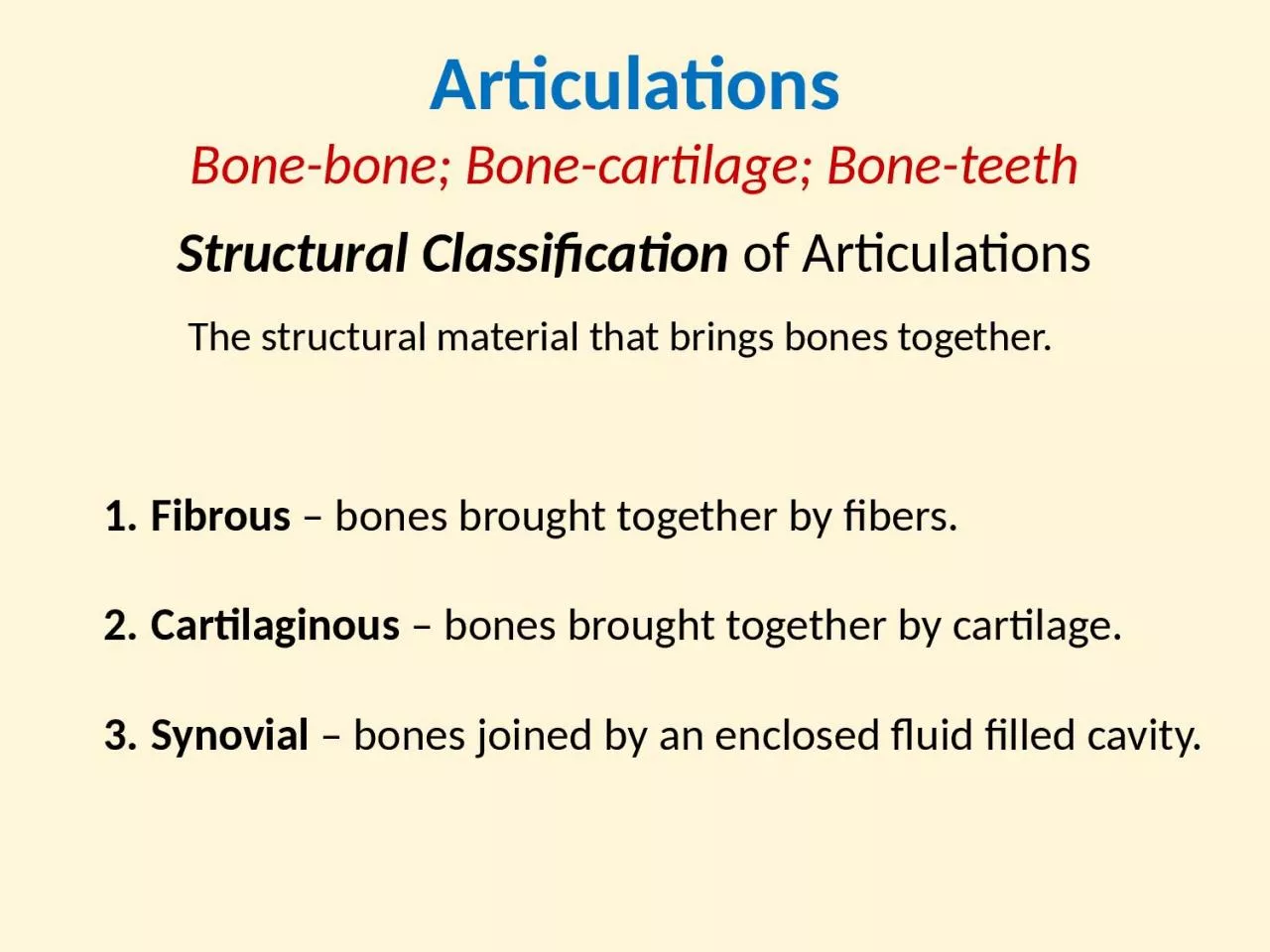
Structural Classification of Articulations Fibrous bones brought together by fibers Cartilaginous bones brought together by cartilage Synovial bones joined by an enclosed fluid filled cavity ID: 1038371

joints joint movement synovial joint joints synovial movement bone head hip ligament knee 1st bones arthritis pivot joined types

Share:

Link:

Embed:

Download Presentation from below link

Download Presentation The PPT/PDF document "Articulations Bone-bone; Bone-cartilage;..." is the property of its rightful owner. Permission is granted to download and print the materials on this web site for personal, non-commercial use only, and to display it on your personal computer provided you do not modify the materials and that you retain all copyright notices contained in the materials. By downloading content from our website, you accept the terms of this agreement.


Presentation Transcript

1. ArticulationsBone-bone; Bone-cartilage; Bone-teethStructural Classification of ArticulationsFibrous – bones brought together by fibers.Cartilaginous – bones brought together by cartilage.Synovial – bones joined by an enclosed fluid filled cavity.The structural material that brings bones together.

2. Fibrous JointsSutureGomphosisSyndesmosisSynostosis

3. Synchondrosis – joined by hyaline cartilage.1st sternocostal jointPubic symphysisintervertebral joints Epiphyseal plate Cartilaginous JointsSymphysis – joined by FibrocartilageEpiphysisDiaphysis

4. Generalized Synovial JointArticular (joint) Capsule Inner ‘cellular’ layer (synovial membrane) Outer ‘fibrous’ layer (dense irregular c.t.)Synovial Joints

5. Functional Classification of ArticulationsSynarthrosis – no movement possible at these joints.Amphiarthrosis – slight movement possible at joint.Diarthrosis – highly moveable at these joints.Degree of movement permitted at articulation:Ranges from: None, to a little bit, to highly movable.

6. Gomphoses – tooth in socketSynarthrosesSutures – immovable joints of the skull

7. Symphysis Slight movement with fibrocartilageAmphiarthrosesSyndesmosis Slightly movable fibrous joint.Pubic symphysis of pelvic girdleCartilaginous joint uniting bodies of adjacent vertebraeInterosseous membrane①②①②

8.

9. Joints characterized by their high mobility and having a joint cavity within a synovial membrane encased in the joint capsule.DiarthrosesDiarthrotic joints are also known as Synovial jointGliding (Planar)HingePivotEllipsoidal/CondyloidSaddleBall and Socket6 Types of Synovial Joints

10. Gliding JointThese joints occur as short boxy bone surfaces move past each other. See at wrist, ankles, and spine.Carpals of the hand Intercarpal jointsTarsal joints (navicular two cuneiforms) Intertarsal jointsVertebral ColumnFacets of intervertebral joints

11. Pivot Joint1) Atlantoaxial joint Formed between the atlas (C1) and the axis (C2) cervical vertebrae of neck. For the “no” gesture, Shaking head left to right.2) Proximal radioulnar jointConsists of the radial head articulating with the radial notch of the ulna. The disc-like head of radius tightly bound by the annular ligament securing it in place.Allows the pivot action of pronation/supination.

12. Hinge JointHumeroulnar joint of the elbow. Interphalangeal joints of the hand. Tibiofemoral join of the knee.

13. Condyloid/Ellipsoid JointMetacarpophalangeal joints II through V. Distal radiocarpal joint of the wrist

14. Saddle Joint1st Carpometacarpal joint – the thumb joint (trapeziometacarpal) Sternoclavicular joint of the thorax Calcaneocuboid joint of the heel

15. Ball and Socket JointHip joint of the pelvic girdle Shoulder joint of pectoral girdle

16.

17. The 6 Types of Synovial Jointsa) Ball and Socket; b) Hinge; c) Saddle; d) Pivot; e) Ellipsoidal/Condyloid; f) Gliding (Planar).a)b)c)d)e)f)

18. Sternoclavicular jointIntercarpal andIntertarsal jointsElbow jointKnee jointAnkle jointInterphalangealjointsAtlantoaxial jointProximalRadioulnar Joint (pronate/supinate)

19. RadiocarpalJointMetacarpophalangealJoints (2-5)TrapeziometacarpalJoint (1st carpometacarpal)ShoulderjointHip joint

20.

21. Movements (Actions) at Articulations

22.

23.

24.

25. Structures that Stabilize Synovial JointsLigamentsMenisciBursaeFat padsTendons

26. Diagrammatic Knee Joint

27. The Knee Joint

28. The Knee Joint

29.

30.

31. Iliofemoral ligaments (n=2)Pubofemoral ligamentIschiofemoral ligamentThe Hip JointPattern: 1st part = name of bone ligament is coming from on os coxa (3 choices). 2nd part = femoral.Plus the don’t forget the ligamentum teres connecting the fovea capitis to the acetabulum

32. Hip Joint

33. Hip Replacement ProcedureA ‘Hip Fracture’ is the breaking of which structure? ______________

34. Shoulder Joint

35. Acromioclavicular ligamentCoracoacromial ligamentCoracoclavicular ligaments(n =2)Segments of articular (joint) capsuleTendon for long head of biceps brachiiThe Ligaments of the Shoulder JointPattern: The coracoid is a big shot and always gets named 1st ; then the acromion; the poor clavicle is always last.

36.

37. Osteoarthritis: “Wear and tear” age-related arthritis, due to use of the joint.Arthritis: Inflammation of a synovial joint, that is often painful and restricts movement at that joint.Rheumatoid Arthritis: Autoimmune disease, in which the body’s defense cells attack synovial joints.Disorders of ArticulationsAnkylosis: Abnormal fusion of a joint, restricting normal movement.

38. Gout (Gouty Arthritis): Uric acid deposits in synovial joints and crystallizes there, causing pain and restricting movement.Bursitis: Inflammation of a bursa, causing pain when ever the associated ligament or tendon moves.Disorders of ArticulationsSprain: When a ligament is stretched beyond normal, to where some collagen fibers are torn.Luxation: Dislocation, when articulating surfaces are forced out of anatomical position.

39.

40. The 6 Types of Synovial Joints