/
Colorectal Cancer Screening and Postoperative Follow-Up Colorectal Cancer Screening and Postoperative Follow-Up

Colorectal Cancer Screening and Postoperative Follow-Up - PowerPoint Presentation

tatyana-admore
tatyana-admore . @tatyana-admore
Follow
387 views
Uploaded On 2018-10-31

Colorectal Cancer Screening and Postoperative Follow-Up - PPT Presentation

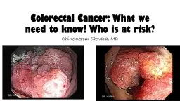
Colorectal Cancer CRC Incidence 2 nd commonest cause of cancer deaths in males 3 rd commonest cause of cancer deaths in females Lifetime risk 2015 Canada 25000 People diagnosed with CRC 45100000 ID: 705050

risk crc screening years crc risk years screening polyps cancer colonoscopy age colorectal syndrome high patients recommendations history adenomatous

Share:

Link:

Embed:

Download Presentation from below link

Download Presentation The PPT/PDF document "Colorectal Cancer Screening and Postoper..." is the property of its rightful owner. Permission is granted to download and print the materials on this web site for personal, non-commercial use only, and to display it on your personal computer provided you do not modify the materials and that you retain all copyright notices contained in the materials. By downloading content from our website, you accept the terms of this agreement.


Presentation Transcript

Slide1

Colorectal Cancer Screening and Postoperative Follow-UpSlide2

Colorectal Cancer (CRC)

Incidence

2

nd commonest cause of cancer deaths in males3rd commonest cause of cancer deaths in femalesLifetime risk2015 (Canada)25,000 People diagnosed with CRC (45/100,000)9,300 Canadians died from CRC (17/100,000)

Lifetime Risk

M

F

Developing CRC (Canada)

6%

7%

Dying from

CRC (Canada)

3.5%

3.1%Slide3

Types of Malignant Colorectal Cancer

Adenocarcinoma

Carcinoid

LymphomaSarcomaSquamous Cell CarcinomaPlasmacytoma

Leukemic Infiltrate

Melanoma – more common anus vs rectum

Metastatic CarcinomasSlide4

Risk Factors for CRC

Age

Sex (M>F)

Family HistoryAssociated IllnessesIBDPolyposis SyndromesGeography

Occupations Associated with Higher Risk

Firemen

Factor Workers (Synthetic Fibers)

Metallic Workers

Asbestos Exposure

Race

Lower risk for Hispanic and Native Indians

Diet/Social Habits

Increased risk with alcohol consumption

No link with smoking

Obesity

Diet – increased with diet high in saturated fats and proteins

Decreased risk with high fiber dietSlide5

“Recommendations on Screening for colorectal cancer in primary care.” Canadian Task Force on Preventative Health Care.

CMAJ Mar 2016, 188(5) 340-348Slide6

Screening For Colorectal Cancer

Rational – Lower deaths and incidence of CRC by

Earlier detection

Removing polyps that may transformIdentifying higher risk patients that require increased surveillanceSlide7

Symptoms ≠ ScreeningSlide8

Symptoms

Rectal Bleeding

Change

in Bowel HabitsAbdominal/Pelvic Pain

Bloating

Weight Loss

Nausea/Vomiting

Anemia/Fatigue

Fistula SymptomsSlide9

Most (“All”) CRCs arise from colon polyps

Polyp to cancer sequence

Slow transformation to invasive carcinoma

Only a limited proportion of polyps develop into CRCSlide10

Polyps – Histologic Types

Neoplastic

Tubular Adenoma (75%)

Tubulovillous Adenoma (15%)Villous Adenoma (10%)Sessile Serrated AdenomasHamartomatousJuvenille PolypsPeutz-Jeghers Syndrome

Cronkhite

-Canada Syndrome

Inflammatory Polyps

Inflammatory

Pseudopolyps

Lymphoid Polyps

HyperplasticSlide11

Neoplastic Polyps

Risk for malignant transformation

Villous>

Tubulovillous>TubularSizeIncreased risk with larger size of polypRare < 1cm Polyp MorphologySlide12

Summers, Ronald M. “Polyp Size Measurement at CT

Colonography

: What Do We Know and What Do We  Need to Know?”

Radiology 255.3 (2010): 707–720. PMC. Web. Slide13

Concerns About Screening for CRC

Detection Rates

Cost Effective

Adverse Effects of TestComplications of procedurePerception/Task to complete procedureFrequency of Screening TestMinimizing False Positive RateSlide14

Screening Tests

Guaiac Fecal Occult Blood Test (FOBT)

Fecal Immunochemical Test (FIT)

Flexible SigmoidoscopyColonoscopyRadiological StudiesCT Colonography

Ba Enema

Other

Fecal DNA Testing

KRAS mutations

Abberant

NDRG4

BMP3 Methylation

Hemoglobin ImmunoassaysSlide15

Not Approved Screening Tests

CEA

Colonoscopy ?Slide16

CRC Screening

2001 Canadian Task Force on Preventative Health Care

Annual or Biennial FOBT and Flexible Sigmoidoscopy every 5 years

(In asymptomatic individual > 50 years old and who are not at high risk)Slide17

CRC Screening

Current Guideline (Provincial Variation)

Age 50-59 (Weak Recommendations)

FIT or FOBT every 2 years ORFlexible Sigmoidoscopy every 10 yearsAge 60-74 (Strong Recommendations)FIT or FOBT every 2 years ORFlexible Sigmoidoscopy every 10 yearsSlide18

CRC Screening Cont.

Age 75+ (Weak Recommendation)

Screening generally not recommended

Decision based on reduced life expectancy in older age groupNo high powered RCTs to show benefit for elderly in lowering mortality/morbidityHigher incidence  ? screening more frequentlyPatient dependant decision*Slide19

“Recommendations on Screening for colorectal cancer in primary care.” Canadian Task Force on Preventative Health Care.

CMAJ Mar 2016, 188(5) 340-348Slide20

High Risk Patients

History of previous adenomatous polyps

Family history of CRC/(Polyps?)

Associated IllnessInflammatory Bowel DiseaseUlcerative ColitisCrohn’s ColitisPolyp SyndromesFamilial Adenomatous Polyps (FAP)Lynch SyndromeOthersAssociated Malignancies Slide21

Inflammatory Bowel Disease (IBD)

Ulcerative Colitis and Crohn’s Colitis: both associated with increased CRC risk

CRC risks - colitis proximal to splenic flexure

Risks CRC5% after 10 years20% after 20 years40% after 25 yearsDecreased risk with left sided disease onlyMinimal risk with ulcerative proctitisIncreased risk in IBD patients with:“severe” inflammationProlonged active disease

More extensive disease (proximal to splenic flexure)

Sclerosing

cholangitis as part of IBD secondary symptoms

Family history CRCSlide22

Recommendations of Screening IBD

Colonoscopy

Every 1-3 years after 8-10 years active disease (Mine every 2 years)

Left sided only - after 15 years?At start of diagnosis of sclerosing cholangitis Random biopsies 4 quadrants every 5 to 10 cmsAttention to – strictures and mass lesionsDifficulties - pseudopolypsHigh risk factors on colonoscopy for IBD

High grade dysplasia (HGD)

Multifocal low grade dysplasia (LGD)

Dysplasia associated with mass lesion (DALM)Slide23

Familial Adenomatous Polyposis (FAP)

<1% of CRC cases

Autosomal dominant syndrome

100’s-1000’s of colorectal polyps50% of patients have polyps by age 1595% by age 35Lifetime risk CRC ≈ 100%Average age of diagnosis of CRC 35-40 yearsSlide24

Familial Adenomatous Polyposis (FAP) Cont.

Screening

Genetic Testing of affected individual

If positive for geneScreening with colonoscopy start as teenagerProphylactic total proctocolectomyAnything less Screen remaining colon/ rectum q yearlyUGI Screening with < EGD Increased risk duodenal/periampullary

cancers

If negative

Higher surveillance until age 40

If normal until then assume normal risk screening protocolSlide25

Lynch Syndrome (Hereditary Nonpolyposis CRC)

Associated with 2-3% of all CRC cases

Autosomal dominant syndrome

Mutation of DNA mismatch repairingLifetime risk of CRC is approximately 70%Presents at earlier ageMore right sided CRCCharacteristic histologic changesIncreased incident synchronous/metachronousHigh risk of other cancers Ovary and uterusStomach

Pancreas

Ureter/Renal Pelvis

Small Bowel

Biliary Tract

BrainSlide26

Screening Recommendations

Colonoscopy starting at 25 (10 years before youngest affected individual)

Repeat every 1-3 years

Screen for other cancersEspecially GUStomachSlide27

Peutz-Jeghers Syndrome

Autosomal dominant syndrome

Polyps anywhere in GI tract

Predominantly small bowel Lifetime risk of CRC ≈ 10%Associated with other cancersStomachPancreasSmall BowelOvaryUterusCervixTesticlePituitary

Screening

Colonoscopy every 3 years

EGD every 2 years

Small bowel investigation every 2 years

GU?Slide28

Screening in Patients with History of Adenomatous CR Polyps

Purposes of surveillance

Detect/Remove new polyps

Detect and remove polyps missed during previous scopesHigh risk factors with adenomatous polypsAdenomas > 10mmMultiple adenomas (>3 per colonoscopy)Advanced HistologyVillous adenoma

High grade dysplasia

Proximally located polyps?

Older age

Family history of CRC in 1

st

degree relative

Large polyps

Increased risk of local recurrenceSlide29

Follow Up Recommendations in Patients With Known CR Polyps

2 or less polyps <10mm adenoma

Colonoscopy 5 years (GI)

Colonoscopy 3 years (ACS)1 Polyp > 10mm Colonoscopy every 3 years3 or more adenomatous polypsColonoscopy every 3 yearsAdvanced histologyColonoscopy every 3 years or lessSlide30

Colorectal Cancer History Follow-Up (My Guidelines)

History and exam every 3 months for 5 years

CBC, LFTs*, CEA*, every 3 months for 5 years

PSA yearly with APR patients CT scan yearly for 5 years *CT Chest, abdomen and pelvisColon cancer – colonoscopy 1 year after resection and if normal every 3 years*Rectal cancer* Flexible sigmoidoscopy 6 months afterColonoscopy at 1 yearThen repeat flex sigmoidoscopy at 18 months/colonoscopy at 2 yearsColonoscopy every 3 years after if normal

*

= only do test if prepared to deal with findings.

Ie

metastatic disease