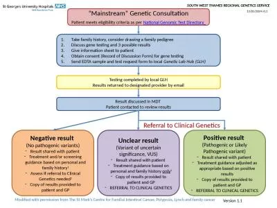
/
*Corresponding author: Vassiliou GS, Haematological Cancer Genetics, W *Corresponding author: Vassiliou GS, Haematological Cancer Genetics, W

*Corresponding author: Vassiliou GS, Haematological Cancer Genetics, W - PDF document

tatyana-admore
tatyana-admore . @tatyana-admore
Follow
380 views
Uploaded On 2015-10-06

*Corresponding author: Vassiliou GS, Haematological Cancer Genetics, W - PPT Presentation

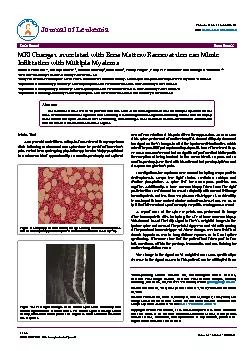
a recent exacerbation of his pain aer a therapy session An MRI scan of his spine performed at another hospital showed diusely abnormal low signal on the T1 images in all of the lumbar vertebral b ID: 151211

recent exacerbation his

Share:

Link:

Embed:

Download Presentation from below link

Download Pdf The PPT/PDF document "*Corresponding author: Vassiliou GS, Hae..." is the property of its rightful owner. Permission is granted to download and print the materials on this web site for personal, non-commercial use only, and to display it on your personal computer provided you do not modify the materials and that you retain all copyright notices contained in the materials. By downloading content from our website, you accept the terms of this agreement.


Presentation Transcript

Volume 1  Issue 4  1000125J Leuk ISSN: 2329-6917 JLU, an open access journal Case Report Paterson et al., J Leuk 2013, 1:4 Volume 1  Issue 4  1000125J Leuk ISSN: 2329-6917 JLU, an open access journal *Corresponding author: Vassiliou GS, Haematological Cancer Genetics,Wellcome Trust Sanger Institute, Wellcome Trust Genome Campus, Hinxton,Cambridge, CB10 1SA, UK, Tel: +44 1223 494908, E-mail: gsv20@sanger.ac.ukDecember 12, 2013 December Paterson SC, Grove C, Crawley C, Scott M, Wright P, et al. (2013) MRI Changes associated with Bone Marrow Reconversion can Mimic In�ltration with Multiple Myeloma. J Leuk 1: 125. doi:10.4172/2329-6917.1000125 This is an open-access article distributed under the terms of the Creative Commons Attribution License, which permits unrestricted use, distribution, and reproduction in any medium, provided the We describe a case of a �t 40-year-old who was referred for investigations to rule out Multiple Myeloma on the basis of an abnormal bone signal on MRI scanning. Haematological investigations including a bone marrow biopsy were normal and upon extended MRI re-scanning, bone changes were identi�ed as those of marrow reconversion a recent exacerbation of his pain aer a therapy session. An MRI scan of his spine, performed at another hospital, showed diusely abnormal low signal on the T1 images in all of the lumbar vertebral bodies, which raised the possibility of myelomatous deposits, hence the referral to us. He was a non-smoker and had no signicant past medical history with the exception of being involved in two car accidents, 10 years and 18 months previously. He lived with his wife and had previously taken oral diazepam 5 mg for back pain.Investigations for myeloma were normal including serum protein electrophoresis, serum free light chains, creatinine, calcium and alkaline phosphatase. A urine test for Bence-Jones proteins was negative. Additionally, a bone marrow biopsy taken from the right posterior iliac crest showed increased cellularity with normal trilineage haematopoiesis and less than 2% plasma cells (Figure 1). Incidentally he was found to have a raised alanine aminotransferase (154, NR 10-44 iu/l) that later resolved spontaneously. Hepatitis serology was normal.A repeat MRI of the spine & pelvis was performed to image other haemopoietic sites including the site of bone marrow biopsy. is showed loss of the fatty signal in the T1-weighted images in the lumbar spine and areas of the pelvis (Figure 2a and 2b) with sparing of the proximal femora (Figure 2c). ese changes are characteristic of chronic hypoxia as seen in long-distance runners. In fact, on further questioning, it became clear that the patient had taken part in two half-marathons within the previous few months and was training for another long-distance race. e change in the signal on T1 weighted MRI scan, specically a decrease in the signal as seen in this patient, can be attributed to an Main TextA 40 year old male tness enthusiast was referred to our myeloma clinic following an abnormal MRI spine done for persistent lower back pain. He had been undergoing physiotherapy for a back injury sustained in a minor accident approximately 18 months previously and suered , Penny Wright, Philip WP Bearcroft and George S VassiliouWellcome Trust Sanger Institute, Cambridge, CB10 1SA, UKCollege of Medical, Veterinary and Life Sciences, Wolfson Medical School Building, University of Glasgow, University Avenue Glasgow, G12 8QQ, UKDepartment of Haematology, Cambridge University Hospitals NHS Foundation Trust, Hills Road, Cambridge, CB2 0QQ, UKDepartment of Histopathology, Cambridge University Hospitals NHS Foundation Trust, Hills Road, Cambridge, CB2 0QQ, UKDepartment of Radiology, Cambridge University Hospitals NHS Foundation Trust, Hills Road, Cambridge, CB2 0QQ, UK Figure 1: Mildly hypercellular marrow for age showing normal haematopoiesis T1 weighted images of the lumbar spine show abnormally dark marrow signal from the vertebral bodies. The marrow signal at this age should be fatty, and should almost parallel the brightness of the subcutaneous fat on Journal of LeukemiaISSN: 2329-6917 Paterson SC, Grove C, Crawley C, Scott M, Wright P, et al. (2013) MRI Changes associated with Bone Marrow Reconversion can Mimic 10.4172/2329-6917.1000125 Page 2 of2 increase in the presence of red (haematopoietic) marrow compared to yellow (fatty) marrow [1]. Normally, red converts to yellow bone marrow during childhood as part of the physiological aging process. It follows a set pattern with conversion occurring in the peripheral bones before the axial skeleton. is process is normally complete by approximately 20-30 years of age [2]. Even within long bones there is a specic pattern of marrow conversion. It occurs rst in the epiphyses and then in the diaphyses before continuing in the distal metaphyses and then the proximal metaphyses [3]. In this case, frequent intermittent hypoxia associated with regular long-distance running was thought to have led to reconversion of the normal yellow marrow back to red marrow leading to the appearances seen on MRI. Such reconversion follows the reverse pattern to that seen with marrow conversion in childhood and occurs in the axial skeleton before the long bones, which explains why the proximal femora were spared. e appearance of these lesions can sometimes be misinterpreted as inltrative disorders aecting the spine such as multiple myeloma as was suspected in this case. As well as frequent exercise-induced hypoxia, these changes can sometimes be seen in association with very heavy smoking, severe obesity and administration of haematopoietic growth factors [1,4,5]. Although uncommon, the possibility of marrow reconversion should be considered in individuals with diuse bony changes on MRI, particularly given the characteristic appearances of this phenomenon.We thank the Haematooncology Diagnostic Service (HODS) at Cambridge University Hospitals NHS Trust for help with processing and analysis of diagnostic GV is funded by a Wellcome Trust Senior Fellowship in Clinical Science. MaA‚kiewicz A, Dziedzic M (2012) Bone marrow reconversion - imaging ofphysiological changes in bone marrow. Pol J Radiol 77: 45-50. Taccone A, Oddone M, Dell’Acqua A, Occhi M, Ciccone MA (1995) MRI “roadmap” of normal age-related bone marrow. Pediatr Radiol 25: 596-606. Seigel, Marilyn J. MRI of Bone Marrow. Saadate-Arab M, Trou�éau P, Stines J, Verhaeghe JL, Rios M, et al. (2002) [MR imaging �ndings of bone marrow reconversion induced by growth factors in 3 Ollivier L, Gerber S, Vanel D, Brisse H, Leclère J (2006) Improving theinterpretation of bone marrow imaging in cancer patients. Cancer Imaging 6: Figure 2b: T1 weighted sequence through the pelvis shows similarly abnormal T1 weighted images through the hips show almost normal fatty marrow within the femoral head, a region characteristically late to undergo