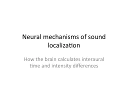
How the brain calculates interaural time and intensity differences Bottom line Calculation of interaural differences in the brain depends on wiring and a balance between neural excitation and inhibition ID: 180143
Download Presentation The PPT/PDF document "Neural mechanisms of sound localization" is the property of its rightful owner. Permission is granted to download and print the materials on this web site for personal, non-commercial use only, and to display it on your personal computer provided you do not modify the materials and that you retain all copyright notices contained in the materials. By downloading content from our website, you accept the terms of this agreement.
Slide1
Neural mechanisms of sound localization
How the brain calculates interaural time and intensity differencesSlide2
Bottom line
Calculation of interaural differences in the brain depends on “wiring” and a balance between neural excitation and inhibition.Slide3
An overview of the auditory pathwaySlide4
The circuit for sound localization starts in the cochlear nucleus
From Pickles (1988)Slide5
Principal cells of the AVCN are spherical or bushy cells
From Pickles (1988)Slide6
Bushy cell and auditory nerve connection
From Ryugo & Fekete (1982)Slide7
Nuclei involved in interaural intensity comparisons
AVCN =
anteroventral
cochlear nucleus
LL = lateral
lemniscus
LSO = lateral superior olive
MNTB = medial nucleus of the trapezoid body
MSO = medial superior olive
TB = trapezoid body
From Webster (1992)Slide8
Lateral superior olive (LSO)
EI
(Excitatory- Inhibitory)
Response
From Pickles (1988)Slide9
Response properties of LSO neurons
Modified from Pickles (1988)Slide10
Layout of LSO (rolled out)
Frequency
IIDSlide11
One frequency row in LSO
1 2 3 4
5 6
7 8 9 10
IID threshold
IID must be around hereSlide12
Pattern of activity gives IID across the spectrum
Frequency
IIDSlide13
If the LSO were a graph, and the x-axis is frequency, then the y-axis is
Intensity
Spectral shape
Interaural intensity difference
Interaural time differenceSlide14
How does response in LSO become specific for IID?Slide15
LSO wiring diagramSlide16
The balance between excitation and inhibition determines response
Response = excitation - inhibition
Ipsilateral
input from AVCN
Contralateral input from MNTB
LSO neuron
If ipsilateral AVCN is responding more than contralateral AVCN (adjusted by MNTB), respond.Slide17
The LSO calculates IID by subtracting the response of the contralateral ear from the response of the ipsilateral ear using inhibition.
By adjusting the amount of inhibition delivered by MNTB, can make different LSO neurons respond over different ranges of
IIDs
.Slide18
If the sound source is close to the right ear, then the LSO neurons on the left side of the brain
respond a lot
respond a little
don’t respond at allSlide19
How about MSO?
From Webster (1992)Slide20
Like LSO neurons, MSO neurons look like they make comparisons
EE
(Excitatory-Excitatory)
Response
From Pickles (1988)Slide21
MSO neurons receive inputs from both AVCNs.Slide22
Branching pattern of AVCN axons is different on ipsilateral and contralateral sides
From Sullivan & Konishi (1986)Slide23
MSO neurons receive a different sort of projection from the 2 AVCNsSlide24
MSO receives the output of a neural delay line
0 .1 .2 .3 .4 ms
Left ear response
delayed by 0.1 ms
Right ear response
Coincidence detectorsSlide25
MSO calculates ITDs by detecting coincident inputs from a delay line constructed from the axons of AVCN neurons.Slide26
IIDs are useful for localizing ____-frequency sounds;
ITDs
are useful for localizing ____-frequency sounds.
high, high
high, low
low, high
low, lowSlide27
The tonotopic organization of the parts of the SOC matches the interaural calculations performed
LSO
MSO
MNTB
From Pickles (1988)Slide28
Conclusions
The neurons of the superior olive calculate interaural differences in intensity and time.
The LSO uses a balance of inhibition and excitation to calculate IIDs.
The MSO uses a circuit established by the axons of AVCN neurons to calculate ITDs.Slide29
Text sources
Pickles, J.O. (1988) An introduction to the physiology of hearing. Berkeley: Academic Press.
Ryugo, D. & Fekete, D. (1982) Morphology of primary axosomatic endings in the anteroventral cochlear nucleus of the the cat: A study of the endbulbs of Held.
J. Comp. Neurol. 210,
239-257.
Sullivan, W. & Konishi, M. (1986) Neural map of interaural phase difference in the owl’s brainstem.
Proc. Natl. Acad. Sci.
83, 8400-8404.
Webster, D.B. (1992). An overview of mammalian auditory pathways with an emphasis on humans. In D.B. Webster, A.N. Popper & R.R. Fay (Eds.) The mammalian auditory pathway: Neuroanatomy. New York: Springer-Verlag.