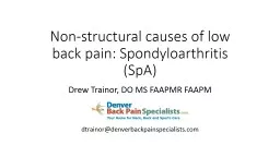
/
Non-structural causes of low back pain: Spondyloarthritis (SpA) Non-structural causes of low back pain: Spondyloarthritis (SpA)

Non-structural causes of low back pain: Spondyloarthritis (SpA) - PowerPoint Presentation

tatyana-admore
tatyana-admore . @tatyana-admore
Follow
386 views
Uploaded On 2018-12-18

Non-structural causes of low back pain: Spondyloarthritis (SpA) - PPT Presentation

Drew Trainor DO MS FAAPMR FAAPM dtrainordenverbackpainspecialistscom We find what we look for and we look for what we know Lecture Outline Case Epidemiology Pathophysiology Clinical manifestations ID: 743553

spondylitis treatment patients ankylosing treatment spondylitis ankylosing patients spa disease spondyloarthritis pain grade axial case arthritis activity therapy 2014

Share:

Link:

Embed:

Download Presentation from below link

Download Presentation The PPT/PDF document "Non-structural causes of low back pain: ..." is the property of its rightful owner. Permission is granted to download and print the materials on this web site for personal, non-commercial use only, and to display it on your personal computer provided you do not modify the materials and that you retain all copyright notices contained in the materials. By downloading content from our website, you accept the terms of this agreement.


Presentation Transcript

Slide1

Non-structural causes of low back pain: Spondyloarthritis (SpA)

Drew Trainor, DO MS FAAPMR FAAPMdtrainor@denverbackpainspecialists.comSlide2
Slide3

We find what we look for and we look for what we knowSlide4

Lecture Outline

CaseEpidemiologyPathophysiologyClinical manifestationsLaboratory findingsImage characteristicsDiagnostic Criteria

Treatment

Questions/DiscussionSlide5

Case

A 42-year-old female presents with a 4-month history of left buttock painDeep and aching with radiation into the posterior thigh and buttockDenies numbness, tingling, weaknessAssociated with >1 hour morning stiffness

Pain improves with physical activity

Pain was significantly reduced with the use ibuprofen (can no longer take NSAIDs)

8-9/10 on NPRSSlide6

Case, continued

Allergies: NKDAMedications: Ambien 5 mg PO QHS PRNPast Medical History: Ulcerative colitis diagnosed 6-months ago after rupture colonPast Surgical History: Partial colectomy on setting of aboveFamily History: Non-contributorySlide7

Case, continued

Other relevant dataMRI L-spineSevere left L4-5 facet degeneration with noted synovial cystPrior proceduresLeft L4-5 facet cyst aspiration and injectionNo benefit in anesthetic or corticosteroid phase

Epidural Steroid Injection (type a location unknown by patient)

Non-diagnosticSlide8

Case, continued

NOTE:

<5 cm excursion indicates lumbar segmental restrictionSlide9

Case, continued

PlanTherapy: patient working 100 + hours/week not logistically possibleMedications: Tylenol 1000 mg PO TID, Tramadol 50 mg PO Q6H PRN, in future consider Celebrex after clearance with GIInjections: left SIJ injection under fluoroscopySurgical considerations: None.

Diagnostics: HLA-B27, ESR, CRPSlide10

Case, continued

75% IMPROVEMENT IN ANESTHETIC PHASESlide11

Case, continued

HLA-B27 – PositiveCRP – ElevatedESR – ElevatedSlide12

Case, continued

PlanRheumatology referral for consideration of TNF inhibitor therapySlide13

Spondyloarthritis (SpA)

Ankylosing Spondylitis (AS)Non-radiographic axial spondyloarthritis (nr-SpA)Psoriatic arthritisArthritis associated with inflammatory bowel disease (IBD-SpA)Ulcerative colitis and Crohn’s diseaseReactive arthritis (ReA)

chlamydia, campylobacter, salmonella, shigella

Undifferentiated Spondyloarthropathy (uSpA)Slide14

Spondyloarthritis (SpA)

More recent classificationAxial PeripheralShare many phenotypical characteristics (discussed later)Slide15

Why do we care?

Higher incidence and prevalence than we realizeEarly identification has consequencesEarly treatment makes a differenceAs surgeons, pain physicians, and physiatrists will likely be the first ones to encounter these patientsBack pain is most common presenting complaintsSlide16

Epidemiology of SpA

Incidence0.48 to 63/100,000Prevalence0.1% to 2.5% in general population4 million people in the U.S. living with SpAEstimated to be 5% in the low back pain population

Should see more than one patient per week

Higher than rheumatoid arthritis (RA)

Int J Environ Health Res. 2015;25(3):322-9. Curr Rheumatol Rep 2013; 15:351. J Rheumatol 1994; 21:2281-2285Slide17

Epidemiology of SpA

Highly HeritableRelative Risk1st degree relative: 75.52nd

degree relative: 20.2

3

rd

degree relative: 3.5Slide18

Pathophysiology of SpA

Not well understoodLikely polygenicSlide19

Pathophysiology of

SpAHLA-B27 by far most commonPresent in 70-90% of patients with ASPresent in 6% of general populationNOT required for diagnosis

3 main hypothesis

Arthritogenic peptide hypothesis

Heavy chain homodimer hypothesis

HLAB27 misfolding hypothesis

Koning, Anoek De, et al. “Pathophysiology of Axial Spondyloarthritis: Consensus and Controversies.”

European Journal of Clinical Investigation

, 2018, doi:10.1111/eci.12913. Slide20

Clinical Manifestations of SpA

Non-musculoskeletalUveitisPsoriasisFeatures of IBDGenital LesionsIn Reactive ArthritisSlide21

UveitisSlide22

PsoriasisSlide23

IBD

Abdominal painVomitingDiarrheaRectal BleedingSevere internal cramps/muscle spasms in the region of the pelvis Weight LossSlide24

Reactive Arthritis

“Can’t see, pee, or climb a tree”ConjunctivitisUrethritisArthritisSlide25

Clinical Manifestations of SpA

Musculoskeletal featuresInflammatory back painPeripheral arthritisEnthesitis (enthesopathy)DactylitisSlide26

Inflammatory Back Pain

Young age at onset, typically seen in those under 40 years oldGradual onset of painSymptoms of back pain improve with exerciseResponse to NSAIDs

Pain does not improve with rest

Pain at night, often waking a person in the second half of the night

Morning stiffness that lasts for more than 30 minutes

Pain lasting for

more than 3 months

Alternating buttock painSlide27

Peripheral Arthritis

Acute OnsetPredominately effects the lower extremitiesAssociated with swellingUsually asymmetricEffects 1 to 3 jointsOligoarthritisSlide28

Enthesitis (enthesopathy)

InflammationLigament to boneTendon to boneJoint capsuleFascia to boneMost commonly manifested as:

Achilles tendinopathy

Plantar fasciitisSlide29

DactylitisSlide30
Slide31
Slide32
Slide33

Laboratory Findings

HLA-B27Positive in 70-90% of patients with ASPositive in 50% to 70% of patients with other forms of SpANote: HLA-B27 positivity is NOT diagnostic in itselfNote even required for the diagnosisSeen in 6% of the general population

Acute Phase Reactants (ESR and CRP)

Elevated in 35% to 50% of patients

Elevated CRP is an indication of:

Radiographic disease progression

Positive response to TNF alpha inhibitorsSlide34

Imaging Findings

Plain RadiographsAxialOnly specific finding is evidence of sacroiliitis (images to follow)May take years to developBridging axial

syndesmophytes

seen on <5% of patients in the absence of sacroiliitis

50% of patients develop syndesmophytes during course of disease

Peripheral

Erosive joint changes

Evidence of EnthesitisSlide35

Sacroiliitis on Plain Radiographs

Graded 0 to 4Grade 0: Normal Grade 1: Suspicious changesGrade 2: Minimal abnormality – Small localized areas with erosions or sclerosis, without alteration in the joint width Grade 3: Unequivocal abnormality – Moderate or advanced sacroiliitis with erosions, evidence of sclerosis, widening, narrowing, or partial ankylosis

Grade 4: Severe abnormality – Total ankylosis

Considered “Positive for Suggestion of SpA” if:

Grade 2 or higher bilateral

Grade 3 or higher unilateralSlide36

Sacroiliitis on Plain Radiographs (Grade 2)

Both sacroiliac (SI) joints show ill-defined margins, sclerosis, and especially at the left SI joint an irregular joint space (grade 2 bilaterally). Slide37

Sacroiliitis on Plain Radiographs (Grade 3)

Sclerosis at the iliac side, widespread erosions, pseudo widening of the joint space, blurring of the joint margins in both sacroiliac (SI) joints (bilateral grade 3). Slide38

Sacroiliitis on Plain Radiographs (Grade 4)

Both sacroiliac (SI) joints show complete ankylosis (grade 4). Slide39

Syndesmophytes on Plain RadiographsSlide40

Differentiation from DISHSlide41

Imaging Findings of EnthesitisSlide42

Evolution in the concepts of SpA

Problems with Radiographic Evidence and AS Decision on radiographs challenging Late Dx Miss opportunity for early treatment TNF-alpha antagonists effective Particularly if start in early stage

Solution: New concepts

Non-radiographic stage of axial SpA identified with MRI

Inflammatory Back Pain Slide43

MRI and SpA

Definition of Sacroiliitis according to the Assessment of SpondyloArthritis International Society (ASAS)Bone marrow edema (BME)short tau inversion recovery (STIR)T2-weighted images with fat suppressionBright

Dark on T1Slide44

MRI and SpASlide45

MRI and SpA

Marrow Edema on T2 weighted imagesSlide46

Diagnostic Criteria- Axial SpASlide47

Diagnostic Criteria- Axial SpASlide48

Diagnostic Criteria- Peripheral SpASlide49

TreatmentSlide50

TreatmentSlide51

Treatment- Medication Management

Non-steroidal Anti-inflammatories (NSAIDs)Strongly Recommend Treatment with NSAIDsConditionally RecommendContinuous treatment with NSAIDs vs. on-demand NSAIDSNo recommendations on any particular NSAID over anotherSlide52

Treatment- Medication Management

NSAIDSMaximum dose is usually required70% to 80% of patients with AS report significant improvementTrials should last at least 4-weeks

Consider more COX-2 selective in addition to GI

PPx

If cardiac risk factors consider naproxenSlide53

Treatment- Medication Management

Tumor Necrosis Factor Inhibitors (TNFi) (adalimumab, etanercept, and infliximab)Strongly recommendTNFi in patients with active AS despite treatment with NSAIDs

Do not recommend one

TNFi

over another, except:

Monoclonal Ab over etanercept in patients with IBD or iritis/uveitisSlide54

Treatment- Medication Management

TNFiCallhof, et al. 2014Systematic Review2400 patients

Statistically significant improvement in

Disease activity

Function

Does timing of treatment matter?

Studies show that patients are more likely to experience remission, if:

Shorter disease duration, which is the best predictor

Elevated CRP

Young age

Callhoff, Johanna, et al. “Efficacy of TNFα Blockers in Patients with Ankylosing Spondylitis and Non-Radiographic Axial Spondyloarthritis: a Meta-Analysis.”

Annals of the Rheumatic Diseases

, vol. 74, no. 6, 2014, pp. 1241–1248., doi:10.1136/annrheumdis-2014-205322. Slide55

Treatment- Medication Management

GlucocorticoidsStrongly recommendAGAINST the use of systemic steroidsNote: did not address procedural interventions on guidelinesLiterature Review on Sacroiliac Joint Injections in AS

Limited to case reportsSlide56

Treatment- DMARDs

DMARDsLittle evidence to support their use in ASSome evidence that Sulfasalazine may be beneficialMore effective in peripheral disease than axial diseaseSlide57

Treatment- Physical Therapy

Strong recommendPhysical Therapy vs. no Physical TherapyConditionally recommendActive physical therapy (supervised exercise) vs. passive therapy interventions (massage, ultrasound, and other thermal modalities)Land based vs. aquatic based exercisesSlide58

Treatment- Physical Therapy

Goalsalleviate painincrease spinal mobilityImprove functional capacityReduce morning stiffnessCorrect postural deformities

Increase mobility

Improve the psychosocial status of the patients

https://www.physio-pedia.com/Ankylosing_SpondylitisSlide59

Treatment- Physical Therapy

Common deformitiesExcessive Thoracic KyphosisCompensatory Cervical LordosisResulting hip flexion contractureDeformity prevention

Proper sleeping posture on a solid, flat bed without pillow. Frequent sleeping or lying in prone position.

Posture exercises with upper back hyperextension (performed with avoidance of lumbar hyperextension).

Breathing exercises to increase or maintain rib cage excursion

, as well as instruction in abdominothoracic breathing.

Range of motion exercises for hips and knees to prevent flexion limitation and contractures.

Periodic rest periods with avoidance of fatigue.

Bracing or corseting (combined with exercises).

https://www.physio-pedia.com/Ankylosing_SpondylitisSlide60

Treatment- Prevention

Conditionally recommendScreening for osteoporosis/osteopenia with DXA scanStrongly recommend againstScreening for cardiac conduction defects with ECGScreening for valvular heart disease with EchoSlide61

Common Laboratory Tests for other Rheumatologic Diseases

Rheumatoid Factor (RF)- only positive in 60% of patients at time of diagnosisAnti-cyclic citrullinated peptide (CCP) antibodies- highly sensitive (76%) and specific (96%) for RAAntinuclear antibody (ANA)- sensitive but not necessarily specific

98% of patients with SLE

40%-to 70% of those with other connective tissue

20% with autoimmune thyroid and liver disease

5% of general population

Anti double-stranded DNA (anti dsDNA)-

specific (95%) but not sensitive (60%) for SLE

Schur, Peter H. “Laboratory Tests in Rheumatic Disorders.”

Oxford Medicine Online

, 2014, doi:10.1093/med/9780199358274.003.0028Slide62

Conclusions

Spondyloarthritis is more common than we realizeIf we don’t look for it we won’t find itEarly diagnosis is important and associated with higher rate of response to treatmentHLA-B27 is common (70-90%) but is NOT required for the diagnosisSpondyloarthritis

is associated with non-musculoskeletal conditions

ASK as part of HPI in patients with no obvious structural cause of back pain (especially if <45 years)

NSAIDs,

TNFi

, and PT are mainstays of treatmentSlide63

Questions?Slide64

References

Braun J, van den Berg R, Baraliakos X, et al. 2010 update of the ASAS/EULAR recommendations for the management of ankylosing spondylitis. Ann Rheum Dis 2011; 70:896.Zochling J, van der Heijde D, Burgos-Vargas R, et al. ASAS/EULAR recommendations for the management of ankylosing spondylitis. Ann Rheum Dis 2006; 65:442.Smolen JS, Braun J, Dougados M, et al. Treating spondyloarthritis, including ankylosing spondylitis and psoriatic arthritis, to target: recommendations of an international task force. Ann Rheum Dis 2014; 73:6.

Ward MM, Deodhar A, Akl EA, et al. American College of Rheumatology/Spondylitis Association of America/Spondyloarthritis Research and Treatment Network 2015 Recommendations for the Treatment of Ankylosing Spondylitis and Nonradiographic Axial Spondyloarthritis. Arthritis Rheumatol 2016; 68:282.

Jang JH, Green CE, Assassi S, et al. The contribution of disease activity on functional limitations over time through psychological mediators: a 12-month longitudinal study in patients with ankylosing spondylitis. Rheumatology (Oxford) 2011; 50:2087.

Calin A, Garrett S, Whitelock H, et al. A new approach to defining functional ability in ankylosing spondylitis: the development of the Bath Ankylosing Spondylitis Functional Index. J Rheumatol 1994; 21:2281.

Bath Ankylosing Spondylitis Functional Index (BASFI) http://basdai.com/BASFI.php (Accessed on February 10, 2014).

Doward LC, Spoorenberg A, Cook SA, et al. Development of the ASQoL: a quality of life instrument specific to ankylosing spondylitis. Ann Rheum Dis 2003; 62:20.

Ankylosing spondylitis quality of life instrument (ASQol) http://ard.bmj.com.proxy.hsl.ucdenver.edu/content/suppl/2002/12/20/62.1.20.DC1/62120Appendices.pdf (Accessed on February 10, 2014).

Garrett S, Jenkinson T, Kennedy LG, et al. A new approach to defining disease status in ankylosing spondylitis: the Bath Ankylosing Spondylitis Disease Activity Index. J Rheumatol 1994; 21:2286.

van der Heijde D, Lie E, Kvien TK, et al. ASDAS, a highly discriminatory ASAS-endorsed disease activity score in patients with ankylosing spondylitis. Ann Rheum Dis 2009; 68:1811.

Vastesaeger N, Cruyssen BV, Mulero J, et al. ASDAS high disease activity versus BASDAI elevation in patients with ankylosing spondylitis as selection criterion for anti-TNF therapy. Reumatol Clin 2014; 10:204.

Assessment of SpondyloArthritis International Society. Ankylosing Spondylitis Disease Activity Score. http://www.asas-group.org/research.php?id=01#null (Accessed on October 15, 2013).

Machado P, van der Heijde D. How to measure disease activity in axial spondyloarthritis? Curr Opin Rheumatol 2011; 23:339.

Assassi S, Weisman MH, Lee M, et al. New population-based reference values for spinal mobility measures based on the 2009-2010 National Health and Nutrition Examination Survey. Arthritis Rheumatol 2014; 66:2628.

Chilton-Mitchell L, Martindale J, Hart A, Goodacre L. Normative values for the Bath Ankylosing Spondylitis Metrology Index in a UK population. Rheumatology (Oxford) 2013; 52:2086.

Sieper J, Rudwaleit M, Baraliakos X, et al. The Assessment of SpondyloArthritis international Society (ASAS) handbook: a guide to assess spondyloarthritis. Ann Rheum Dis 2009; 68 Suppl 2:ii1.