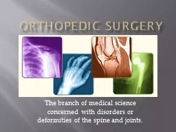
/
 Orthopedic Surgery The branch of medical science concerned with disorders or deformities  Orthopedic Surgery The branch of medical science concerned with disorders or deformities

Orthopedic Surgery The branch of medical science concerned with disorders or deformities - PowerPoint Presentation

tatyana-admore
tatyana-admore . @tatyana-admore
Follow
343 views
Uploaded On 2020-04-03

Orthopedic Surgery The branch of medical science concerned with disorders or deformities - PPT Presentation

Orthopedic Terminology Position and Movement Abduction move a part away from body Adduction move a part toward the body Dorsiflexion bend or flex foot toward leg Plantar flexion ID: 775021

bone joint bones continued bone joint bones continued tissue fracture procedures extremity prep skin movement leg joints shoulder hip

Share:

Link:

Embed:

Download Presentation from below link

Download Presentation The PPT/PDF document " Orthopedic Surgery The branch of medica..." is the property of its rightful owner. Permission is granted to download and print the materials on this web site for personal, non-commercial use only, and to display it on your personal computer provided you do not modify the materials and that you retain all copyright notices contained in the materials. By downloading content from our website, you accept the terms of this agreement.


Presentation Transcript

Slide1

Orthopedic Surgery

The branch of medical science concerned with disorders or deformities of the spine and joints.

Slide2

Orthopedic Terminology“Position and Movement”

Abduction

move a part away from body

Adduction

move a part toward the body

Dorsiflexion

bend or flex foot toward leg

Plantar flexion

extend foot with toes pointed down (as when depressing the gas pedal)

Flexion

to bend a part

Extension

make a limb straight

Eversion

turn outward

Inversion

turn inward

Distal

farthest away from point of origin

Proximal

closest to point of origin

Medial

nearest mid-line

Lateral

away from the midline

Slide3

Orthopedic Terminology“Position and Movement”

Valgus -abnormal displacement of part of a limb away from the midline of the body – distal from the affected joint – knees togetherVarus - a deformity in which part of a limb is turned inward to an abnormal degree – distal from the affected joint – knees apart

Slide4

Orthopedic Terminology

Acetabulum

hollowed area of pelvis that receives head of femur

Acromioclavicular (AC) joint

where clavicle joins acromion process of scapula

Arthritis

inflammation of a joint

Arthrodesis

surgical fusing or fixation of a joint

Arthroplasty

surgical reconstruction of a joint

Arthroscopy

visualization of a joint through an endoscope (diagnostic or operative)

Arthrotomy

surgical incision into a joint

Articulation

joint movement

Atrophy

muscle wasting from lack of use

Bone marrow

found in medullary canal of long bones and

porosites

of cancellous bone

Cartilage

elastic, strong, dense connective tissue

Compact bone

hard outer covering of bone

Cortical bone

hard bone that forms shell of bones/acts as supporting structure

Condyle

rounded part of a bone where ligaments articulate with adjacent bones

Curvature

normal or

abnormal bending

Slide5

Orthopedic Terminology

Diaphysis

shaft of a long bone

Dislocation

displacement of a joint

Dysplasia

abnormal tissue growth

Endosteum

inside lining of bones where new bone forms (marrow)

Epiphysis

two ends of a long bone

Exostosis

bony growth arising from a bone’s surface

Fibroma

tumor composed of fibrous or connective tissue

Foramen

normal bone opening through which nerves, vessels, etc. pass

Foramen magnum

occipital bone opening where spinal cord passes to vertebral column

Fossa

shallow depression of a bone

Fracture

break or crack of a bone

Hallux

big toe

Implant

implantation of graft (synthetic or tissue)

Slide6

Orthopedic Terminology

Lamina

flat layer or plate of a bone

Ligament

connective tissue that joins bone surfaces

Malleolus

rounded bone process (ankle)

Malunion

faulty union of a fractured bone

Nonunion

failure of fractured bone to unite

Osteogenesis

origination/development of bone (ossification)

Osteomyelitis

inflammation of bone tissue

Osteoporosis

diminished calcium in a bone

Osteotomy

surgical cutting of bone

Slide7

Orthopedic Terminology

Pelvic Girdle

bony structure that supports the trunk and provides attachment for the legs

Periosteum

membrane surrounding bone (contains blood vessels)

Polydactylism

more than normal number of digits

Scoliosis

abnormal curvature of spine

Syndactylism

webbing between digits

Synovial membrane

lining of a joint capsule

Tendon

fibrous tissue that connects muscle to bone

Traction

force placed on bones or muscles to align or immobilize parts

Slide8

Primary purposes of orthopedic surgery

Repair, revision, reconstruction, reattachment or removal any of the 206 bones of the skeletal structure and surrounding tissue

Bones, joints and affected muscle tissue

Tendons, ligaments or cartilage

Accurately classify treatment

Axial skeletal procedures

Upper extremity procedures

Lower extremity procedures

Limb reattachment procedures

Amputation procedures

Slide9

Purposes (continued)

Investigate, preserve and restore form and function to the musculoskeletal structures and associated tissues of the extremities and also the spine. Treatment depends on the type of injury and the duration of necessary immobility.

Stages of treatment

Investigation – diagnosis of structural issues

External

Internal

Preservation

Restoration

Types of treatment

Fracture management

Reduction

Immobilization

Rehabilitation

Corrective surgeries

Bone grafts

Implants

Internal and external fixation

Slide10

The Skeleton

Slide11

Function of Skeletal System

SupportProtectionMovementStorageHematopoiesis - the formation of blood cells in the living body (especially in the bone marrow)

Slide12

Bone Histology

Bone is a type of connective tissue2 Types of Bone:Dense/Compact Bone/ Corticalhard on outside/canal on insidecomposed of Haversian Units or OsteonSpongy/Cancellous Bone

Slide13

Bone Formation

Osteogenesis

is bone formation

Two Types:

Intramembranous

Endochondral

More Terms

:

Osteoblast

:

a cell from which bone develops

Osteocytes

: a star-shaped cell, is the most abundant cell found in bone. They are

osteoblasts

that have completed their bone-forming function and have become trapped in new bone tissue, evolving into structural bone cells and is involved in the maintenance of that bone. A mature bone cell.

Osteoclasts

: cells break down and assimilate bone. They are located in minute, bony chambers called lacuna.

Slide14

Intramembranous Ossification

Sheets of primitive connective tissue form at site of future bone

Primitive connective cells collect around blood vessels in these layers

Connective tissue cells differentiate into osteoblasts, which deposit spongy bone

Osteoblasts become osteocytes when bony matrix surrounds them (lacunae)

Connective tissue on surface of each developing structure forms a periosteum

Osteoblasts on the inside of periosteum deposit compact bone

Slide15

Endochondral Ossification

Masses of hyalin cartilage form models of future bones

Cartilage tissue breaks down/Periosteum develops

Blood vessels and differentiating osteoblasts from the periosteum invade the disintegrating tissue

Osteoblasts form spongy bone in space occupied by cartilage

Osteoblasts become osteocytes when bone matrix completely surrounds them

Osteoblasts beneath periosteum deposit compact bone around spongy bone

Slide16

Relevant anatomy

Skeletal system – articulated skeleton comprised of 206 bones

Axial skeleton – skull, spine and ribs

Skull – includes cranial and maxillofacial bones

Cranial bones – 8 cranial bones

Facial bones – 13 facial bones

Middle ear bones – 6 middle ear bones

Mandible – one jaw bone

Hyoid bone

Vertebral column – 26 backbones

Verebral – 24 backbones: 7 cervical, 12 thoracic, and 5 lumbar

Sacrum – one sacrum bone

Coccyx – one tailbone

Thoracic cage – 25 thoracic bones

Sternum one cartilaginous bone that supports most ribs

Rib cage – 24 rib bones; 12 pair posteriorly attached to the spine

Slide17

Relevant anatomy - Cervical

C-1 : a.k.a. Atlas: like Atlas man (Greek mythology), the bone supporting the skull. C-2 Axis: bone that allows the head to pivot.

Slide18

Axial Skeleton

Slide19

Appendicular Skeleton

The appendicular skeleton consists of 126 bones in the human body which make motion possible and protects the organs of digestion, excretion, and reproduction. The word appendicular refers to an appendage or anything attached to a major part of the body, such as the upper and lower extremities.The appendicular skeleton has four major regions:Pectoral Girdles(4 bones) Upper Limbs (60 bones) Pelvic Girdle(2 bones) Lower Limbs(60 bones)

Slide20

Anatomy (continued)

Appendicular SkeletonUpper extremities –shoulder, arm and hand bones of the appendicular skeletal systemPectoral girdle – four pectoral or collar bonesScapula – two posterior collar bonesGlenoid fossaCoracoid processAcromion processClavicle – two anterior collar bonesUpper limbs –arm, wrist and hand bonesHumerusRadiusulna

Slide21

Anatomy (continued)

Lower extremities –hip and leg bones of the appendicular skeletal systemPelvic girdleIliumPubisIschiumLower limbs – leg and foot bonesFemurPatellaTibiaFibulaTarsalsMetatarsalsphalanges

Slide22

Anatomy of a Bone

Slide23

Bone Marrow

Within the long bones are two types of bone marrow: red marrow and yellow marrow. The yellow marrow is fatty tissue. During starvation, the body uses the fat in yellow marrow for energy.

Slide24

Bone Marrow

The red marrow of some bones is an important site for blood cell production. Here all red blood cells, platelets, and white blood cells form in adults. Red blood cells carry oxygen and nutrients to the body tissues. Platelets help in blood clotting.White blood cells help fight disease and infection.

Slide25

Muscles

Slide26

Slide27

Anatomy (continued)

Muscular anatomy

Neck muscles – sternocleidomastoid, platysma and trapezius

Torso muscles – deltoid, pectoralis, serratus anterior, latissimus dorsi, transverse abdominus, rectus abdominus and levator ani

Arm muscles – biceps brachii, triceps brachii, brachialis, brachioradialis, carpi, digitorium and pollicis

Leg muscles – gluteus, sartorius, quadriceps femoris, adductor, hamstring, gastrocnemius, tibialis anterior and digitorium, both flexor and extensor

Slide28

Muscles

Functional unit of a muscle is the

sarcomere

3 Types:

Skeletal

voluntary/conscious movement

striated in appearance

found along-side skeletal system

Cardiac

involuntary/unconscious movement

found only in myocardium of heart

Smooth

involuntary/unconscious movement

found in the viscera

Slide29

4 muscles of the Rotator Cuff

Slide30

Knee anatomy

Anterior

Posterior

Slide31

Knee Anatomy

Slide32

Foot

Extensor digitorum longus (EDL) – MUSCLE -The EDL extends or lift the toes

Slide33

Hand

Slide34

Bone composition types

Membranous bone – highly specialized connective osseous tissue that originally is membrane, then ossifies to bone

Cranial

Facial – maxilla (upper jaw), mandible (lower), nasal and

lacrimal

bones

Cartilaginous bone

Long bones

Flat bones

Irregular bones

Short bones

Sesamoid

bones

Slide35

Bone Types

Slide36

Types of Joints

Slide37

Joint Classification

Functionally

based on degree of movement

*

synarthroses

-no movement

*

amphiarthroses

-slight movement

*

diarthroses

-freely moveable

Structurally

based on type of connective tissue and type of joint cavity

*fibrous-no movement, no joint cavity, dense fibrous connective tissue,

synarthoses

*

cartiligenous

-slight to no movement, can be

synarthroses

or

amphiarthroses

*synovial-joint cavity,

diarthroses

Slide38

Diarthroses Joints

The 6 types of

diarthroses

joints

:

Ball-and-Socket

Condyloid

Saddle

Pivot

Hinge

Gliding

Slide39

Ball-and-Socket Joint

The ball-shaped end of one bone fits into a cup shaped socket on the other bone allowing the widest range of motion including rotation. Examples include the shoulder and hip.

Slide40

Condyloid Joint

Oval shaped condyle fits into elliptical cavity of another allowing angular motion but not rotation.

Slide41

Saddle Joint

This type of joint occurs when the touching surfaces of two bones have both concave and convex regions with the shapes of the two bones complementing one other and allowing a wide range of movement.The only saddle joint in the body is in the thumb.

Slide42

Pivot Joint

Rounded surfaces of one bone fit into a ring of one or tendon allowing rotation.An example is the joint between the axis and atlas in the neck.

Slide43

Hinge Joint

A hinge joint allows backward and forward movement in only one direction, much like a door opening and closing.ExamplesKnee jointElbow joint

Slide44

Gliding Joint

Flat surfaces move against each other allowing sliding or twisting without any circular movement

Slide45

Joints and surrounding tissue

Joints – points of articulation where movement between bones can occur

Axial skeleton

Skull

Cranial and facial sutures

Temporomandibular

Vertebral column

Atlanto-occipital

Intervertebral

Ribs and sternum

Sternoclavicular

sternocostal

Slide46

Joints and surrounding tissue (continued)

Upper extremities

Pectoral girdle

Acromioclavicular

Shoulder (glenohumeral or humeroscapular)

Elbow

Hand

Wrist (radiocarpal)

Digit

Lower extremities

Pelvic girdle

Sacroiliac

Pubic symphysis

Hip

Knee (tibiofemoral and femoropatellar)

Slide47

Joints and surrounding tissue (continued)

Tibiofibular (proximal and distal)

Ankle

Foot

Intertarsal

Metatarsophalangeal

Toe (interphalangeal)

Joint structure

Articular hyaline cartilage

Fibrous capsule

Fat pad

Articular joint disc

Ligaments – connecting bone to bone

Tendons – connect muscle to the bone

Synovial membrane and fluid

Slide48

Joints and surrounding tissue (continued)

Joint articulation types

Synovial – allow free movement/have a joint cavity

Cartilaginous – allow little movement/no joint cavity

Fibrous – allow no movement/No joint cavity

Surrounding soft tissue

Circulatory – blood vessels

Peripheral nerves

Foramen

muscles

Slide49

Pathology

Pathologic

Congenital

Dysplasia – abnormal tissue growth

Hip dislocation

Polydactylism

Scoliosis, kyphosis and lordosis – abnormal curvature of the vertebral column

Syndactylism – webbing between digits

Acquired disease

Arthritis – inflammation of a joint

Osteoarthritis (OA)

Rheumatoid arthritis (RA)

Bursitis – inflammation of the synovial fluid

herniation

Slide50

Pathology (continued)

Infection

Osteomyelitis – inflammation of bone tissue

Calcium disorders

Rickets – vitamin D and calcium deficiency

Osteomalacia – soft bones

Osteoporosis – fragile and porous bones

Tumors

Osteochondroma – generally benign

Osteoma – benign tumor of the bone

Fibroma – composed of fibrous tissue

Osteosarcoma – malignant tumor of the bone

Myeloma – cancer in the bone marrow

Chondrosarcoma – tumors of the hyaline cartilage, often malignant

Volkmann’s contracture

Strain – stretching of joint tendons

Slide51

Pathology (continued)

Traumatic

Damaged or dislocated joints

Fracture

Closed (simple) – bon does not protrude the skin

Open (compound)

Complete or incomplete

Multiple

fragmentation

Slide52

Bone fracture pathology

Fractured bones

Simple (closed)

Compound (open)

Compression – bone is crushed

Comminuted – bone breaks into more than 2 pieces

Depressed – bone forced inward

Greenstick – partially bent or broken

Impacted – driven into another bone fragment

Fracture geometry

Longitudinal (linear) – fracture line runs along the length of the bone

Slide53

Slide54

Slide55

Bone fracture pathology (continued)

Oblique – fracture line lies at an angle

Spiral

Transverse

Stages of bone healing after fracture

Hematoma or hemorrhage (stage 1)

Granulation (stage 2)

Bony callus formation (stage 3)

Consolidation, calcification and bone remodeling (stage 4)

Osteogenesis – bone growth stimulated by use of electrical impulses

Complications in bone healing

Delayed union of bone

Mal-union of bone

Non-union of bone

Slide56

Fracture management methods

Closed reduction (CR) procedures

Closed reduction via external fixation (CREF) – manipulation of fracture of bone using external devices such as casts or traction

Closed reduction via internal fixation (CRIF) externally manipulated fracture of bone using internal devices such as pins or rods

Open reduction (OR) procedures

Open reduction with external fixation (OREF)

Open reduction with internal fixation (ORIF)

Slide57

External Fixation

Slide58

External Fixation

Slide59

External ManipulationTraction Techniques

Closed Reduction Via External Fixation

Slide60

Fracture management stabilization devices

External fixation

Casts

Plaster (fast, medium, slow-setting)

Fiberglass

Types

Shoulder spica

Minerva jacket

Body cast

Short arm/leg

Long arm/leg

Hip spica

Cylinder cast

Slide61

Slide62

Hip Stabilization

Slide63

Goals of Casting/Splinting

Relieve pain

Augment healing

Stabilize fracture

Prevent further injury

Splinting is better if practical because it is easier to manage swelling considering the entire limb is not isolated by a circumferential cast

Slide64

Casting Considerations

Casts

Proper placement of cast brings patient safety issues

Patient’s limb should be elevated

Webril should be placed so no wrinkles are in cotton to cause pressure sores

As plaster or fiberglass is placed, assistant must

not

make marks in plaster as it dries—these may cause pressure sores

Reflective materials will reflect heat given off by

casting material if fiberglass and may burn patient’s limb

Tip of limb should be cleaned of all prepping solution so patient may be monitored for signs of circulatory disruption: increasing pain, pain that progresses into numbness, cyanotic skin, cold skin, poor capillary refill

Slide65

Casting Differences

Plasterwebril firstwet casting with warm water before applicationprimarily used on children or where a lot of swelling is anticipated because can split if necessary (poor circulation due to swelling)

Fiberglass

webril first

can wet with warm or cold water

cannot split if needed/must be removed and reapplied

Slide66

Combo Casting

“Orthoglass”Outer soft sleeve (sock-like on outside)Inside composed of moldable fiberglassWet, apply, wrap with ace

Slide67

Fracture management (continued)

SplintsAbduction splintBracesFrames and external fixation devicesTractionBuck’s traction – skin tractionSkeletal traction

Slide68

Fracture management (continued)

Grafts – human material used to stabilize boneBone graftsAutogenous graft – bone from own bodyCotrical graft – “matchsticks” or small narrow slices of cortical boneCancellous graft – spongy boneHomogenous graft – donor bone from another human

Slide69

Fracture management (continued)

Orthopedic implantsMetal, ceramic, silicone or high-density molecular plastic prostheticsHumeral endoprosthesis for shoulderUlnar prosthesis for elbowSilastic implant for finger jointsFemoral endoprosthesis for hipKnee arthroplasty implants – total kneePress-fit implants – secured to area without cementFixation options – cemented or non-cemented

Slide70

Common diagnostics performed prior to surgery

Lab studies – blood cultures, urine samples, spinal fluid or synovial fluid tests

Biopsy, bone marrow

Erythrocyte sedimentation rate (ESR) – measures rate of RBC fall, since inflammations cause them to fall faster than normal

Serum alkaline phosphates (SAP) – check for increased levels of SAP, which indicated multiple kinds of bone disease

Slide71

Diagnostics (continued)

Diagnostics

Arthrocentesis - procedure of using a syringe to collect synovial fluid from a joint capsule. It is also known as joint aspiration. Arthrocentesis is used in the diagnosis of gout, arthritis, and synovial infections.

Arthrography – injection of gas or contrast media for inspection of cartilage and ligaments surrounding joints

Arthroscopy

Bone densitometers – measuring bone density

Computerized tomography (CT)

Magnetic resonance imaging (MRI)

X-rays

Slide72

The Operating Room

Slide73

OR Beds and Positioners

Chic Table

Blue Allen

Slide74

OR Beds and Positioners

Jackson Flat Top

Jackson FX table

Slide75

OR Beds and Positioners

Andrews Table

Cloward

Slide76

OR Beds and Positioners

Beach chair

McConnel head rest

Slide77

OR Beds and Positioners

Jackson Sling

Vac Pac or Bean bag

Slide78

OR Beds and Positioners

Wilson Frame

Hana Table for Anterior Approach Total Hips

Slide79

OR Beds and Positioners

Mayfield head rest

Peg board positioner

Slide80

Special considerations

General considerations

Preoperative considerations

Aseptic technique

Full 10 minute scrub (varies per institution/surgeon)

Additional scrub attire

Protective attire

Extra drapes

Laminar air flow

Ultraviolet irradiation

Cast rooms are separated from operating rooms to reduce plaster dust contamination. If a cast room is not available, preoperatively bivalve the cast in patient’s room or holding area, then remove in OR

Slide81

Special considerations (continued)

Intraoperative considerations

Use antibiotic irrigation solution

Magnetic mat may be used for placement of instruments

Postoperative considerations

Elevate the extremity

Cooling apparatus

Slide82

Special considerations (continued)

Surgery-specific considerations

Implants – require proper selection, handling and application

Methyl methacrylate

Handling of implant

Casts

Tourniquets

Endoscopic equipment

Powered equipment

Compressed gas cylinders or wall units

Slide83

Basic orthopedic supplies

Beanbags, sandbags and pillows

Sutures

Surgical steel

Ethibond

,

Prolene

and

Nurolon

– used in attaching tendons, ligaments, bones

Vicryl

– used for work with

periosteum

and closure

Drapes

Fixative (bone cement) – Methyl methacrylate or

polymethyl

methacrylate (PMMA)

Agents

Anti-inflammatory agents – Cortisone

steriods

Hemostatic agents

Avitene

– applied dry directly to bone surface

Bone wax

Gelfoam

thrombin

Slide84

Basic orthopedic equipment

Arthroscopic support equipment

Bone stimulator

Braces, casts and other immobilizers

Coblation – a new cauterization method that is non-heat driven

Fluoroscope (C-arm)

Mandatory lead apron

Irrigation

Specialty positioning devices

Fracture table

Andrews frame – maintains patient in modified knee-chest position

Wilson frame – prone position

Tourniquet

Traction devices

Slide85

Basic orthopedic instrumentation

Basic sets

Bone sets

Small bone set – used on extremities such as hands and feet

Large bone – long bones and joints

Hip set

Knee set

Shoulder set

Bone graft set

Minimally invasive surgery

Arthroscope

Support instrumentation

Slide86

Basic instruments (continued)

Bone cutting

Curettes, bone

Cutters, bone

Single or double-action

Chisels

Hibbs

Elevators, periosteal

Files, bone

Drills

Gouges

Hooks, bone

mallets

Slide87

Basic instruments (continued)

Knives, orthopedic

Amputating knife

Smillie meniscus knives

Osteotomes

Rasps

Reamers

Rongeurs

Single or double action

Slide88

Basic instruments (continued)

Bone manipulation tools

Bone clamps

Lowman

Lane

Bone hooks

Retractors

Bennett

Hohmann

Saws

Gigli saw

Amputation saw

Tendon pulling forceps

Tendon strippers

Powered instruments – includes power drills, reamers, and oscillating and reciprocating saws

Slide89

Basic instruments (continued)

Bone piercing tools – generally used to insert fixation devices

Cutters

Kirschner wires, Rush rods, Steinman pins, screws, plates

Plates

Pins (pin cutter must be available)

Steinman pins are smooth or threaded

Sizes 1/32”, 1/16”, 3/32”, 1/8”, 5/32”, 3/16” and ¼”

Slide90

Basic instruments (continued)

Screws

Cortical screws

Cancellous screws – common diametes are 32 mm and 64 mm

Malleolar screws

Rods or intermedullary nails

Kirschner rod or intermedullary nail

Rush rod

Vertebral column rod (Harrington)

Wires

Kirschner wires (K-wires) – available smooth or threaded

Sizes – 0.032, 0.045, 0.062

Rush awl reamer

Screwdrivers

Traction bow

Slide91

Basic instruments (continued)

Bone measuring devices

Screw gauges

Bone screw gauge

Depth gauge – used to determined length of screw needed

Calipers

rulers

Slide92

Relevant positions, skin prep and draping

General information

Position – varies greatly, depending on surgical area

Skin prep – generally one joint above and one joint below operative site. Shaving may be required. 10-minute skin prep with Betadine scrub and paint is most commonly used

Draping – while still holding the extremity in a raised position, place the “down” sheet, an impervious flat sheet, under the extremity. Apply the impervious stockinette, covering the entire extremity. A variety of large incision sheet may be used (extremity sheet, U-drape, split sheet or laparotomy sheet)

Slide93

Relevant positions, skin prep and draping (continued)

Upper extremities

Shoulder and upper arm surgeries

Position

Supine or modified supine

Fowler’s or “Beach-chair”

Skin prep – prep entire arm and shoulder, requires additional person

Draping – apply impervious “down sheet” tucked under shoulder and axillary area. Follow with sterile stockinette from the fingers to the shoulder. Coban may be used to secure the stockinette. Place split-sheet around the shoulder. Drape the arm free

Elbow, forearm and hand surgeries

Position – supine with armboard

Skin prep – elevate and prep entire hand and arm to tourniquet

Draping – apply impervious “down sheet” over armboard. Follow with stockinette and extremity sheet.

Slide94

Relevant positions, skin prep and draping (continued)

Lower extremities

Hip surgeries

Positions – varies according to procedure

Supine with a rolled towel-covered sand bag placed under the thigh

Full lateral with bean bag

Supine or lateral on fracture table

Skin prep – elevate affected leg, enlisting additional personnel if needed. Prep entire leg and foot, prepping toes and groin areas separately and last. When fracture table is used, prep affected side of hip from umbilical line to knee

Draping – isolate perineum with adhesive sterile plastic U-drape. Tuck impervious “down sheet” under hip joint and extend the length of OR table, then apply laparotomy sheet or U-drape

Slide95

Relevant positions, skin prep and draping (continued)

Knee and lower leg surgeries

Position – modified supine with knees at table break, which is lowered to 90- degrees

Skin prep – support affected leg by the foot for entire prep. Prep from tourniquet on upper thigh to foot and toes

Draping – place impervious “down sheet” under affected leg, covering opposing leg. Apply stockinette over leg and foot to tourniquet, then place extremity sheet or split sheet

Ankle, foot and toe surgeries

Position – supine

Skin prep – support affected leg using leg holder or personnel. Prep from knee, including the foot and toes

Draping – apply impervious “down sheet”. Apply stockinette over foot to tourniquet, then place extremity sheet

Slide96

Common axial skeletal procedures

Craniofacial – maxillofacial or Le Fort fractures (usually performed by plastic surgeon)

Vertebral column

Laminectomy

Disectomy

Spinal fusion

Trauma

scoliosis

Slide97

Common joint reconstruction procedures

Arthrodesis – surgical fixation or fusion of a joint.

Arthrotomy – incision into a joint

Arthroscopy – direct visualization into a joint

Arthroplasty – surgical repair of a joint

Repair of joint dislocations

Slide98

Common upper extremity procedures

Clavicle surgery

Acrominoclavicular

(AC) separation repair – reattach the ligaments at the joint between the clavicle and the acromion

Acromioplasty – relieve the impingement of soft tissue in the joint

Shoulder joint

Glenohumeral dislocation repair

Bristow procedure – the coracoid process (a long, curved projection from the scapula) with its muscle attachments is transferred to the neck of the scapula and creates a muscle sling at the front of the glenohumeral joint

Rotator cuff repair

Bankart procedure performed for recurrent dislocation of the shoulder

Putti-Platt procedure – detachment of the subscapularis tendon and the capsule

Arthroplasty of the total shoulder – total replacement of the shoulder

Slide99

Common upper extremity procedures (continued)

Humerus (supracondylar, epicondylar, intercondylar) and elbow – for all procedures distally of the humerus, a tourniquet is usually applied high on the affected arm. The entire hand and arm to the tourniquet will be prepped and draped

ORIF of the humeral head

Arthroplasty of the humeral head

Fractured humerus

Supracondylar, epicondylar, intercondylar fracture

Arthroplasty of the total elbow

Fracture olecranon

Slide100

Common upper extremity procedures (continued)

Radius and ulna

Excision of the radial head

Fractures of the radius and ulna

Ulnar nerve transposition – anterior ulnar nerve is brought to the posterior position after damage from elbow trauma

Colles fracture of the distal radius near the wrist joint

Excision of ganglionic cyst

Wrist

Fractures of the carpals

Arthroplasty of the wrist

Slide101

Common upper extremity procedures (continued)

Hand – involves metacarpals and phalanges

Fractures of the metacarpal and/or phalange

Arthroplasty of the metacarpal phalangeal joint (MPJ)

Arthroplasty of the phalangeal joints – similar to MPJ procedure with silicone implants

Palmar fasciectomy (Dupuytren’s release) – prevents full extension of finger, usually ring and little fingers

Syndactyly release – requires a split-thickness skin graft

Slide102

Common lower extremity procedures

Hip and femur procedures

Congenital hip dislocation reduction, open and closed

Fractured hip

Intertrochanteric fracture – very common fracture; located in the area between the greater and lesser trochanteres

Femoral head fractures

Subcapital fracture or near the proximal area of the femoral neck

Arthroplasty of the total hip

Fractured femoral shaft

Closed

ORIF of femur

Slide103

Common lower extremity procedures (continued)

Knee procedures

Arthroscopic procedures

Diagnositc

Shaving of articular cartilage fragments

Synovectomy

Medial or lateral meniscectomy

Removal of loose bodies

Repair of the anterior (ACL) and posterior cruciate ligaments (PCL) with autogenous or homogenous grafts

Open knee surgery

Arthroplasty of the total knee

Baker’s cyst excision – located in the posterior popliteal fossa

Patellectomy – removal of entire patella

Slide104

Common lower extremity procedures (continued)

Tibia and fibual procedures – this area is prone to open fractures

Fractured tibia

Ligament repairs connecting the femur

Tibial osteotomy – performed to re-align the tibia

Fractured fibula

Fractured ankle joint

Slide105

Common lower extremity procedures (continued)

Ankle and/or foot procedures

Arthrodesis

Arthrodesis, ankle

Arthrodesis, triple – fusion of the talocalcaneal, talonavicular and calcaneocuboid joints

Arthroplasty of the total ankle

Arthroplasty of the tarsals

Fractured metatarsals and phalanges

Bunionectomy – excision of exostosis of the metatarsal-phalangeal joint of the great toe

Hammer toe deformity correction

Slide106

Common tendon and ligament repairs

Tendon repairs (

Tenorrhaphy

)

Achilles tendon – most powerful tendon in the foot

Tibial

tendon

Extensor tendon of the forearm

Flexor tendon of the forearm

Ligament repairs – reconstruction of ligaments may require non-absorbable sutures, wires, staples, and grafts. Grafts may be autographs,

allografts

, or synthetic

Gamekeeper’s thumb

Release of trigger finger

Slide107

Limb reattachment procedures

General background

Involves reattachment of severed extremity; every case is different

Extremely delicate and lengthy procedure; often 12 to 24 hours

Involves many specialists

Exchange in personnel to avoid extensive fatigue

May involve two teams

Requires extensive, detailed positioning of patient and affected areas

Basic sequence of events

Bones – anatomically aligned and stabilized

Vasculature and nerves

Restructuring – plastic surgeon completes restructuring process

Slide108

Amputation procedures

Disarticulation – amputation through a joint

Above-elbow (AE) amputation

Below-elbow (BE) amputation

Above-knee (AK) amputation

Below-knee (BK) amputation

Transmetatarsal amputation – dissection through the metatarsals

Single toe amputation

Slide109

Any questions Bone Heads?