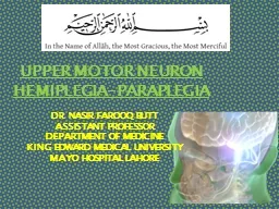
DR NASIR FAROOQ BUTT ASSISTANT PROFESSOR DEPARTMENT OF MEDICINE KING EDWARD MEDICAL UNIVERSITY MAYO HOSPITAL LAHORE UPPER MOTOR NEURONS UMN start from cerebral cortex passes through internal capsule ID: 774791
Download Presentation The PPT/PDF document " UPPER MOTOR NEURON HEMIPLEGIA-PARAPLEG..." is the property of its rightful owner. Permission is granted to download and print the materials on this web site for personal, non-commercial use only, and to display it on your personal computer provided you do not modify the materials and that you retain all copyright notices contained in the materials. By downloading content from our website, you accept the terms of this agreement.
Slide1
UPPER
MOTOR NEURON HEMIPLEGIA-PARAPLEGIA
DR NASIR FAROOQ BUTTASSISTANT PROFESSORDEPARTMENT OF MEDICINE KING EDWARD MEDICAL UNIVERSITYMAYO HOSPITAL LAHORE
Slide2UPPER MOTOR NEURONS
(
UMN) start from cerebral cortex, passes through internal capsule,
corticospinal
tracts, and ends at cranial nerve nuclei in brain stem or anterior horn cells of spinal cord at different levels
. The axons of upper motor neurons are contained within the pyramidal system, which is composed of the
corticospinal
(pyramidal) and
corticobulbar
tracts.
LOWER MOTOR NEURONS
(LMN) start from nuclei of cranial nerves or anterior horn cells of spinal cord, extend through cranial /spinal nerves and ends at motor end plate of concerned muscle.
Slide3.
.
Slide4UPPER MOTOR
NEURONS LESION (UMNL)
.
Slide5Pyramidal Drift Of An Upper Limb
Normally
, the outstretched upper limbs are held symmetrically, when the eyes are closed. With a pyramidal lesion, when both upper limbs are held outstretched, palms uppermost, the affected limb drifts downwards and medially. The
forearm tends to pronate and the fingers flex
slightly. This sign is often first to emerge, sometimes before weakness and/or reflex changes become apparent.
Slide6Pyramidal Drift Of An Upper Limb
Slide7Weakness And Loss Of Skilled Movement
A unilateral pyramidal lesion
above the
decussation
in the medulla causes
weakness of the opposite limbs
.
When
acute and complete
, this weakness will be immediate and total, a hemiplegia, e.g. following an
internal
capsule infarct
.
With
slowly progressive
lesions (e.g. a hemisphere
glioma
) a characteristic pattern of weakness emerges – a hemiparesis.
There is also
loss of skilled movement
– fine finger and toe control
diminishes
.
Slide8Changes In Tone And Tendon Reflexes
An
acute
lesion of one pyramidal tract (e.g. internal capsule stroke) causes
initially
FLACCID
paralysis with loss
of
tendon reflexes.
Increase in tone follows, usually within several days due to loss of inhibitory effects of the
corticospinal
pathways and an increase in spinal reflex
activity.
SPASTICITY
is
characterized by sudden changing resistance to passive movement – the clasp-knife effect.
Relevant
TENDON REFLEXES become exaggerated
;
CLONUS
may
emerge.
Slide9Slide10Changes In Superficial Reflexes
Abdominal (and cremasteric) reflexes are abolished on the side affected.The Babinski sign.
Slide11Patterns of UMN disorders
There are three main patterns
:
HEMIPARESIS
means weakness of the limbs on one side; it is usually caused by a lesion in the brain and occasionally in the cord.
PARAPARESIS
means weakness of both lower limbs and usually diagnostic of a cord lesion; bilateral brain lesions occasionally cause
paraparesis
.
TETRAPARESIS
(syn.
quadriparesis
) means weakness of four limbs.
Slide12Extrapyramidal system
The extrapyramidal system is a general term for
BASAL GANGLIA
motor
systems, i.e. corpus striatum (caudate nucleus +
globus
pallidus
+ putamen),
subthalamic
nucleus,
substantia
nigra
and parts of the thalamus. In basal ganglia/ extrapyramidal disorders, two features (either or both) become apparent, in limbs and axial muscles
:
Reduction in speed (
bradykinesia
, meaning slow movement) or
akinesia
(no movement), with muscle
rigidity
Involuntary
movements
(e.g. tremor, chorea,
hemiballismus
,
athetosis
, dystonia).
Slide13Extrapyramidal system
Extrapyramidal
disorders are classified broadly
into:
Akinetic
-rigid
S
yndromes
where
poverty of movement
predominates
D
yskinesias
where there are involuntary
movements.
The
most common extrapyramidal disorder is
Parkinson’s disease.
Slide14Click to add title
Click to add text
Slide15Case Scenario
An
83-year-old right-handed woman was eating dinner when she suddenly slumped to the right and her speech became slurred and
nonfluent
. Her vascular risk factors were hypertension and dyslipidemia, and the only other past medical history of interest was long-term hypothyroidism and a curative mastectomy for breast cancer. The patient was on two antihypertensive medications and a statin, and was not receiving any antiplatelet medication. Her husband immediately called the emergency medical services.
Slide16Case Scenario
On Examination Pulse is 84/min (regular), BP: 180/105 mm Hg.
Angle of mouth is deviated towards left (UMN type Right 7
th
CN)
Tone is decreased in right upper and lower limb,
Power is 2/5 in right
right
upper and lower
limb,
Reflexex
are absent in right
right
upper and lower
limb.
Right Planter is
upgoing
.
Tone, power, reflexes are normal on left side with
downgoing
planter
What is your Diagnosis????
Slide17STROKE
This patient has had STROKE causing Right sided uncrossed Hemiplegia with UMN type Facial Nerve Palsy
which can be caused by a vascular event such as thrombosis, embolism or hemorrhage or by
neaplasm
of brain (
aetiology
). The patient is limited by hemiplegia (functional status)
Slide18Stroke
DEFINITION
: Stroke
is
defined
as a syndrome of
rapid onset
of cerebral deficit (usually focal) lasting >24
h or
leading to death, with no cause apparent
other than
a vascular
one.
Slide19Slide20Stroke
Ischaemic
stroke/infarction (80%)
Thrombotic
Cardio-embolic
Large
artery stenosis
Small
vessel disease
Hypoperfusion
Haemorrhagic
stroke
(17
%)
Intracerebral
hemorrhage
(12
%)
Subarachnoid hemorrhage
(5
%)
Other
(3%),
e.g. arterial dissection, venous
sinus thrombosis
,
vasculitis
Slide21Types of Stroke
Depending upon the rate of developing and reversibility, stroke can be of following types:
Transient Ischemic Attack (TIA):
means a brief episode of neurological dysfunction due to temporary focal cerebral or retinal
ischemia
without infarction, e.g. a weak limb, aphasia or loss of vision, usually lasting seconds or minutes with complete recovery
.
Stroke-in-Evolution
:
If neurological deficit increases by step wise progression or there is slow uninterrupted progression, it is called stroke-in-evolution. It may take 1-2 weeks to complete
Complete Stroke
: It means complete hemiplegia
RIND (Reversible
Ischemic Neurological
Deficit):
It is a term that is not widely used anymore.
It
means a stroke that the patient was able to fully recover from within a few weeks.
Slide22Types of Stroke
Depending upon the blood supply of affected area of brain, stroke can be of following types:
ANTERIOR CIRCULATION STROKE:Carotid systemPOSTERIOR CIRCULATION STROKE:Vertebrobasilar system
Slide23Slide24Slide25Slide26Risk Factor For Ischemic Stroke
Hypertension
Diabetes
Smoking
Lifestyle
Alcohol
High cholesterol
Atrial fibrillation
Obesity
Severe carotid
stenosis
Sleep
apnoea
Slide27Rarer Causes Of Stroke
Hyperviscosity
states.
Thrombocythaemia
,
Polycythaemia
Thrombophilia
Anti-
cardiolipin
and lupus anticoagulant antibodies
oral contraceptive.
Migraine
Vasculitis
Systemic lupus
erythematosus
(SLE),
Polyarteritis
Giant
cell arteritis
Granulomatous
CNS
angiitis
:
Slide28Rarer Causes Of Stroke
Amyloidosis
.
Hyperhomocysteinaemia
Neurosyphilis
Mitochondrial
disease
Drugs
Sympathomimetic drugs
cocaine
Vasoconstrictors
neuroleptics
CADASIL (cerebral dominant
arteriopathy
with subcortical
infarcts and
leucoencephalopathy
)
Slide29Clinical Features of Stroke
SYMPTOMS:
Weakness of one half of body
Onset – Sudden, can be gradual
Headache, vomiting, fits
Unconciousness
Past H/O TIAs
H/O
smooking
, HTN, DM or IHD
Family H/O Stroke
Slide30Clinical Features of Stroke
SIGNS:
PULSE
: Irregular (At. Fib), Slow (Raised ICP)
BP
: High
Unconciouness
Pupils
: Small or Dilated
EYES:
Devited
towards on side (conjugate gaze)
Cranial Nerves
may be involved
Dysphasia, Dysarthria
Initially limbs may be
flaccid
with absent reflexes (Spinal Shock). This may persist for days, weeks or months and then Classical UMNL signs develop
Slide31Clinical Features of Stroke
SIGNS:
Hemiplegic
weakness
of
upper
limb
affects
the
shoulder
abductor,
elbow
extensors
,
wrist
and
finger
extensors
and
small
musles
of
hand
Hemiplegic
weakness
of
lower
limb
affects
hip
flexors
,
knee
flexors
and
dorsiflexors
and
evertors
of
the
foot
Slide32Slide33Clinical Features of Stroke
SIGNS:
Signs
of
meningial
irritation
(SOMI) –
Subarachnoid
Hemorrhages
Fundoscopy
:
Diabetic
or
Hypertensive
Retinopathy
Carotid
Pulsation
:
weak
,
bruit
CVS
Exam
:
Murmur
eg
Mitral
stenosis
Homonymous
hemianopia
and
sensory
inattention
Speech
defects
Slide34Slide35Click to add title
Click to add text
Slide36Amaurosis
fugax
R
efers
to a transient loss of vision in one or both eyes
due to embolism in retinal artery.
This is a feature of TIA in carotid circulation
Slide37ISCHEMIC STROKE
Cerebral Thrombosis:
Occurs when patient is asleep
Atherosclerotic narrowing of blood vessel
Middle cerebral art is commonly involved
Patient usually remain conscious
HTN is a major risk factor
Clinical Features:
Pt
can not move one half of body
Dysphasia
Uncrossed hemiplegia
High BP
Carotid Bruit
Slide38ISCHEMIC STROKE
Cerebral Embolism:
Sudden onset
Past history of TIAs
Source of Embolism
Carotid artery atheroma (weak carotid
pulse,bruit
)
Atrial Fibrillation or other
arrythmias
Recent Myocardial Infarction
Valvular
Heart Disease
Infective Endocarditis
Clinical Features:
Sudden loss of power in one half of body
Conciouness
may be lost
Dysphasia
Uncrossed hemiplegia
Irregular Pulse
Signs of
valvular
heart disease
Carotid Bruit
Slide39HEMORRHAGIC STROKE
Primary
Intracerebral
Hemorrhage:
H/O HTN
Internal capsule is most common site. Other sites: subcortical white matter, pons, and cerebellum.
O
ccurs when
pt
is
engaged in some activity
Causes: HTN
,
Saccular
Aneurysm,
AV
Malformation, Bleeding Disorders
Clinical Features:
Sudden loss of power in one half of body
Headache, vomiting and loss of
Conciouness
Uncrossed hemiplegia
Fundoscopy
: Papilledema
Slide40HEMORRHAGIC STROKE
Subarachnoid Hemorrhage:
Primary:
Bleeding occurs directly into subarachnoid space.
Causes
:
Saccular
Aneurysm, AV Malformation, Bleeding
Disorders,
Vasculitis
Clinical Features:
Sudden onset of severe Headache with or without vomiting
SOMI are present
Focal neurological signs
Fundoscopy
:
Subhyloid
hemorrhage, Papilledema
Secondary
:
Extension
of
intracerebral
hemorrhage
into
subarachnoid
space
Slide41SPACE OCCUPYING LESION
Lesion which occupy the cranial cavity and displace or
destroy existing structures
Causes
:
Tumour
, Abscess, Hematoma,
Tuberculomas
Symptoms:
H/O gradual onset of weakness developing over weeks to months.
Headache and vomiting
There may be H/O trauma (Hematoma)
H/O waxing and waning of
concious
level (Subdural Hematoma)
H/O fever and ear discharge suggest an abscess.
Signs:
In addition to motor deficit, following signs may be present:
Bradycardia
Fundoscopy
……Papilledema
Investigations:
CT scan (with contrast), MRI
Slide42Localization of Lesion
UNCROSSED HEMIPLEGIA
:
Cranial nerves involvement and hemiplegia are on the same side
Lesion is above brainstem on the side opposite to the hemiplegia
CROSSED
HEMIPLEGIA
:
Cranial nerves
are involved on one side
and hemiplegia
is
on the
other
side
Lesion is
in
brainstem on the side
of cranial nerve involvement.
HEMIPLEGIA WITHOUT CRANIAL NERVES INVOLVEMENT:
Lesion is in spinal cord above C5 on the side of paralysis.
Slide43Localization of Lesion
UNCROSSED HEMIPLEGIA
:
Lesion can be in Motor cortex, Corona
Radiata
or Internal Capsule
Motor cortex:
Specific manifestation due to lesion of various lobes:
Parietal Lobe
Temporal Lobe
Occipital Lobe
Frontal Lobe
Slide44Localization of Lesion
PARIETAL LOBE
:
Sensory cortex (
Precentral
Gyrus
)
demage
only affects special sensory functions
Loss of cortical sensations:
Two point discrimination
Astereogonosis
(inability to identify common objects by palpation with one hand while senses are intact)
Homonymous lower
quadrantanopia
Apraxia
(inability to perform a learned act in the absence of significant motor weakness.
Additional Features of Dominant Hemisphere
Gerstmann’s
synd
: Inability to calculate, write, distinguish between right and left, to recognize different fingers.
Alexia (inability to read), Bimanual
astereognosis
,
Sensory dysphasia
Slide45Localization of Lesion
PARIETAL LOBE
:
Additional
Features
of Non-
Dominant
Hemisphere
Sensory inattention (when both side are touched separately patient can recognize them, but when they are touched simultaneously, side opposite to affected parietal lobe is ignored)
Indifference to illness
Anosognosia
(
pt
is unaware of paralysis)
Autopagnosia
(
pt
ignores paralyzed body)
Spatial disorientation (
Pt
is unable to find his way around)
Slide46Localization of Lesion
TEMPORAL LOBE
:
Auditory or Olfactory hallucination
Auditory or Visual hallucination
Homonymous upper
quadrantanopia
of opposite side
Deja
vu phenomenon (feeling of undue familiarity with surroundings)
Automatism (lip smacking, swallowing, walking aimlessly or unconscious performance of highly skilled activity like driving a car)
OCCIPITAL LOBE
:
Visual
hallucination
Homonymous hemianopia of opposite side
Slide47Localization of Lesion
PRE-FRONTAL LOBE :
Lack of initiative and concentration
Lack of concern of consequences of any action
Micturition
disturbance
Grasp Reflex is +
ve
PRECENTRAL GYRUS (FRONTAL LOBE):
Weakness of
of
opposite half of the body
Jacksonian
fits
Expressive, motor or non-fluent dysphasia if lesion is in the dominant hemisphere.
Slide48Localization of Lesion
UNCROSSED HEMIPLEGIA
:
CORONA RADIATA:
Although Weakness is more marked in one limb, whole half is involved to some extent
INTERNAL CAPSULE:
Motor and sensory
fibere
are very closely packed…so lesion cause
Dense complete hemiplegia
Facial nerve palsy of same side
Cortical sensory loss and hemianopia may occur
Slide49Localization of Lesion
CROSSED HEMIPLEGIA
:
Midbrain:
3
rd
/4
th
CN are involved on one side and hemiplegia of opposite side
Pons:
5
th
, 6
th
, 7
th
and 8
th
CN nuclei are present in pons.
One or more of these CNs are paralyzed on one side and hemiplegia is on the other side.
Pupils are pinpoint but reactive to light
Hyperpyrexia may be present
Medulla Oblongata:
9
th
,
10
th
,
11
th
and
12
th
CN nuclei are present in
medulla.
One or more of these CNs are paralyzed on one side and hemiplegia is on the other side.
Slide50Localization of Lesion
SPINAL CORD.
Brown
-
Sequard
syndrome
(BSS)
is a rare neurological condition characterized by a lesion in the spinal cord which results in weakness or paralysis (
hemiparaplegia
) on one side of the body and a loss of sensation (
hemianesthesia
) on the opposite
side.
Causes:
Trauma
Non-traumatic: Tumor
(primary or
metastatic), Multiple sclerosis, Disk herniation,
Epidural
hematoma, Vertebral
artery
dissection, Transverse myelitis,
etc
Click to add title
Click to add text
Slide53MIDDLE CEREBRAL ARTERY
Supplies lateral surface of
frontal,frontal
,
perital
and temporal lobes
Most of the motor and sensory cortex
Ecxept
the part on medial side and control lower limb, internal capsule
Total Occlusion
:
Uncrossed hemiplegia
Hemianesthesia
Global Dysphasia
Homonymous hemianopia
Apraxia,
Agnosia
Partial occlusion
:
Any of the above features.
Lower limb is either not or minimally involved
Slide54ANTERIOR CEREBRAL ARTERY
Supplies MEDIAL surface of hemisphere
M
otor and sensory cortex on medial side which control lower limb
Soon its origin it gives a penetrating branch (
Heubner’s
artery) which supply internal capsule (containing fibers of upper limb and face)
Occlusion at the origin
:
Features are similar to total occlusion of MCB
Occlusion of
Heubner’s
artery
:
Weakness of upper limb and face
Occlusion
after the origin of
Heubner’s
artery
:
Weakness of
lower limb
Loss of cortical sensory function in lower limb
Slide55POSTERIOR
CEREBRAL ARTERY
Branch of Basilar artery
Supplies occipital lobe which include visual cortex
Occlusion
:
Visual field loss
Homonymous
hemianopia
Visual
agnosia
Disorders
of reading
Disorders of color vision
Memory impairment
Motor dysfunction
Slide56Click to add title
Click to add text
Slide57Click to add title
Click to add text
Slide58LATERAL MEDULLARY SYNDROME
Also called posterior
inferior cerebellar artery (PICA) thrombosis and
Wallenberg’s syndrome
)
A
common
example of brainstem infarction presenting as acute vertigo with cerebellar
and other
signs. It follows
T
hromboembolism
in the PICA or its branches, vertebral artery thromboembolism or dissection.
Features
depend on the precise structures damaged.
MEDIAL MEDULLARY SYNDROME
C
haracterized
by a set of clinical features resulting from occlusion of the anterior spinal artery. This results in the infarction of medial part of the medulla oblongata.
Slide59Slide60Slide61Investigations - STROKE
The purpose of investigations in stroke is:
To
confirm the clinical diagnosis
and distinguish between
hemorrhagic
and thromboembolic infarction;
To look for
underlying causes
and to direct therapy;
To
exclude other causes
, e.g.
tumour
.
Sources of embolus should be sought
Slide62Investigations of Stroke
Brain
Computed
Tomography (CT) Scan
Magnetic resonance imaging (MRI
)
Computed Tomography Arteriogram and Magnetic Resonance
Arteriogram:
Aneurysm, AV
malformation
Carotid
Ultrasound
ECG and
Echocardiography
Lumber Puncture:
Subarachnoid
Hemorrhage
Slide63Investigations - STROKE
Routine bloods (for ESR,
polycythaemia
,
infection,
vasculitis
, thrombophilia, syphilitic serology, clotting studies, autoantibodies)
Blood Sugar Level
Fasting
Lipid profile
Chest X-ray
ECG and Echocardiography
Carotid Doppler studies
Lumber
Puncture:
Subarachnoid
Hemorrhage
Slide64Investigations - STROKE
Imaging in Acute Stroke.
Non-contrast CT:
will
demonstrate
haemorrhage
immediately but cerebral infarction is often not detected or only subtle changes are seen initially.
Slide65Slide66Slide67Slide68Slide69Slide70Slide71CASE SCENARIO
A 40 year old male presented in OPD with C/O gradually progressive weakness of both legs for the last one month. He is also C/O backache. There is no H/O Trauma.
There is H/O Numbness
and
paresthesias
in
legs. He is unable to control his urine.
Slide72CASE SCENARIO
On Examination Pulse is 84/min (regular), BP: 180/105 mm Hg.
Bulk is normal in both legs
Tone
is
increased
in
both legs,
Power is
3/5
in right
leg
and
2/5 in left leg.
Knee and Ankle
Reflexex
are
Brisk
bilaterally
.
B
oth Planters are
upgoing
. Ankle Clonus is present on both sides.
Upper limbs are normal.
Tenderness at back of chest.
What is your Diagnosis????
Slide73This patient has SPASTIC PARAPLEGIA (Lesion) at T10 spinal level due to Cord compression (
aetiology
). It is complicated by bladder involvement (functional status)
Slide74SPASTIC PARAPLEGIA
HISTORY:
Ask about onset, duration and course of symptoms
Back pain: whether localized
Ask about radicular pain
Numbness and
parasthesia
particularly below the level of lesion
Weakness: whether gradual or sudden
Sphincter control and bladder sensation
Functional status: wheelchair transfers, walking aids, orthotic shoes and whether house has been modified for the patient’s disability
Take a family history (hereditary spastic paraplegia)
Take a history of birth anoxia (cerebral palsy)
History of urinary infections, pressure sores and deep venous
thromboses
.
Slide75SPASTIC PARAPLEGIA
EXAMINATION:
Increased tone in both lower
limbs,
Hyperreflexia
, Ankle
clonus
Weakness in both lower
limbs
Then
proceed to:
Check
the sensory level
Examine
the spine (spinal tenderness or deformity
)
Tell the examiner that you would like to do the
following:
Check sacral sensation
Examine
the hands to rule out involvement of upper limbs
Check
for cerebellar signs (multiple sclerosis,
Friedreich’s
ataxia).
Slide76SPASTIC PARAPLEGIA
EXAMINATION:
Try to
localize the level of lesion
using the following
:
UMNL signs in
lower
limb and LMNL signs in upper
lims
:
lesion of
C5–T1.
UMNL signs in lower limb and
Wasting of small muscles of hand:
lesion of
C8–T1
).
Spasticity of the lower limb alone:
lesion of thoracic cord (T2–L1).
Flaccid
Paraplegia:
Lesion
below L1
Slide77SPASTIC PARAPLEGIA
Paraplegia means UMN type weakness of both lower limbs.
CAUSES:
Spinal Cord Compression
Transverse Mellitus
Subacute
combined
degenration
of spinal cord
Friedreich’s
Ataxia
Amyotrophic Lateral Sclerosis (MND)
Spinal cord infarction
Hereditary
Spaatic
parapresis
Tabes
dorsalis
(
Neurosyphilis
)
Syringomyelia
Intracranial Lesions
Parasagital
Meningioma
Thrombosis of Sagittal Sinus
Slide78SPINAL CORD COMPRESSION
CAUSES
A. EXTRADURAL:
Vertebral Traumatic Fracture
Myeloma
Tuberculosis of Spine
Metastases (breast, prostate, lungs)
Traumatic and degenerative lesions of disc
B. EXTRAMEDULLARY
:
Meningioma
AV Malformation
C. INTRAMEDULLARY
:
Spinal Cord Tumors
Slide79SPINAL CORD COMPRESSION
Symtoms
:
H/O Backache,
agg
revated
by
coughing, sneezing and straining
Pain in the distribution of nerve root
H/O Trauma
Weakness is usually gradually progressive except in trauma when it is of sudden onset.
Numbness and
paresthesia
in lower limbs
Micturation
-urgency or
hesistancy
Signs:
Local examination of the vertebral column may reveal deformity or tenderness
Sensory loss with definite upper limit
Slide80SPINAL CORD COMPRESSION
Investigations:
X- Rays of vertebral column may show reduce disc space, bony
errosions
,
osteolytic
or
osteosclerotic
lesions, vertebral collapse or fracture.
MRI
Routine Investigations
Slide81TRANVERSE MYELITIS
Inflamation
of spinal cord
Can present with paraplegia or quadriplegia
Respiratory paralysis can
occur
Causes:
Unknown 60 % (
viral
infections or
post-infectious??)
Remaining 40 % associated
with autoimmune disorders such as multiple sclerosis,
neuromyelitis
optica
, systemic lupus erythematous,
Sjogren’s
syndrome
and
sarcoidosis
TRANVERSE MYELITIS
4 classical
Symptoms:
Backache/discomfort is first symptoms
Rapidly progressive weakness of lower limbs
Sensory symptoms such as numbness or tingling
Sphincter disturbances (Bladder/bowel) are
common
Signs
:
UMN signs in lower limbs
Sensory
Level
Investigations
:
Lumber Puncture with CSF Exam Lymphocytosis
X-Ray and MRI to rule out Cord compression
Slide83Subacute
Combined Degeneration Of Spinal Cord
Due to
Vit
B12 Deficiency
Degeneration of Posterior columns and pyramidal tract
Peripheral Neuropathy
Symptoms:
Symptoms of anemia
Gradual progressive weakness of legs
Numbness, tingling and
hyperesthesias
Signs:
Pallor
Paraplegia, Ankle Jerk Absent, Up going planters.
Sense of position and vibration are lost
Slide84Subacute
Combined Degeneration Of Spinal Cord
Investigations:
Peripheral Blood Film:
Macrocytosis
(MCV is raised),
Hypersegmented
Neutrophils
Pancytopenia
Mild unconjugated
hyperbilirubinemia
(du to ineffective
erythropoisis
.
Serum B12 is low. Elevated LDH,
Bonemarrow
biopsy:
Megaloblasts
Schilling Test
Causes of
Vit
B12 Deficiency:
Nutritional
def
,, Pernicious anemia, Bacterial overgrowth and blind loop syndrome, Diverticulosis,
Diphyllobothrium
latum
infestation, diseases of terminal ileum (
Crohn’s
disease,
ileocecal
TB)
Slide85Friedreich’s
Ataxia
Familial Autosomal Recessive
Slowly progressive degeneration of
P
osterior
columns,
P
yramidal
tracts,
S
pinocerebellar
tracts and
P
eripheral
nerves
Age of onset: 8 – 16 years
Slowly progressive weakness of legs and unsteadiness on walking
UMN type weakness of legs
Absent Ankle jerk
Planters
up going
Sense of position and vibration are lost
Slide86Friedreich’s
Ataxia
Sensory Ataxia, Romberg’s sign may be +
ve
Cerebellar Signs (
nystagmus
, scanning speech, intension tremors) may be present
Associated features:
Pes
cavus
(high arched foot)
Kyphoscoliosis
Spina
Bifida
Hypertrophic Obstructive
Cardiomuopathy
(HOCM)
Slide87Pes
Cavus
Causes:Hereditary Motor and Sensory Neuropathy Type 1 (Charcot-Marie-Tooth disease) Friedreich's Ataxia
Slide88Causes Of Absent
Ankel
Jerk And Up Going Planters
:
Friedreich's
ataxia
Subacute
combined degeneration of the spinal cord
Motor
neurone
disease
Syphilis
(
taboparesis
)
Combined
conus
medullaris
and
cauda
equina
lesions
Combination
of common problems e.g. diabetic peripheral neuropathy and cervical
spondylosis
Slide89CASE SCENARIO
A
65
year old
male
presented in OPD with complains of
slowly
progressive weakness of legs
with numbness
, tingling and
hyperesthesias
. He is a strict vegetarian. On
examination
he has
spastic
parapresis
with absent ankle jerk and
upgoing
planters. Sense of position and vibration are
lost.
His hemoglobin
is 6
gm
/dl.
What
is most likely diagnosis?
Slide90CASE SCENARIO
Answer:
Subacute
Combined Degeneration Of Spinal Cord
Slide91