/
M. Chakrabarti et al. - Ocular Contusion Injury421 M. Chakrabarti et al. - Ocular Contusion Injury421

M. Chakrabarti et al. - Ocular Contusion Injury421 - PDF document

tawny-fly
tawny-fly . @tawny-fly
Follow
388 views
Uploaded On 2017-03-07

M. Chakrabarti et al. - Ocular Contusion Injury421 - PPT Presentation

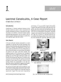
Lacrimal Canaliculitis A Case Report Dr Bindu N Das Dr Sisira lacrimal canaliculus which is often overlooked andtreated unsatisfactorily Bacteria fungi and viruses mayall produce such infectio ID: 523118

Lacrimal Canaliculitis Case Report Dr.

Share:

Link:

Embed:

Download Presentation from below link

Download Pdf The PPT/PDF document "M. Chakrabarti et al. - Ocular Contusion..." is the property of its rightful owner. Permission is granted to download and print the materials on this web site for personal, non-commercial use only, and to display it on your personal computer provided you do not modify the materials and that you retain all copyright notices contained in the materials. By downloading content from our website, you accept the terms of this agreement.


Presentation Transcript

M. Chakrabarti et al. - Ocular Contusion Injury421 Lacrimal Canaliculitis, A Case Report Dr. Bindu N. Das , Dr. Sisira lacrimal canaliculus which is often overlooked andtreated unsatisfactorily. Bacteria, fungi and viruses mayall produce such infection , the most common agentsreported being actinomyces. Here we report a case ofCase Reportleft eye of 1 year duration, pain and swelling of the lefteyelids for 2 months. On examination his vision wasleft eye showed a tender fusiform swelling in the medialone third of left upper and lower eyelids. Thelacrimal sac area. The upper and lower puncti werefound to be pouting with expression of tenacious puson pressure over the swelling. The right eye also showedpouting of lower puncta though it did not showswelling. Patient was asymptomatic in the right eye.Rest of the anterior and posterior segment was normal.The punctum was split under topical anaesthesia usingtenacious purulent material was expressed from theirrigation with penicillin solution (100, 000 units). Thewere sent for microbiological investigations. Repeatedinoculated on blood agar and brain heart infusion broth.On Gram staining the organisms appeared as gram- Dept of Ophthalmology, Kozhikode Medical College, Kozhikode Fig. 3. The casts removed after 3-snip procedureFig. 2a-b showing thick tenacious purulent material expressedfrom canaliculusFig. 1. showing conjunctivalinflammation CASE 422Kerala Journal of OphthalmologyVol. XX, No. 4(Fig.4) The patient recovered rapidly and was left withpatent. Post-operatively, the swelling, conjunctivitis and species is a gram-positive, cast-bacillus. Its filamentous growth and mycelia likecolonies have a striking resemblance to fungi. They are(eg, wet hay, straw). It is primarily a commensalcrypts, in dental plaques, and in caries teeth and entersthe lacrimal system through the nasal passage orCervico-facial actinomycosis has also been reported.Canaliculitis usually presents as chronic watering,redness and discharge from eye. A pouted punctum is50 % of all patients who are affected. Typically, theCanalicular discharge and canaliculiths may be sent forCulture and sensitivity (ie, blood agar, Sabouraud,TreatmentFailure of resolution of canaliculitis by topical treatmentsystem and removal of any casts. Extensive surgery isnot always required. A 2-snip punctoplasty, castremoval, curettage, and probing is usually done.Subsequent lacrimal irrigation with 1 MU of penicillinReferences1.Jordan DR. Dacryoadenitis, Dacryocystitis, andMosby). 1997;687-693. 2.Richards WW. Actinomycotic lacrimal canaliculitis.American J Ophthalmol 1975;75:155-157. 3.Pine L, Hardin H, Turner L, Roberts SS. Actinomycotic4.Sridhar MS, Gopinathan U, Garg Pthe cornea. Surv Ophthalmol 2001;45:361-378. 5.Sharma S. Ocular Microbiology. 1st ed. (Aravind EyeHospitals Publication, Madurai) 1988:79-84. Fig. 5.P