/
CGH Assessment: Within the Context of  Cervical Spine Examination CGH Assessment: Within the Context of  Cervical Spine Examination

CGH Assessment: Within the Context of Cervical Spine Examination - PowerPoint Presentation

test
test . @test
Follow
355 views
Uploaded On 2018-11-21

CGH Assessment: Within the Context of Cervical Spine Examination - PPT Presentation

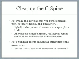
Cervical Treatment Based Classification Fritz amp Brennan 2007 Physical Examination Objectives Identify c ervical contribution to HAs Is there a comparable sign Identify Impairments that may be directly or indirectly contributing to HAs ID: 731290

head cervical upper side cervical head side upper spine rotation flexion extension patient assessment posture left angle ligament motion

Share:

Link:

Embed:

Download Presentation from below link

Download Presentation The PPT/PDF document "CGH Assessment: Within the Context of C..." is the property of its rightful owner. Permission is granted to download and print the materials on this web site for personal, non-commercial use only, and to display it on your personal computer provided you do not modify the materials and that you retain all copyright notices contained in the materials. By downloading content from our website, you accept the terms of this agreement.


Presentation Transcript

Slide1

CGH Assessment: Within the Context of Cervical Spine ExaminationSlide2
Slide3

Cervical Treatment Based Classification

Fritz & Brennan (2007)Slide4

Physical Examination Objectives

Identify

c

ervical contribution to HA’s

Is there a comparable signIdentify Impairments that may be directly or indirectly contributing to HA’s

Develop Prognosis

SINSS, Contributing factors, Psychosocial Issues Slide5

Age of onset and duration

MOI- history of trauma including MVA, manipulations, falls, quick

mvts

, pregnancy.

Nature and quality of HA’s (unilateral, bilateral, throbbing, pulsating, constant, intermittent, duration)

Associated Symptoms – nausea, photo or

phonobia

, “5 D’s”

Aggravating and alleviating factorsPosture, Stress, Response to medication.How are symptoms changingPrevious Treatments

History – Important QuestionsSlide6

Assessment & Biomechanics of the Upper Cervical SpineSlide7

C0-C1

Flexion/Extension

35 degrees ;10 flexion/25 extension (

Sizer

2005)

Axis through External Auditory

Meati

Occipital

condyles roll in same direction, glide opposite (1,2)

Unilateral limitations in flexion result in deviation to opposite side (3)

Limitation in R OA flexion, chin will deviate to left with OA flexion.

Unilateral limitations in extension result in deviation to same Limitation in R OA extension, head will tilt to the right

Assessment & Mechanics of Upper Cervical Spine

Greater amounts of Upper cervical flexion achieved in Cervical retraction, extension with protraction.Slide8

C0-C1

Side-Bending

Axis through the nose

Occipital

condyles

roll to same side and slide opposite

Obligatory motion of the Atlas* (Paris &

Sizer

) Translate to same side and rotate opposite ( SBR, atlas will translate right and rotate left).

Obligatory motion at C2-3*

Rotation to same side as SB (due to

Alar ligament)OA will not SB if C2 cannot rotate on C3 to same side. (1)C2-3 “Keystone to Upper Cervical motion” (1)

Assessment & Mechanics of Upper Cervical SpineSlide9

C1-C2

40-45 degrees rotation to each side

With right rotation the right C1 facets slides posterior to C2 facet and the left C1 facet slides anterior to left C2 facet

The

occiput

will SB opposite direction of rotation

(1)

Absence of this will produce an obvious

ipsilateral SB with rotation

Assessment & Mechanics of Upper Cervical SpineSlide10

Observe Posture

AROMCervical physiologicAA Rotation

OA SB

Cranial Nerves

Palpation of Sub-Occipital TriangleUpper Cervical Ligamentous

Testing

Transverse

Alar

ASSESSMENT LAB - SittingSlide11

Subcranial

Posterior Rotation & Anterior head Translation leads to a decrease in

Craniovertebral

Angle

O/A and AA Functional spaces Altered

Compression of

subcranial

structures including the vertebral arteries and their sympathetic nerves, the first two cervical nerves, and soft tissue. (1)

Hypomobility about the

u

pper cervical spine and upper thoracic spine (1,2)

Mid-Cervical hypermobility (3)Alterations in muscle length tension relationships and muscle function (Upper Cross Syndrome) (3)

Forward Head PostureSlide12

Observation / Postural

Examination

View patient’s posture from the side

Assess

:

Forward head posture

Shoulder carriage

Typical patterns include

:

Sub-Cranial Posterior Rot.

Flexed (rounded) T1-T2

Extended (flat) T3-T7Flexed (rounded) T8-T12Slide13

Weakened Muscles Shortened Muscles

Deep Cervical Flexors Sub-Occipitals

Lower and Mid

Trapezius

Upper

Trapezius

Serratus

Anterior Pectorals

Forward Head Posture – Upper Cross Syndrome (3)Slide14

Measured

Craniovertebral

Angle by measuring the angle formed by horizontal line through C7 and a line form C7 to the Tragus of the Ear.

Smaller angle associated with CTTH (4,5)

Craniovertebral

Angle –

Fernández

-de-

las

-

Peñas

C et al (2007)Slide15

Visual Observation

Sitting

Manubrium

to

Mentonian

Symphysis

(lowest point on mandible) to Malar Bone

Position of SCM (60 deg angle) (structure changes function)

Palpate C0-C2 space

CV Angle Ability to correctStandingHead to Wall (measure).

Forward Head Posture - AssessmentSlide16

Brame

M. Headaches and the Upper Cervical Spine. Course Handout. North American Seminars 2005

CranioMandibular

Sytem

. On-Line Course Material. University of St. Augustine for Health sciences 2010.

Lau et al. Clinical measurement of

craniovertebral

angle by electronic head posture instrument: A test of reliability and validity.

Manual Therapy 2009; 14:363–368

Moore M. Upper Crossed Syndrome and its Relationship to Cervicogenic Headache. Journal of Manipulative and Physiological Therapeutics 2004;27:414-20

Fernandez-de-las-Penas

C.

Performance of the

Craniocervical

Flexion Test, Forward Head Posture, and Headache Clinical Parameters in Patients With Chronic Tension-Type Headache

: A Pilot Study. JOSPT 2007;37(2):33-39

ReferencesSlide17

Cranial Nerve ExamSlide18

Cranial Nerve ExamSlide19

Cranial Nerve ExamSlide20

Upper Cervical

Ligamentous

TestingSlide21

Transverse Ligament (1)

Prevents separation of C1 and C2Prevents tipping of the Dens into brainstem and spinal cord

Alar

Ligament

Assists Transverse LigamentTaught in extension, SB and ipsilateral rotation

Responsible for coupled motions

Upper Cervical

Ligamentous

TestingSlide22

Upper Cervical

Ligamentous

Testing – Sharp Purser

Purpose:

Position of Atlas and Dens (Transverse Ligament)

Patient:

Sitting

Technique:

The palm of one hand is placed on the patient’s forehead while the spinous process of the axis is held by a pinch grip of th opposite hand. Then the head and neck are the gentlyflexed. Through palmar

pressure on the forehead, the

occiput

and atlas are translated posteriorly.Positive: Decrease symptoms or clunk. Mintken P

et al . JOSPT 2008;38(8):465-475Slide23

Patient seated in upright posture

Stand at patients side and achieve pincher grip of SP of C2 (you many need to flex cervical spine if patient has significant FH)

Side-bend head to one side

Test: You should feel an

obilgatory movement of the SP of C2 moving away from the side the side –bending is occurring. This is due to obligatory rotation to same side with intact Alar

Ligament.

Upper Cervical

Ligamentous

Testing – Alar LigamentSlide24

Base of

Occiput

to TP of Atlas

TP of Atlas to SP of C2

C2 to Base of

Occiput

Note texture of tissue

and provocation.

Palpation of Sub-Occipital TriangleSlide25

PROM

OA flexion , extension and SB

AA

Rotation

with

flexion and/or SB

C2-3 Accessory Glides

General Upper Thoracic (PA)

Palpation (length)Trapezius

SCM

Sub-occipitals

Splenius Muscle Performance (Motor Control)DCF with or without biofeedback

ASSESSMENT LAB - SupineSlide26

OA Extension and Flexion

Patient supine with cervical spine in neutral. Cradle head with both hands with thumbs resting on temporal

region. Gently

nod

occiput

forward and backward around

a transverse axis

through

the

External Auditory

Meati

. Bias flexion to the right

or left by rotating head 20-30 degrees in same direction. Alternate technique is to place one hand on forehead and use a coupling motion with both hands to induce flexion/extension

Cradle patients head with both hands. Use the radial border of your second phalanx to lift the

occiput

anteriorly

. Bias extension towards the right by lifting up on the left, assessing the left side

.Slide27

Upper Cervical

Ligamentous

Testing – Anterior Shear Test

Purpose

:

Transverse Ligament

Patient:

Supine

Position: Head is supported with second index fingers resting between occiput and C2

Technique:

Head and C1 are lifted

anteriorlyPositive: Produces nystagmus, paresthesias of lips, hands toes, increase patients symptoms. Note end feelMintken

P et al . JOSPT 2008;38(8):465-475.Slide28

OA -

Sidebending

Patient supine with head in neutral.

Grasp head with both hands with hand/thumb on side where SB to occur on mandible.

Use coupled motion to induce SB through

subcranial

region.

Can use abdomen to perform comfortable axial load to stabilize cervical spine.

10-15 degrees is normalSlide29

AA Rotation with Flexion

Cervical Spine is fully flexed with patients head supported by clinicians abdomen.

Cervical Spine is rotated fully to the both sides.

Note range of motion, end-feel and patient response

. Slide30

AA Rotation with SB

Cervical spine is resting on pillow in neutral flexion/extension.

SB to one side to first barrier. Rotate head gently to opposite side

Important: No more than 40-45 degrees should be available. Assess range, quality and pain. Do not lose SBSlide31

Palpation and

Uglide

of C2-3 (R)

Patient supine with heads resting on pillow

Palpate the

articular

pillar of C2 with your finger tips and slide right index finger down along pillar to approximate the middle phalanx.

Rotate head and neck minimally to the right

without

feeling motion

takng

palce at C2-3. Add slight SB to left using mostly your trunkUse your contact point to provide a “lifting” motion in a 45 degree plane toward patients left eyeSlide32

O’Leary S et al 2009

Motor Performance of the DCF

With Biofeedback:

Cervical Spine is in neutral. Inflate cuff to 20 mm hg. Instruct patient to perform nodding movement (yes) to 22 mm hg for 10

secs

. Provide 10 sec rest and move up to 30 in increments of 2 if patient able to perform. Should achieve 26-30 mm hg.

Without Biofeedback:

Retract neck and perform chin tuck. Lift head one inch. Maintain tucked chin and hold head up.

Neck pain: 24 Without: 38

Childs JD et al 2008Slide33

Unilateral PA’s

C0-1

C2-3

C1-2

T2-4

Apophyseal

and CT joints

Assessment Lab - Prone

Tip: In these techniques utilize shoulder adductors and trunk to grade force while relaxing the thumbs. Slide34

C2-3 Unilateral PA

Head and neck are in neutral. Take up slack in soft tissue. PA is applied to the

articular

pillar of C2 assessing further rotation of C1 on C2

.

Using arms (pectorals) and trunk to impart pressure which is mild. Note resistance and reproduction of pain.

Without rotation assess C2-3. Can be a treatment technique with graded oscillationsSlide35

C1-C2/ Unilateral PA

With Permission – Fearonphysicaltherapy.com

Head is rotated 30 degrees to the side tested. Take up slack in soft tissue. PA is applied to the

articular

pillar of C2 assessing further rotation of C1 on C2

.

Using arms (pectorals) and trunk to impart pressure which is mild. Note resistance and reproduction of pain.

Can

be a treatment technique with graded oscillations. Slide36

Sizer

PS et al. Diagnosis and Management of

Cervicogenic

Headache.

Tuitorial

. Pain Practice 2005; 5(3): 255-274

Paris SV. S3 Seminar manual. University of St. Augustine.

Patris

, Inc 4th

Edition 2000.

Cervico

-Thoracic Integration. Course Manual. Institute of Physical Art 2002. References