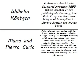
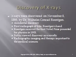
Accidental discovery First radiograph of Mrs Roentgens hand Roentgen received the first Nobel Prize presented for physics in 1901 Public viewed discovery as a novelty Radiographic imaging and therapy important to the medical sciences ID: 775158
Download Presentation The PPT/PDF document " Discovery of X-rays X-rays were discove..." is the property of its rightful owner. Permission is granted to download and print the materials on this web site for personal, non-commercial use only, and to display it on your personal computer provided you do not modify the materials and that you retain all copyright notices contained in the materials. By downloading content from our website, you accept the terms of this agreement.
Slide1
Discovery of X-rays
X-rays were discovered on November 8, 1895, by Dr. Wilhelm Conrad Roentgen.
Accidental discovery
First radiograph of Mrs. Roentgen's hand
Roentgen received the first Nobel Prize presented for physics in 1901.
Public viewed discovery as a novelty
Radiographic imaging and therapy important to the medical sciences
Slide2X-rays as Energy
A form of electromagnetic radiationBehave both like waves and like particlesMove in waves that have wavelength and frequencyWavelength and frequency are inversely relatedX-rays also behave like particles and move as photons
Slide3Properties of X-rays
X-rays are invisible.
X-rays are electrically neutral.
X-rays have no mass.
X-rays travel at the speed of light in a vacuum.
X-rays cannot be optically focused.
X-rays form a polyenergetic or heterogeneous beam.
X-rays can be produced in a range of energies.
X-rays travel in straight lines.
Slide4Properties of X-rays (cont.)
X-rays can cause some substances to fluoresce.
X-rays cause chemical changes to occur in radiographic and photographic film.
X-rays can penetrate the human body.
X-rays can be absorbed or scattered by tissues in the human body.
X-rays can produce secondary radiation.
X-rays can cause chemical and biologic damage to living tissue.
Slide5Birth of Radiology
Dry plate- used to record x-ray images
exposure required were extremely long.
Glass plate easily broken.
Thomas Edison developed the first intensifying screen
WWI x-ray film was produced
the cellulose nitrate film base and was highly flammable and a fire hazard
Slide6X-ray Production
The production of x-rays requires a rapidly moving stream of electrons that are suddenly decelerated or stopped.
The negative electrode (cathode) is heated, and electrons are emitted (thermionic emission).
The electrons are attracted to the anode, move rapidly towards the positive electrode, and are stopped or decelerated.
Slide7X-ray Tube Housing
Metal or glass envelopeNegatively charged electrodePositively charged electrode
Slide8Cathode
FilamentSource of electronsFilament currentThermionic emissionCoiled tungsten wireLarge and smallFocusing cupSpace charge effect
Slide9Anode
Rotating anodeRequires a stator and rotor to rotateTungsten metalHigh melting pointEfficient x-ray production TargetDecelerates and stops electronsEnergy converted to heat and x-raysBremsstrahlung and characteristic interactions
Slide10Target Interactions
Bremsstrahlung interactions
Braking or slowing down radiation
85% of x-ray beam
Characteristic interactions
Projectile electron energy at least 69.5 keV
Inner shell electron ionized
15% of x-ray beam
X-ray properties the same
Slide11Bremsstrahlung- electrons interacts with the atomic nucleus, more energy is lost and a stronger x-ray is produced; account for the majority of the x-ray beam.Characteristic Radiation - electrons interact with an orbital electron from the atom, the pulling down of another electron from an outer shell causes an x-ray to be produced; account for a small majority of the x-ray beam
Review of Interactions in the x-ray tube
Slide12Target Interactions (cont.)
Slide13Contains low energy rays which will be absorbed by the x-ray tubeAverage energy of the beam is 1/3 of the maximum energyX-rays are an inefficient process99% heat, 1% converted to x-rays
Heterogeneous Beam
Slide14Primary Radiation (PR)- portion of beam from tube to the patient; radiation before it enters the patientRemnant Radiation (RR)- radiation emerging from patient’s body to expose the film; image forming radiation
X-ray Beam
Slide155% of primary beam passes through the patient without any interactions15% of the primary beam interacts with atoms and produce secondary radiation, they make it out of the patient and expose the film.80% will be totally absorbed by patient
Primary Beam Distribution
Slide1620% or 1/5 of the intensity of the original beam exposes the filmWith remnant radiation about 75% to 80% of the beam is made up of secondary radiation
Distribution of Remnant Radiation
Slide17mA- milliampereS- seconds timekVp- kilovoltage peakSID- source to image distanceThese are all controlled by the technologist
Prime Factors of Radiography
Slide18X-ray Emission Spectrum
The range and intensity of x-rays emitted changes with different exposure technique settings on the control panel.
Slide19Kilovoltage
Creates potential differenceDetermines the speed of the electrons in tube currentGreater speed results in greater quantity and quality of primary beamIncreasing electron speed will increase x-ray beam penetrability
Slide20Milliamperage
Unit to measure tube current or number of electrons flowing per unit timemA directly proportional to quantity of x-rays producedDouble the mA will double the number of x-ray photons produced
Slide21Milliamperage and Time
Exposure time determines the length of time x-rays are produced.
Increasing time will increase the total number of x-rays produced.
Exposure time and x-ray quantity are directly proportional.
Slide22Beam Filtration
Aluminum filtration added to x-ray beam to absorb low-energy photonsTotal filtrationInherentAddedReduces patient exposure
Slide23Compensating Filtration
Added to primary beam to alter its intensityWedge filterTrough filterUsed to image non-uniform anatomic areasThicker part of filter lined up with thinner part allowing fewer x-ray photons to reach anatomic area
Slide24Image Formation
Differential absorption
Anatomic tissues absorb and transmit x-rays differently based on their composition (atomic number and tissue density).
Bone absorbs more x-rays than muscle.
Attenuation: the primary x-ray beam loses some of its energy (number of photons) as it interacts with anatomic tissue.
Absorption
Scattering
Slide25Differential Absorption
Slide26X-ray Beam Absorption
During absorption, the energy of the primary beam is deposited within the atoms comprising the tissue.
Photoelectric effect: complete absorption of the incoming photon
X-ray ionizes atom
Low energy secondary x-ray photon created
Probability of photoelectric effect dependent on the energy of the incoming x-ray photon and tissue atomic number
Slide27Determining Attenuation of the Beam
Three essential aspects of tissues will determine their attenuation properties and the resulting subject contrast:
Tissue Thickness
Tissue density
Tissue atomic number
Slide28X-ray interaction with matter
When the primary x-ray beam interacts with anatomic tissues. Three processes occur during attenuation of the x-ray beam:
Absorption
Scattering
Transmission
Slide29Transmission
If the incoming x-ray photon passes through the anatomic part without any interaction with the atomic structures, it is called transmission.
The combination of absorption and transmission of the x-ray beam will provide an image that represents the anatomic part.
Slide30Scattering
The Compton effect occurs when an incoming photon loses some but not all of its energy, then changes its direction.
It can occur within all diagnostic x-ray energies and is dependent only on the energy of the incoming photon, not the atomic number of the tissue.
Higher kVp reduces the number of interactions overall, but the number of Compton interactions increases in comparison to the number of photoelectric interactions.
Slide31Absorption versus Scattering
Slide32Photoelectric Effect
The secondary x-ray photon does not reach the film.
The photoelectric effect is crucial to the formation of the radiographic image.
The photoelectric effect is responsible for the production of contrast on the radiographic image.
Slide33Photoelectric Effect
During attenuation of the x-ray beam, the photoelectric effect is responsible for total absorption of the incoming x-ray photon.
Slide34Scattering/ Compton Effect
The Compton photon may be scattered in any direction.
Scatter refers to any x-ray photon which has changed direction from the direction of the primary beam.
The Compton Effect may be considered as scatter, since 99% of all scattered x-ray photons originate from Compton interactions in the patient.
Slide35Where do interactions occur
Compton interactions occur only in the outer shells of an atom.
Photoelectric interactions occur only in the inner most shell of an atom.
Slide36Factors Affecting Beam Attenuation
Tissue thickness
X-rays are attenuated exponentially and generally reduced by ~ 50% for each 4 to 5 cm (1.6" to 2") of tissue thickness.
Type of tissue
Tissues composed of a higher atomic number will increase beam attenuation.
Tissue density
Increasing the compactness of the atomic particles will increase beam attenuation.
X-ray beam quality
Higher kVp increases the energy of the x-ray beam and will decrease beam attenuation.
Slide37Exit Radiation
Remnant or exit radiation is composed of transmitted and scattered radiation.The varying amounts of transmitted and absorbed radiation create an image that structurally represents the anatomic area of interest.Scatter radiation reaching the image receptor creates unwanted exposure called fog.
Slide38Secondary Radiation vs. Scatter Radiation
Secondary Radiation refers to any radiation resulting from interactions within the patient.
Scatter radiation refers only to that secondary radiation which has been emitted in a direction different than the original x-ray beam.
Most secondary radiation is scattered.
Slide39Radiographic Quality
A quality radiographic image accurately represents the anatomic area of interest, and its information is well visualized for diagnosis.
Visibility of anatomic structures
Density
Contrast
Accuracy of structural lines (sharpness)
Resolution or recorded detail
Distortion
Slide40Density
A film image is evaluated by the amount of density or overall blackness after processing.
A radiographic image must have sufficient brightness or density to visualize the anatomic structures of interest.
Slide41Image Contrast
The radiograph must exhibit differences in the brightness levels or densities (image contrast) in order to differentiate among the anatomic tissues.
Slide42Subject Contrast
Subject contrast is a result of the absorption characteristics of the anatomic tissue radiographed and the quality of the x-ray beam.
The ability to distinguish among types of tissues is determined by the differences in brightness levels or densities in the image or contrast.
Contrast resolution describes an imaging receptor's ability to distinguish between objects similar in subject contrast.
Gray scale: number of different shades of gray that can be stored and displayed in a digital image
Scale of contrast: the range of densities visible on film
Slide43Scale of Contrast
Short scale or high contrast
Long scale or low contrast
Slide44Recorded Detail
Anatomic details must be recorded accurately and with the greatest amount of sharpness.
Recorded detail
refers to the distinctness or sharpness of the structural lines that make up the recorded film image.
All radiographic images have some degree of
unsharpness
.
Slide45Distortion
Radiographic misrepresentation of either the size or shape of the anatomic part
Size distortion or magnification is an increase in the object's image size compared to its true or actual size.
SID and OID affect magnification.
Shape distortion is a misrepresentation of an object's image shape.
Elongation and foreshortening
Central ray (CR) alignment of the x-ray tube, part, and image receptor affect distortion.
Slide46Scatter
Unwanted exposure to the image receptor resulting in fog
A result of Compton interactions
Provides no useful information
Scatter or fog decreases image contrast.
Slide47Radiographic film is composed of two main partsBaseEmulsion
Modern x-ray film
Slide48Consists of emulsion of finely precipitated silver bromide crystals Crystals are suspended in a gelatin and is coated on both sides with a transparent blue tinted polyester support called the base
Film Construction
Slide49Visible image you see when the film is processedWhat you see as your final radiograph
Manifest Image
Slide50Film-screen Image Characteristics
Film used as medium for acquiring, processing, and displaying the radiographic image
Film emulsion: active layer of film that contains the crystals that serve as latent imaging centers
Intensifying screens: used to convert exit radiation intensities to visible light and expose the emulsion crystals
Film is chemically processed to display the range of densities created as a result of the x-ray attenuation characteristics of anatomic structures.
Slide51Primary Exposure Factors
Milliamperage (mA)
Directly proportional to radiation quantity
Inversely related to exposure time to maintain exposure to image receptor (IR)
Exposure time (time)
Directly proportional to radiation quantity
Inversely related to mA to maintain exposure to IR
mA × time (seconds) = mAs
Kilovoltage (kVp)
Directly related to radiation quality and quantity
Inversely related to radiographic contrast
Slide52Beam Attenuation
As the primary beam passes through the patient it will loose some of its’ original energy.
This reduction in the energy of the primary beam is known as attenuation.
Slide53Image Receptor Exposure
To increase exposure to image receptor, increase mA, exposure time, or kVp.
To decrease exposure to image receptor, decrease mA, exposure time, or kVp.
To maintain exposure to image receptor
Increase mA and proportionally decrease time
Increase time and proportionally decrease mA
Increase kVp 15% and decrease mAs by half
Decrease kVp 15% and increase mAs by two times
Slide54Changing Kilovoltage
Kilovoltage affects
X-ray beam quality and quantity
X-ray beam penetration and absorption in anatomic tissues
Increasing kVp increases penetration and decreases absorption.
Decreasing kVp decreases penetration and increases absorption.
Subject contrast
Increasing kVp decreases subject contrast.
Decreasing kVp increases subject contrast.
Slide55kVp and Wavelength
KVP increases, wavelength gets shorter; penetrating ability increases.
KVP increases, wavelength decreases, indirect relationship
KVP increases, penetration increases, direct relationship
Slide56kVp and Wavelength
shorter the wavelength, stronger the penetration, higher the kVp
Higher the kVp, shorter the wavelength
Lower the kVp, longer the wavelength
Slide57Primary Factors: Film-screen
Kilovoltage and mAs have a direct effect on radiographic density for film-screen imaging.
Repeating a radiograph for a density error requires a change in mAs by a factor of 2 or a change in kVp by 15%.
For insufficient density multiple the mAs by 2 or increase kVp by 15%.
For excessive density divide the mAs by 2 or decrease kVp by 15%.
The best factor to change for density errors is mAs, because kVp also affects radiographic contrast.
Slide58OID
Distance between the anatomic part and image receptor will affect
Radiation intensity reaching the image receptor
Amount of scatter radiation reaching the image receptor
Magnification
Recorded detail/spatial resolution
Slide59OID (cont.)
An air gap will decrease the intensity of radiation and scatter reaching the image receptor.
Increasing the OID will decrease the exposure to the image receptor, increase contrast and magnification, and decrease recorded detail/spatial resolution.
Decreasing the OID will increase the exposure to the image receptor, decrease contrast and magnification, and increase recorded detail/spatial resolution.
Slide60OID (cont.)
Distance created between the object and image receptor will reduce the amount of scatter radiation reaching the image receptor.
Slide61Magnification Factor
Magnification Factor (MF) = SID . SODSOD = SID – OIDObject size = image size MF
Source-to-object distance is the distance between the source of the x-ray and the object radiographed.
Slide62Shape Distortion
Any misalignment of the CR among these three factors—tube, part, or image receptor—will alter the shape of the part recorded on the image receptor.
Slide63Grids
Limiting the amount of scatter radiation that reaches the image receptor improves the quality of the radiograph. The effect of less scatter or unwanted exposure on the image is to increase the radiographic contrast.
Much of the scatter radiation toward the image receptor will be absorbed when a grid is used.
Slide64Grids (cont.)
Grids also absorb some of the transmitted radiation exiting the patient and therefore reduce the amount of radiation reaching the image receptor. Grid conversion formula mAs1 = grid ratio1 mAs2 grid ratio2
Slide65Beam Restriction
A larger field size (decreasing collimation) increases the amount of tissue irradiated, causing more scatter radiation to be produced and increasing the amount of radiation reaching the image receptor. The increased amount of scatter reaching the image receptor results in less radiographic contrast.
A smaller field size (increasing collimation) reduces the amount of tissue irradiated and reduces both the amount of scatter radiation produced and the amount of radiation reaching the image receptor. The decreased amount of scatter radiation reaching the image receptor results in higher radiographic contrast, but it requires an increase in mAs.
Slide66Generator Output
Generators with more efficient output, such as three-phase units or high frequency, require lower exposure technique settings to produce an image comparable to those of single-phase units.
Slide67Patient Factors
Body habitus
Hypersthenic, sthenic, hyposthenic, asthenic
Part thickness affects
Beam attenuation
Exposure reaching image receptor
Scatter production and image contrast
Pediatric patients
Small size may require a reduction in exposure
Quicker exposure times may be necessary
Slide68Body Habitus
Hypersthenic: The hypersthenic body is of massive build with a broad and deep thorax. The diaphragm is high and the stomach and gallbladder also occupy high positions. An extreme body type, the hypersthenic classification accounts for only about 5% of all people (large- stocky build).
Sthenic: Means active or strong. The sthenic body is the one we usually associate with the athletic type. The body is rather heavy with large bones. The sthenic body type is the predominant type, with about 50% all people falling into this classification( normal or average build).
Slide69Body Habitus
Hyposthenic: Slender and light in weight with the stomach and gallbladder situated high in the abdomen. About 35 %of all people fall into this classification( slender, taller build).
Asthenic: Extremely slender, light build, with a narrow, shallow thorax, and the gallbladder and stomach situated low in the abdomen. An extreme type, the asthenic classification accounts for only about 10% of all people.
Slide70Special Considerations
Projections and positions
Casts and splints
Pathology
Soft tissue imaging
Contrast media
Slide71The 5 X-ray densities
Low density material such as air is represented as black on the final radiograph. Very dense material such as metal or contrast material is represented as white. Bodily tissues are varying degrees of grey, depending on density, and thickness.
Air –fat-soft tissue-bone-metal
Slide72Positive Contrast Agents
Iodine or barium
Because of their high atomic number(ability to attenuate the beam) not there density, viscosity, or weight
Iodine is 53
Barium 56
Slide73Rules to Remember
Changes in the average thickness of your patient still exist, and the changes in part thickness affects the x-ray absorption of photons in an exponential way.
For every 4 centimeter change in patient thickness, change the mAs by a factor of 2.
Scatter radiation exists in every radiograph, it increases as patient thickness increases
Slide74Diseases which affect Radiographs
Degenerative disease
This disease breaks bone down; you need less kvp to penetrate it
Arthritis, emphysema
Additive disease
With this disease, you may need more kvp to penetrate it.
pneumonia, pleural effusion- fluid retention
Slide75exposure latitude
With higher kvp’s you have greater exposure latitude.
With larger body parts there is greater exposure latitude.
thicker the body part, the greater the kvp
As kvp decreases, the exposure latitude decreases making it narrow latitude.
Slide76Scatter Radiation
Scatter radiation is detrimental to radiographic quality, because it adds unwanted exposure (fog) to the image without adding any patient information.
Radiographic contrast for both film-screen and digital images will be decreased.
Increased scatter radiation either produced within the patient or higher energy scatter exiting the patient will affect the exposure to the patient and anyone within close proximity.
Slide77Scatter Production
Increasing the volume of tissue irradiated results in increased scatter production.
Patient thickness
Increased thickness will increase the volume of tissue.
X-ray beam field size
Increased field size will increase the volume of tissue.
Higher kVp increases the energy of scatter radiation exiting the patient.
Slide78Scatter Control
Beam restriction
Beam-restricting devices decrease the x-ray beam field size and the amount of tissue irradiated, thereby reducing the amount of scatter radiation produced.
Grids
Radiographic grids are used to improve radiographic image quality by absorbing scatter radiation that exits the patient, reducing the amount of scatter reaching the image receptor.
Slide79Beam Restriction
Limits patient exposure
Reduces the amount of scatter radiation produced within the patient.
A beam-restricting device changes the shape and size of the primary beam.
Collimation
Increasing collimation means decreasing field size, and decreasing collimation means increasing field size.
Less scatter radiation is produced within the patient, and less scatter radiation reaches the image receptor.
To maintain exposure to the image receptor, mAs must be increased.
Slide80Types of Beam Restrictors
Aperture diaphragm
A flat piece of lead (diaphragm) that has a hole (aperture) in it and is placed directly below the x-ray tube window
Cones and cylinders
An aperture diaphragm that has an extended flange attached that varies in length and shape
Collimator
Located immediately below the tube window, has two or three sets of lead shutters that limit the x-ray beam
Slide81Radiographic Grids
A device that has very thin lead strips with radiolucent interspaces, intended to absorb scatter radiation emitted from the patient
Construction
Radiolucent interspace material
Grid frequency
Grid ratio
Grid ratio = h/D
Slide82Grid Pattern
Linear grid pattern
Crossed/cross-hatched grid pattern
Slide83Grid Focus
Comparison of transmitted photons passing through A, a parallel grid and B, a focused grid
Slide84Grid Focus (cont.)
Imaginary lines drawn above a linear focused grid from each lead strip meet to form a convergent point. The points form a convergent line along the length of the grid.
The convergent line or point of a focused grid falls within a focal range.
Slide85Reciprocating Grids
Stationary grids produce visible grid lines on the radiography.
Slightly moving the grid during the x-ray exposure will blur the grid lines, which will therefore be less visible.
Reciprocating grids are a part of the Potter-Bucky diaphragm.
Grid motion is controlled electronically.
Slide86Grid Conversion
Grid conversion factor (GCF) = mAs with grid . mAs without grid mAs1 = GCF1 mAs2 GCF2
Slide87Grid Cutoff
A decrease in the number of transmitted photons that reach the image receptor because of some misalignment of the grid
Upside-down focused grid
Off-level grid
Off-center grid
Off-focus grid
Slide88Grid Selection
Used for anatomic parts 10 cm (4") or larger
Examinations requiring higher than 60 -70kVp
Higher ratio grids will
Increase scatter absorption
Increase patient exposure
Increase potential for grid cutoff
Slide89Film-screen Image Receptors
Radiographic film serves as the medium for image acquisition, processing, and display.
Double emulsion screen film is placed between two intensifying screens, which will allow patient exposure to decrease.
Sensitivity specks within the film's emulsion serve as the focal point for the development of the latent image centers. These latent image centers appear as radiographic density on the manifest image after processing.
Slide90Film Characteristics
Film speed: the degree to which the emulsion is sensitive to x-rays or light
The greater the film speed, the more sensitive it is.
Increased film speed will require less exposure to produce a given density.
Film contrast: the ability of radiographic film to provide a level of contrast (density differences)
Film can be manufactured to display low, medium, or high contrast
Exposure latitude: the range of exposure needed to produce diagnostic densities
Films manufactured to display high contrast have a narrow exposure latitude compared to low-contrast films having a wider exposure latitude.
Slide91Film-screen Imaging
Radiographic film must be sensitive to the light emission of the intensifying screen.
Spectral matching: correctly matching the color sensitivity of the film to the color emission of the intensifying screen
Spectral sensitivity: the color of light to which a particular film is most sensitive
Spectral emission: the color of light produced by a particular intensifying screen
Slide92Intensifying Screens
A device found in radiographic cassettes that contains phosphors to convert x-ray energy into visible light, which exposes the film
Phosphor layer: active layer that absorbs the transmitted x-rays and converts them to visible light
Luminescence: the emission of light from the screen when stimulated by radiation
Fluorescence: the ability of phosphors to emit visible light only while exposed to x-rays
Purpose is to intensify the action of the x-rays and permit lower patient radiation exposure
Slide93Intensifying Screen Speed
Intensifying screens can be manufactured at different speeds (their capability to intensify the action of the x-rays).
Faster speed screens emit more light for the same x-ray intensity.
Patient exposures will decrease.
Recorded detail decreases.
Slide94Intensifying Screen Speed (cont.)
Factors
Absorption efficiency
Conversion efficiency
Thickness of phosphor layer
Size of phosphor crystal
Presence or absence of a reflecting layer, an absorbing layer, or dye in the phosphor layer
Slide95Slide96Slide97FILM-SCREEN Image Quality
Slide98Variables affecting quality
Electrical
Geometrical
Patient status
IR system
Processing variable
Viewing conditions
Slide993 cardinal rules
Time
Distance
shielding
Slide100Image Instensification Tube
Slide101Conventional Fluoroscopy
Allows imaging of the movement of internal structures
Uses a continuous beam of x-rays to create images of moving internal structures
Uses a low mA (0.5 to 5)
Deadman type switch
5-minute timer
Fluoroscopic images viewed on a monitor
Slide102Fluoroscopy/tomography
Please review PowerPoints from this semester.
Any
questions email me