/
Hirofumi Oyama et al.the common carotid artery, obstruction could be a Hirofumi Oyama et al.the common carotid artery, obstruction could be a

Hirofumi Oyama et al.the common carotid artery, obstruction could be a - PDF document

test
test . @test
Follow
393 views
Uploaded On 2016-02-24

Hirofumi Oyama et al.the common carotid artery, obstruction could be a - PPT Presentation

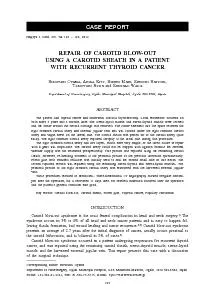
REPAIR OF CAROTID BLOWOUT strictly controlled after the operation and no antiplatelet drugs were administeredThreedimensional CT angiography showed irregular stenosis in the right common carotid ID: 229429

REPAIR CAROTID BLOW-OUT strictly controlled

Share:

Link:

Embed:

Download Presentation from below link

Download Pdf The PPT/PDF document "Hirofumi Oyama et al.the common carotid ..." is the property of its rightful owner. Permission is granted to download and print the materials on this web site for personal, non-commercial use only, and to display it on your personal computer provided you do not modify the materials and that you retain all copyright notices contained in the materials. By downloading content from our website, you accept the terms of this agreement.


Presentation Transcript

Hirofumi Oyama et al.the common carotid artery, obstruction could be avoided even after the thrombus was formed around the repaired sites. This procedure can be one option when an unexpected rupture of the carotid artery occurs.1)Pathak KA, Viallet NR, Nason RW. Sternocleidomastoid muscle interposition to prevent carotid artery blowout. J Surg Oncol, 2008; 98: 565–566.2)Mika H, Bumb P, Günther R. Rupture, emergency ligature and elective ligature of the carotid artery. Laryngol Rhinol Otol (Stuttg), 1982; 61: 634–638. [Article in German]Powitzky R, Vasan N, Krempl G, Medina J. Carotid blowout in patients with head and neck cancer. Otol Rhinol Laryngol, 2010; 119: 476–484.Kebebew E, Clark OH. Locally advanced differentiated thyroid cancer. Surg Oncol, 2003; 12: 91–99.Hayashi S. Histology of the human carotid sheath revisited. Okajimas Folia Anatomica Japonica, 2007; 84: 6)Chaloupka JC, Roth TC, Putman CM, Mitra S, Ross DA, Lowlicht RA, Sasaki CT. Recurrent carotid blowout syndrome: diagnostic and therapeutic challenges in a newly recognized subgroup of patients. J Neuroradiol, 1999; 20: 1069–1077.7)Okamura HO, Kamiyama R, Takiguchi Y, Kimizuka K, Ishikawa N, Kishimoto S. Histopathological examination of ruptured carotid artery after irradiation. ORL J Otorhinolaryngol Relat Spec, 2002; 64: 226–228.Zidar N, Ferluga D, Hvala A, Popovi M, Soba E. Contribution to the pathogenesis of radiation-induced injury to large arteries. J Laryngol Otol, 1997; 111: 988–990.9)Hoppe H, Barnwell SL, Nesbit GM, Petersen BD. Stent-grafts in the treatment of emergent or urgent carotid artery disease: review of 25 cases. J Vasc Interv Radiol, 2008; 19: 31–41. REPAIR OF CAROTID BLOW-OUT strictly controlled after the operation, and no anti-platelet drugs were administered.Three-dimensional CT angiography showed irregular stenosis in the right common carotid artery just after the re-operation (Fig. 3). Digital subtraction angiography also showed irregular stenosis in the right common carotid artery 5 days after the re-operation (Fig. 4). However, the stenosis was found to recover on magnetic resonance angiography 11 days later. No cerebral infarction occurred after the operation and the patient’s general condition was good. The pathological specimen revealed the papillary carcinoma which showed prominent invasion of the nervous system. The patient is in good condition without neurological complication, and recurrence of the tumor 22 months after the operation.Thyroid cancer frequently invades the recurrent laryngeal nerve, strap muscles and trachea, and it is followed by invasion of the esophagus, internal jugular vein and carotid artery. Surgical resection is the primary treatment for locally advanced thyroid cancer, and the carotid sheath invaded by a tumor must be excised with the thyroid tumor. During this procedure, the adventitia that encircles the carotid artery and jugular vein can easily be damaged.Carotid blow-out syndrome patients present with sentinel bleeding, but 60% of patients will develop a life-threatening hemorrhage requiring emergent intervention. The incidence is higher due to the lack of supporting healthy tissues if there is a history of radical neck dissection.Other risk factors for carotid blow-out syndrome include radiotherapy, ap necrosis, mucocutaneous stula, wound infection, and recurrent tumor invasion. A sevenfold increased incidence of carotid blow-out syndrome is found after radiotherapy. Injury to the vasa vasorum and necrotizing vasculitis in all three layers is an important factor in the development of radiation-induced vasculopathy of large arteries. Bacterial inammation can also cause thrombosis of the vasa vasorum and arterial wall injury. The carotid rupture in the current case was related to the previous radical neck dissection followed by the recurrent tumor invasion.Over 90% of patients with carotid blow-out syndrome are treated with endovascular therapy (embolization 56%, stenting 36%), and surgical ligation (7%) is rarely indicated. Carotid artery embolization has a 15% to 20% neurologic morbidity rate and a lower incidence of associated mortality. Endovascular embolization can also help predict cerebrovascular complications with temporary balloon occlusion and collateral cerebral blood ow analysis. Stent-grafts may be useful in the treatment of carotid blow-out syndrome and show a high procedural success rate in selected cases. However, stent placement has a higher risk of recurrent carotid blow-out syndrome (44%) than embolization therapy (10%).Emergency ligation of the carotid artery causes neurological casualties in 30% of the patients.Other studies of carotid blow-out syndrome treated with surgical ligation report an average mortality rate of 40% and an average morbidity rate of 60%. Although trapping with ligation was considered in the current case, trapping of the right common carotid artery was not done, because the cerebral vascular supply was not assessed preoperatively. Instead, reconstruction was done with the remaining carotid sheath and muscle aps. Fibrin glue was used to seal the space between the carotid artery and the surrounding covering ap. We thought that it was important to seal the ruptured portion in multiple layers using vital, vascularized tissue. Thus, the reconstruction was reinforced by covering the common carotid artery with the harvested external jugular vein and sternocleidomastoid muscle. This surgical technique was an emergency measure following the sudden rupture of the common carotid artery. We nally managed to control hemorrhaging without occluding the common carotid artery. Given the large diameter of Hirofumi Oyama et al. However, re-bleeding occurred spontaneously at the proximal portion of the previous laceration during the dissection of the opposite side. Fibrin glue with oxidized cellulose was initially used to seal the second small hole in this lesion (Fig. 2C). The ruptured section was then covered with the remaining sterno-thyroid muscle as the rst layer and the sterno-hyoid muscle as the second layer (Fig. 2C, D). These procedures resulted in hemostasis. The proximal portion of the right common carotid artery was covered with the harvested external jugular vein because there were no vascularized tissues surrounding this artery (Fig. 2E). Finally, the repaired right common carotid artery was widely covered with the sternomastoid muscle. Blood pressure was Fig. 3 Axial view of the enhanced CT scan just after operation. The carotid sheath used for the repairment was partially pushed into the arterial cavity (white arrow).Fig. 4 Right carotid angiography 5 days after operation. The irregularity of the wall still remained after the repair (black arrow). REPAIR OF CAROTID BLOW-OUT Fig. 2A: The carotid sheath (arrows) was sutured to the right common carotid artery at the distal end of the lacerated portion (arrowhead). Fig. 2B: The lacerated portion was covered with the remaining carotid sheath surrounding the right common carotid artery. Fig. 2CRe-bleeding occurred at the proximal portion (black arrow) of the previous laceration. Fibrin glue with oxidized cellulose (arrow head) was initially used to seal the second small hole (black arrow) in this lesion. The ruptured section was then covered with the remaining sterno-thyroid muscle (white arrow) as the rst layer. Fig. 2DThe ruptured section was then covered with the remaining sterno-hyoid muscle (white arrow) as the second layer. Black arrow shows the right common carotid artery. White arrowhead indicates the right vagus nerve. Black arrowhead shows the right internal jugular vein.Fig. 2EThe proximal portion of right common carotid artery (black arrow) was covered with the harvested external jugular vein (white arrow).Fig. 2Intraoperative view