/
Molecular diagnosis of hereditary nonpolyposis colorectal cancer Lync Molecular diagnosis of hereditary nonpolyposis colorectal cancer Lync

Molecular diagnosis of hereditary nonpolyposis colorectal cancer Lync - PDF document

thomas
thomas . @thomas
Follow
344 views
Uploaded On 2022-10-14

Molecular diagnosis of hereditary nonpolyposis colorectal cancer Lync - PPT Presentation

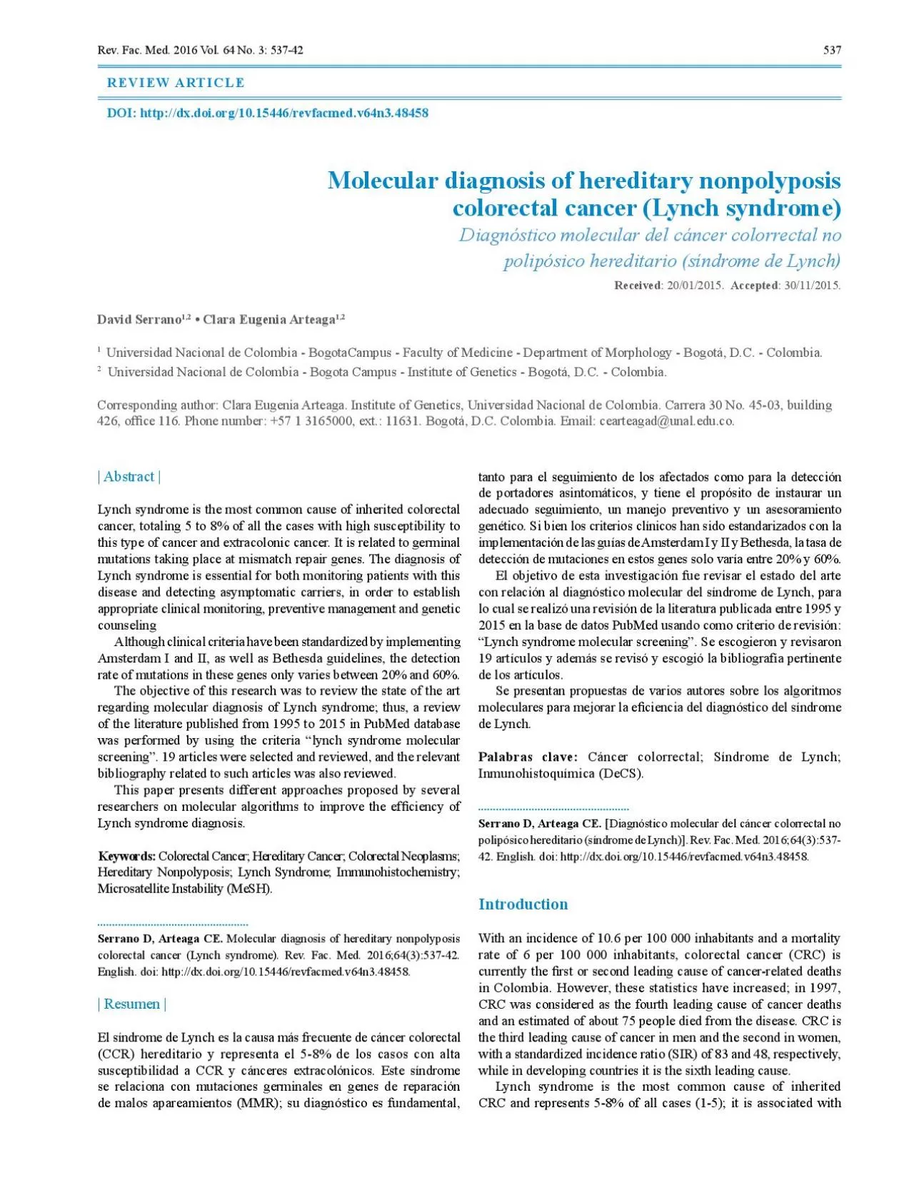
537 Abstract Lynch syndrome is the most common cause of inherited colorectal cancer totaling 5 to 8 of all the cases with high susceptibility to this type of cancer and extracolonic cancer It is ID: 960050

lynch cancer http syndrome cancer lynch syndrome http org doi colorectal genes mutations msh2 x00660069 mlh1 msi mmr microsatellite

Share:

Link:

Embed:

Download Presentation from below link

Download Pdf The PPT/PDF document "Molecular diagnosis of hereditary nonpol..." is the property of its rightful owner. Permission is granted to download and print the materials on this web site for personal, non-commercial use only, and to display it on your personal computer provided you do not modify the materials and that you retain all copyright notices contained in the materials. By downloading content from our website, you accept the terms of this agreement.


Presentation Transcript

537 Molecular diagnosis of hereditary nonpolyposis colorectal cancer (Lynch syndrome)Diagnóstico molecular del cáncer colorrectal no polipósico hereditario (síndrome de Lynch): 30/11/2015. • Clara Eugenia Arteaga Universidad Nacional de Colombia - BogotaCampus - Faculty of Medicine - Department of Morphology - Bogotá, D.C. - Colombia.Corresponding author: Clara Eugenia Arteaga. Institute of Genetics, Universidad Nacional de Colombia. Carrera 30 No. 45-03, building | Abstract |Lynch syndrome is the most common cause of inherited colorectal cancer, totaling 5 to 8% of all the cases with high susceptibility to this type of cancer and extracolonic cancer. It is related to germinal mutations taking place at mismatch repair genes. The diagnosis of Lynch syndrome is essential for both monitoring patients with this disease and detecting asymptomatic carriers, in order to establish appropriate clinical monitoring, preventive management and genetic Although clinical criteria have been standardized by implementing tanto para el seguimiento de los afectados como para la detección de portadores asintomáticos, y tiene el propósito de instaurar un adecuado seguimiento, un manejo preventivo y un asesoramiento genético. Si bien los criterios clínicos han sido estandarizados con la implementación de las guías de Amsterdam I y II y Bethesda, la tasa de detección de mutaciones en estos genes solo varía entre 20% y 60%. Rev. Fac. Med. 2016 Vol. 64 No. 3: 537-42 538 mutations in the germline of MMR genes including hMLH1, hMSH2, hMSH6, hPMS2, hPMS1 and hMLH3, which generate microsatellite instability in most cases. About 90% of the mutations identi�ed in this group correspond to hMLH1 (50%) and hMSH2 and MSH2 genes in Colombian families, the detection rate for this Clinical featuresLynch syndrome is a hereditary cancer syndrome with an autosomal dominant inheritance pattern. 40-60% of families who meet the clinical criteria for this disease have mutations in MMR genes. The risk of developing cancer among mutation carriers is 80% at age 70, and the average age for the onset of a neoplastic lesion, either in the colon or outside the colon, is 45, much earlier than in sporadic cancer; however, the risk of cancer and age of onset is different for each of the genes involved (8,9). Some studies suggest that mutations in MLH1 are at increased risk of CRC in MSH2, which have increased risk of extracolonic cancers. Mutations in MSH6, when compared to MLH1 and MSH2, show a lower expression of From the anatomopathological point of view, adenocarcinomas framed in this syndrome are characterized for being solid, poorly differentiated, mucinoide-like, with signet ring cells and peritumoral lymphocytic in�ltration —similar to the in�ltration observed in Crohn’s disease—, which is currently considered as a prognostic marker. The most common site of lesion is the proximal colon and the number of adenomas varies slightly with villous growth (8,11).Extracolonic malignancies that occur in Lynch syndrome include endometrial, stomach, ovary, ureter, renal pelvis, brain, small intestine, hepatobiliary and skin (sebaceous adenoma) cancers. These tumors can be synchronous or metachronous. Regarding extracolonic tumors, endometrial cancer predominates in Western countries and gastric cancer in Eastern countries (12); less frequently, cases of breast cancer have also been reported (13).In clinical practice, the diagnosis of Lynch syndrome is mainly based on Amsterdam I criteria. Selecting families using these criteria allows a mutation detection rate of about 60% (14) (Table 1).Table 1. Amsterdam I Criteria. At least three members of the family should be affected with CRC and the following criteria must be met:First degree of consanguinity in at least two affected Tumor verification through pathology testsCRC: colorectal cancer. Source: Own elaboration based on data obtained from Lynch Amsterdam II criteria were proposed later, in 1999, because the �rst classi�cation did not include extracolonic tumors, which are part of the phenotype of hereditary nonpolyposis colorectal cancer (HNPCC) (Table 2). Today, the revised Bethesda criteria, established by the US National Cancer Institute, are also taken into account, which allows selection of patients by determining the microsatellite instability (15) (Table 3).Table 2. Amsterdam II criteria. At least three members of a family with hereditary nonpolyposis or associated (endometrial, small bowel, ureter or renal pelvis cancer) colorectal cancer and the following criteria must be met:First degree of consanguinity in at least two of the Tumors verification through histopathology testsCRC: colorectal cancer.Source: Own elaboration based on data obtained from Allen . (18).Table 3. Tumors of individuals should be screened for microsatellite instability in the following situations:Presence of synchronous, metachronous or other HNPCC-CRC: colorectal cancer; HNPCC: hereditary nonpolyposis colorectal cancer; MSI: Microsatellite instability.Source: Own elaboration based on data obtai

ned from Asad Umar (15).Repair systemsRepair systems are crucial for maintaining the integrity of the genome. The mismatch system repair increases �delity of replication by a factor of 1 000 correcting errors generated during this event. The process begins by recognizing the alteration of DNA and continues with the repair of the defect. The mismatches are caused by errors during replication and recombination, through the generation of small insertions or deletions or physical damage of DNA caused by deamination or cytosine methylation. The best studied system is mutHLS in Escherichia coli; here, mismatch recognition is performed by the MutS protein with ATPase function (16-19).The MMR has evolved to correct errors that are beyond 3’→ 5’ exonuclease correction activity of DNA polymerases. The process begins with the recognition of the mismatch caused by the binding of the hMSH2/hMSH6 heterodimer, also known as hMutSa; this complex undergoes a conformational change promoted by ATP which turns it into a clamp, which is displaceable through the DNA strand and, then, recruits the hMLH1 / hPMS2 heterodimer, also This ternary complex can move in any direction along DNA, and when it encounters the broken chain that is subjected to PCNA loading an exonuclease 5 ‘→ 3’ (EXO1), degradation of the thread starts towards the site where the mismatch is located. The chain that is not degraded, is stabilized by replication protein A (RPA) preventing the action of EXO1. When the lesion is removed, the 539 degraded region is again synthetized by DNA polymerase then the ends are joined by DNA ligase action (20,21) (Figure 1). Mismatch repair process. Source: Own elaboration based on data MLH1 gene is located in the chromosomal region 3p21.3, with a length of 2752pb and 19 exons, and encodes a protein of 756 amino acids with a conserved region of 300 amino acids in its N-terminal end and 27 splicing variants. Location of protein is intranuclear MutL and hMLH1 are members of the GHKL ATPase/kinase superfamily including gyrase, type II topoisomerase, Hsp90 and histidine kinase. The hMLH1 protein has three domains of importance: a ATPase domain at the N-terminal region, an interaction domain with MutS that has another �exible and poorly preserved hinge region, and a domain in the C-terminal region (CTD) involved in homo- and heterodimerization (17). Protein produces heterodimers with hPMS2 proteins, forming the MutLa and hMLH3 complex, and also the MutLb The hMLH1 protein is part of the surveillance genome complex known as BASC, which includes BRCA1, BLM and ATM proteins, and RAD50-MRE-NBS1, MSH2, MSH6 and MLH1 complexes. They intervene as control points during the cell cycle in the presence of DNA damage (23).MSH2 gene is located in the region 2p21, is 3307pb long and has 16 exons, encodes a protein of 934 amino acids and has 13 splicing variants. The localization of the protein is intranuclear (22) and contains �ve domains: domain I DNA binding, domain II interaction with hMSH3 and hMSH6, domain III of ATP binding, domain IV of interaction with homologous MutL, and domain V of ATPase function (24). The protein forms two heterodimers: MutSa, Mismatch Displaceable clampExonucleaseResynthesisG5’3’T 3’ Recognition ADPADPATPATPhMSH2hMSH65’3’3’ PCNADNAPolymerase5’3’ GT 5’5’3’ TA Exonuclease IDivided nucleotides5’5’3’ consisting of MSH2/MSH6, which is more involved in the repair As mentioned above, the MSH2 protein is also part of the genome surveillance complex and acts as a potential damage sensor Microsatellite instabilityMicrosatellites correspond to short repetitive sequences of 1-4 nucleotides, generally adjacent to coding regions; they are also known as short tandem repeats (STR). Microsatellite instability (MSI) is de�ned as a change in length of the repeat units due to an insertion/deletion of one or more of these units. This can occur in a microsatellite tumor tissue when compared to normal tissue from the same patient (25). When there is damage in MMR, microsatellites tend to change the number of repetitions. About 70-90% of cases of HNPCC show positive MSI, which is even higher in families with mutation in any of the MMR genes, whereas, in sporadic colorectal tumors, it is only observed in 10-15% of cases (26); therefore, instability is a relatively In 1998, the National Cancer Institute of the United States proposed a panel of �ve markers for MSI analysis: mononucleotide in repeats BAT25 and BAT26 and dinucleotide repeats in D2S123, D5S346 and D17S250. A tumor is graded as high MSI (MSI-H), if two or more markers are altered; mild or low microsatellite instability (MSI-L), if a marker is altered, and stability (MSS) if markers are not altered (Table 4).Table 4. Criteria interpretation of microsatellite instability. Number of unstable markersPercentage of unstable markersInterpretationSource: Own elaboration based on data obtained from Boland BAT26 is extremely sensitive for detecting tumors with instability and shows an insignif

0069;cant size variation, either between two alleles Colorectal tumors with MSI-H are found predominantly in the proximal colon, have histopathologically mucinous appearance and may be resistant to cytotoxicity induced by chemotherapeutic agents. Microsatellite instability varies from adenoma to adenocarcinoma and, then, to metastatic tumor. MSI testing has 80-91% sensitivity in patients with mutations in the MLH1 or MSH2 gene and of 55-77% in MSH6 or PMS2; MMR gene and somatic mutations in Lynch syndromeThe MMR genes have a role in genome maintenance. The presence of a mutation in some of these genes, mainly MLH1 or MSH2, triggers a cascade of events that affect genes with tandem repeats in their sequence. The repetitive sequences are highly susceptible to misalignment during the replication process, resulting in an increase Repetitive sequences are dispersed throughout the genome; a large number of human genes have mononucleotide repeats, therefore, they are possible targets of change in the frameshif during replication, which, in turn, generates truncated proteins. These genes, 540 which include MMR, MED-1 and RAD50, are usually involved in signal transduction (TGFβ-RII, IGFIIR, PTEN), apoptosis and in�ammation (BAX, caspase-5), transcription regulation (E2F4, Among the most important genes susceptible to mutations, gene TGFβRII (receptor II transformer of growth factor β) is found, which contains an adenine (A) repetition tract (7) found in 75-90% of patients with MSI, for both HNPCC and sporadic colon cancer. If the inactivation of one of the receptors occurs, the cells lose their responsiveness to TGFβ and cell growth, which represents an important milestone in tumorigenesis of various cancers such as stomach, neck and prostate. In colon cancer, this inactivation corresponds to an event that occurs early during the transition from adenoma to carcinoma. The inactivation of TGFβRII occurs frequently in MSI+ gastric tumors, but Insertion/deletion mutations in repetitive mononucleotide regions are located in BAX (G8), TCF-4 (A9), IGFIIR (G8) and hMSH6 (C8) genes. Such mutations also occur at a signi�cant rate in MSI+ colorectal tumors. Other genes such as caspase-5 (A10), hMSH3 (A8) and RAD50 (A9) are inactivated, with a lower frequency, in primary tumors, but show a high incidence of frameshift mutations In a study by Yamaguchi et al., the frequency of frameshift mutations in genes ACVR2 (activin receptor 2) and TGFβ -RII was between 70-95% for HNPCC. The signaling pathway of TGFβRII downstream includes Smad 2, 3 and 4 proteins, with subsequent inhibition of cell growth; this pathway may be disrupted by mutations in ACVR2. The Wnt pathway is also affected by disruption caused by mutations in APC. Other genes less involved in the early stages of tumor development are PTHLH, MARCKS, hMSH3, TCF4, CASP5, Molecular analysis for the diagnosis of Lynch syndromeStudies have been performed worldwide to determine the presence of mutations that predispose to Lynch syndrome, and about 400 genes in all MMR have been identi�ed; 90% are in MLH1 and MSH2. The �rst publications about Lynch syndrome indicate that age 44 to 45 is the average for CRC installation. However, it is now clear that not all cases appear at such a young age and this can vary according to the type of mutation which may not differ from sporadic cases, and could explain the lack of sensitivity of The most frequent mutations in MLH1 and MSH2 genes are nonsense, missense, frameshift and splicing site changes, while the proportion of genome rearrangements varies in each population from 5% to 20% on average; there are also low frequency cases (1.5%) and, others with higher frequency due to a founder effect (12,31,32), Conventional methods have been used for detecting mutations as single stranded conformational polymorphism (SCPP), denaturing gradient gel electrophoresis (DGGE), conformation sensitive gel electrophoresis (CSGE), denaturing high performance liquid chromatography (DHPLC) and direct sequencing of the gene. Since large genome rearrangements are not detected by these methods, the analysis by MLPA (Multiplex Ligation-Dependent Probe Ampli�cation) is used, which allows semi-quantitatively assess the change number of copies in a speci�c region of the gene. The combination of techniques allows a better characterization of the Given the high cost of molecular screening for all MMR genes in suspected families for Lynch syndrome, several pretest strategies, such as determining the degree of instability in tumors, have been suggested. The MSI phenotype is a speci�c marker useful in HNPCC. However, this can also be observed in sporadic cancers, since about 15% of them are caused by somatic mutations, loss of heterozygosity of MMR genes and promoter methylation of the gene MLH1; this situation is also observed in a study of hypermethylation of MLH1 promoter, predominantly in women The promoter methylation status is suggested as a marker to distinguish sporadic tumors from hereditary tumors. The discrimination of these

two subtypes may improve detection strategy, therapy and prevention. In order to standardize the MSI tests, a working group from the National Cancer Institute in the United States recommended the use of the Bethesda panel with �ve markers, two single nucleotide and three dinucleotide (38,39). In this regard, Pedroni (40) demonstrate that a two mononucleotide markers panel (BAT 25 and BAT 26) is more ef�cient in detecting tumors with high MSI in the absence of MMR proteins compared with the Bethesda panel (93% vs. 54%). Another initial approach method is the demonstration of the absence of MMR protein expression through immunohistochemical staining (IHC). It has been proved that IHC for MMR proteins (MLH1, PMS2, MSH2 y MSH6) provides a faster, cost-effective, sensitive and very speci�c screening technique for MSI. Some studies compare methods for determining MSI based on PCR and IHC assessment for MMR protein expression is performed on tissue sections containing both tumor tissue and normal colonic mucosa. Monoclonal antibodies of mice are used in different dilutions for the total length of proteins. Normal tissue and lymphocytes adjacent to the respective tumor are used as positive internal controls and the loss of protein expression is de�ned as the complete absence of nuclear staining in tumor cells, but maintained (20) described a group of patients with symptoms compatible with Lynch syndrome in absence of IHC expression of the MSH2 protein and had no detectable mutations in this gene; individuals had deletion of epithelial cell adhesion molecule gene (EpCAM) located upstream of MSH2. In this study, it was also established that this deletion leads to somatic hypermethylation of MSH2 and loss of protein expression. The silencing of MSH2 gene promoter by deletions of gene EPCAM causes Lynch syndrome in 20-25% of patients with IHC negative for MSH2 and in whom no mutation is found in the germline, which corresponds to 2-3% of all patients with this syndrome (20). Studies have shown that a negative IHC for EpCAM with negative MSH2 indicates deletion Lynch (46) suggest that patients with suspected Lynch syndrome should initially have a test for MSI and then immunohistochemistry tests for MMR proteins, and molecular tests for the negative gene of IHC. In addition, the authors recommend that patients with clinical criteria for Lynch syndrome, even in the absence of germinal mutations for MMR, In 2011, a survey conducted by the National Society of Genetic Counselors to evaluate screening programs for Lynch syndrome and barriers for their implementation showed that more than 50% of �rst case of colon or endometrial cancer in the family appeared. Screening methods in tumor tissue varied in 64.2% when the study initiated with IHC testing, 20.8% with MSI testing and 15% with both tests simultaneously. Also, with the results of the survey, the cost of testing and the lack of medical information were deemed as 541 Moreover, taking into account that only up to 80% of germline mutations are detected in despite of the Amsterdam and Bethesda clinical criteria, there are increasingly strong trends that support universal screening of all newly diagnosed cases with CRC and The debate still lingers regarding the methods used to start screening for Lynch syndrome, as several authors propose IHC as the �rst test due to its cost-effectiveness and because the absence of protein expression can be detected in both CRC as endometrial cancer. However, as mentioned above, a percentage of tumors with absence of MLH1 and MSH2 expression may relate to somatic events such as promoter hypermethylation or BRAF V600E mutation. Cost-effectiveness data suggest that the best strategy for these cases is to follow IHC test with BRAF mutation or hypermethylation of MLH1 (49) suggest that, for patients with high microsatellite instability, somatic BRAF V600E mutation should be considered as a pre-molecular study of MMR genes, because this alteration is much more related to sporadic tumors and, thus, Lynch syndrome can be discarded. Similarly, the use of multiplex ligation-dependent probe ampli�cation (MLPA) to detect large rearrangements (50) propose a diagnostic strategy for Lynch syndrome that starts with �nding MSI, uses the panel of �ve mononucleotide markers, and searches for speci�c high prevalence mutations, comprehensive determination of mutations in the MLH1 and MSH2 by sequencing and techniques for large MLPA rearrangements. All of this should be done before searching in other genes, including MSH6 and PMS2, through the MLPA technique (50).The molecular diagnosis of Lynch syndrome is essential to locate affected individuals and carriers in families and to provide adequate monitoring and genetic counseling. It is necessary to gather evidence on the cost-effectiveness of making universal screening on CRC or to start the process with the use of clinical guidelines, determining the degree of microsatellite instability and IHC, and with this result, determining the next step f

or sequencing and search of large rearrangements in the MLH1 and MSH2 genes, and subsequently Con�ict of interestsThis study was supported through the call for encouragement to research projects and research in graduate programs of the Faculty of Medicine of Universidad Nacional de Colombia (health research References Jemal A, Siegel R, Xu J, Ward E.CA Cancer 2010;60(5):277-300. http://doi.org/bhfrgh.Peto J. Cancer epidemiology in the last century and the next decade. Nature. 2001;411(6835):390-5. http://doi.org/cxcvxb.Ferlay J, Shin HR, Bray F, Forman D, Mathers C, Parkin DM. GLOBOCAN 2008: Cancer incidence and mortality worldwide. International Lagerstedt Robinson K, Liu T, Vandrovcova J, Halvarsson B, Clendenning M, Frebourg T, Lynch syndrome (hereditary nonpolyposis colorectal cancer) diagnostics. http://doi.org/cgsq7t.Giraldo A, Gómez A, Salguero G, García H, Aristizábal F, Gutiérrez tary nonpolyposis colorectal cancer (Lynch syndrome)–description of four novel mutations. Fam. Cancer. 2005;4(4):285-90. http://doi.org/crkvqg.Montazer Haghighi M, Radpour R, Aghajani K, Zali N, Molaei Four novel germline mutations in the MLH1 and PMS2 mismatch repair genes in patients with hereditary nonpolyposis colorectal cancer. Int. J. Colorectal Dis. 2009;24(8):885-93. http://doi.org/b5fz2f.Gómez A, Salguero G, García H, Aristizábal F, Gutiérrez O, Ángel LA, et al. Detección de mutaciones de los genes hMLH1 y hMSH2 del sistema de reparación de malos apareamientos del ADN en familias colombianas sospechosas de cáncer colorrectal no polipósico hereditario (síndrome de Lynch). . 2005;25(3):315-24. http://doi.org/bm49.Vasen HF. Review article: the Lynch syndrome (hereditary nonpolyposis colorectal cancer). Aliment. Pharmacol. Ther. 2007;26(Suppl 2):113-26. http://doi.org/cq93d2.Lindor NM, Petersen GM, Hadley DW, Kinney AY, Miesfeldt S, Recommendations for the care of individuals with an inherited predisposition to Lynch syndrome: a systematic review. JAMA. 2006;296(12):1507-17. http://doi.org/dkcdvz.Lynch HT, Boland CR, Gong G, Shaw TG, Lynch PM, Fodde R, Phenotypic and genotypic heterogeneity in the Lynch syndrome: Eur. J. Hum. 2006;14(4):390-402. http://doi.org/ch3tdb.11.Lynch HT, Smyrk T, Lynch J. An update of HNPCC (Lynch syndrome). 1997;93(1):84-99. http://doi.org/b9cmk2.Tang R, Hsiung C, Wang JY, Lai CH, Chien HT, Chiu LL, Germ line MLH1 and MSH2 mutations in Taiwanese Lynch syndrome families: characterization of a founder genomic mutation in the MLH1 2009;75(4):334-45. http://doi.org/ft4gds.Bianchi F, Raponi M, Piva F, Viel A, Bearzi I, Galizia E, intronic mutation in MLH1 associated with familial colon and breast cancer. Fam. Cancer. 2011;10(1):27-35. http://doi.org/fmtt2v.Vasen HF, Watson P, Mecklin J, Lynch HT. New clinical criteria for hereditary nonpolyposis colorectal cancer (HNPCC, Lynch syndrome) proposed by the International Collaborative group on HNPCC. Gastroenterology. 1999;116(6):1453-6. http://doi.org/d7g7rn.Umar A, Boland CR, Terdiman JP, Syngal S, de la Chapelle A, Rüs Revised Bethesda Guidelines for hereditary nonpolyposis colorectal cancer (Lynch syndrome) and microsatellite instability. J. Natl. 2004;96(4):261-8. http://doi.org/b55tnc.Guerrette S, Acharya S, Fishel R. The interaction of the human MutL homologues in hereditary nonpolyposis colon cancer. J. Biol. Chem. 1999;274(10):6336-41. http://doi.org/fq534h.Kosinski J, Steindorf I, Bujnicki JM, Giron-Monzon L, Friedhoff P. Analysis of the quaternary structure of the MutL C-terminal domain. 2005;351(4):895-909. http://doi.org/dr7zd4.Allen DJ, Makhov A, Grilley M, Taylor J, Thresher R, Modrich P, et al. MutS mediates heteroduplex loop formation by a translocation 1997;16(14):4467-76. http://doi.org/b4sv2c. 542 Jacob S, Praz F. DNA mismatch repair defects: role in colorectal carci 2002;84(1):27-47. http://doi.org/dmkr8m.Stojic L, Brun R, Jiricny J. Mismatch repair and DNA damage signaDNA Repair. 2004;3(8-9):1091-101. http://doi.org/bn2kxv.Hsieh P. Molecular mechanisms of DNA mismatch repair. Mutat. Res. Repair. 2001;486(2):71-87. http://doi.org/bjvtrh.Ensembl Genome Browser. Cambridge: Ensembl; [cited 2015 Jun 23]. Available from: http://goo.gl/vm8zth. Wang Y, Cortez D, Yazdi P, Neff N, Elledge SJ, Qin J. BASC, a super complex of BRCA1-associated proteins involved in the recognition and repair of aberrant DNA structures. Genes Dev.Lamers MH, Perrakis A, Enzlin JH, Winterwerp HH, de Wind N, Sixma The crystal structure of DNA mismatch repair protein MutS binding to a G·x T mismatch. Nature. 2000;407(6805):711-7. http://doi.org/dc5qt2.Boland CR, Thibodeau SN, Hamilton SR, Sidransky D, Eshleman JR, Burt RW, A National Cancer Institute Workshop on Microsatellite Instability for cancer detection and familial predisposition: development of international criteria for the determination of microsatellite instability in colorectal cancer. Loukola A, Eklin K, Laiho P, Salovaara R, Kristo P, Järvinen H, et al. Microsatellite marker analysis in screening for hereditary nonpolyposis 2001;61(11):4545-9. Evaluation of Genomic Applications in Practice and Prevention

(EGAPP) Working Group. Recommendations from the EGAPP Working Group: genetic testing strategies in newly diagnosed individuals with colorectal cancer aimed at reducing morbidity and mortality from Lynch syndrome 2009;11(1):35-41. http://doi.org/drhv5s.Yamaguchi T, Iijima T, Mori T, Takahashi K, Matsumoto H, Miyamo Accumulation profile of frameshift mutations during development and progression of colorectal cancer from patients with hereditary nonpolyposis colorectal cancer. 2006;49(3):399-406. http://doi.org/bw6xqj. Current Lynch Syndrome Tumor Screening Practices: A Survey of Genetic Counselors. J. Genet. Counsel. 2014;23(1):38-47. http://doi.org/bm5b.Castellví-Bel S, Castells A, Strunk M, Ferrández A, Piazuelo E, Milà Genomic rearrangements in MSH2 and MLH1 are rare mutacancer. 2005;225(1):93-8. http://doi.org/b65cqv.Grabowski M, Mueller-Koch Y, Grasbon-Frodl E, Koehler U, Keller G, Vogelsang H, et al. Deletions account for 17% of pathogenic germline alterations in MLH1 and MSH2 in hereditary nonpolyposis colorectal cancer (HNPCC) families. Genet. Test. 2005;9(2):138-46. http://doi.org/cst6b3.Martínez-Bouzas C, Ojembarrena E, Beristain E, Errasti J, Viguera N, Tejada-Minguéz MI. High proportion of large genomic rearrangements in hMSH2 in hereditary nonpolyposis colorectal cancer (HNPCC) families of the Basque Country. 2007;255(2):295-9. http://doi.org/dqbgxk.Zavodna K, Krivulcik T, Bujalkova MG, Slamka T, Martinicky D, Partial loss of heterozygosity events at the mutated gene in tumors from MLH1/MSH2 large genomic rearrangement carriers. BMC Cancer. 2009;9(1):405. http://doi.org/b7n5fq.Fridrichova I. New aspects in molecular diagnosis of Lynch syndrome Perez-Cabornero L, Velasco E, Infante M, Sanz D, Lastra A new strategy to screen MMR genes in Lynch Syndrome: HA-CAE, MLPA and RT-PCR. Eur. J. Cancer. 2009;45(8):1485-93. Thodi G, Fostira F, Sandaltzopoulos R, Nasioulas G, Grivas A, Boukovinas I, et al. Screening of the DNA mismatch repair genes MLH1, MSH2 and MSH6 in a Greek cohort of Lynch syndrome suspected families. BMC Cancer. 2010;10(1):544. http://doi.org/bxqjpp.Chamorro ME. Metilación del gen MLH1 e inestabilidad de microsatélites en una serie de pacientes con cáncer colorrectal [Tesis de Maestría]. Tranø G, Sjursen W, Wasmuth HH, Hofsli E, Vatten LJ. Performance of clinical guidelines compared with molecular tumour screening methods in identifying possible Lynch syndrome among colorectal cancer patients: a Norwegian population-based study. Br. J. Cancer.http://doi.org/cdxtwz.Velasco E, Infante M, Durán M, Pérez-Cabornero L, Sanz DJ, Esteban-Cardeñosa E, Heteroduplex analysis by capillary array electrophoresis for rapid mutation detection in large multiexon genes. Nat. Protoc. 2007;2(1):237-46. http://doi.org/b6r45d.Pedroni M, Roncari B, Maffei S, Losi L, Scarselli A, Di Gregorio C, et A mononucleotide markers panel to identify hMLH1/hMSH2 germline 2007;23(3):179-87. http://doi.org/bm5t. Era of universal testing of microsatellite instability in colorectal cancer. World J. Gastrointest. Oncol. 2013 [cited 2015 Jun 23]; 5(2):12-19 Available from: URL: http://goo.gl/ZfkKOf.Bartley AN, Luthra R, Saraiya DS, Urbauer DL, Broaddus RR.Identification of cancer patients with Lynch syndrome: clinically significant discordances and problems in tissue-based mismatch repair testing. Cancer Prev. Res. (Phila Pa). 2012;5(2):320-7. http://doi.org/b8h6qp.Rigau V, Sebbagh N, Olschwang S, Paraf F, Mourra N, Parc Y, Microsatellite instability in colorectal carcinoma. The comparison of immunohistochemistry and molecular biology suggests role for hMLH6 Arch. Pathol. Lab. Med.Musulen E, Blanco I, Carrato C, Fernandez-Figueras MT, Pineda et al. Usefulness of epithelial cell adhesion molecule expression in the algorithmic approach to Lynch syndrome identification. 2013;44(3):412-6. http://doi.org/bm5v.Ligtenberg MJ, Kuiper RP, Chan TL, Goossens M, Hebeda KM, Voorendt M, Heritable somatic methylation and inactivation of MSH2 in families with Lynch syndrome due to deletion of the 3′ exons of TACSTD1. 2009;41(1):112-7. http://doi.org/fw4jkz.Lynch HT, Lynch JF, Lynch PM, Attard T. Hereditary colorectal cancer syndromes: molecular genetics, genetic counseling, diagnosis Fam. Cancer. 2008;7(1):27-39. http://doi.org/fv6pcf.Hampel H, Frankel WL, Martin E, Arnold M, Khanduja K, Kuebler P, Feasibility of Screening for Lynch Syndrome Among Patients With Colorectal Cancer. J. Clin. Oncol. 2008;26(35):5783-8. http://doi.org/fb869f.Gudgeon JM, Williams JL, Burt RW, Samowitz WS, Snow GL, Williams MS. Lynch Syndrome Screening Implementation: Business Analysis by a Healthcare System. Am. J. Manag. Care. 2011;17(8):e288-300.Shi C, Washington K. Molecular testing in colorectal cáncer: diagnosis Am. J. Clin. Pathol. 2012;137(6):847-59. http://doi.org/bm5w.Liu Y, Chew MH, Goh XW, Tan SY, Loi CT, Tan YM, et al. Systematic Study on Genetic and Epimutational Profile of a Cohort of Amsterdam Criteria-Defined Lynch Syndrome in Singapore. PloS One. 2014;9(4):e94170. http://doi.org/bm5z. Molecular diagnosis of Lynch syndrome: 537-42 Rev. Fac. Med. 2016 Vol. 64 No. 3: 53