/
Key Area 1.3 Membrane Proteins Key Area 1.3 Membrane Proteins

Key Area 1.3 Membrane Proteins - PowerPoint Presentation

tracy
tracy . @tracy
Follow
64 views
Uploaded On 2024-01-29

Key Area 1.3 Membrane Proteins - PPT Presentation

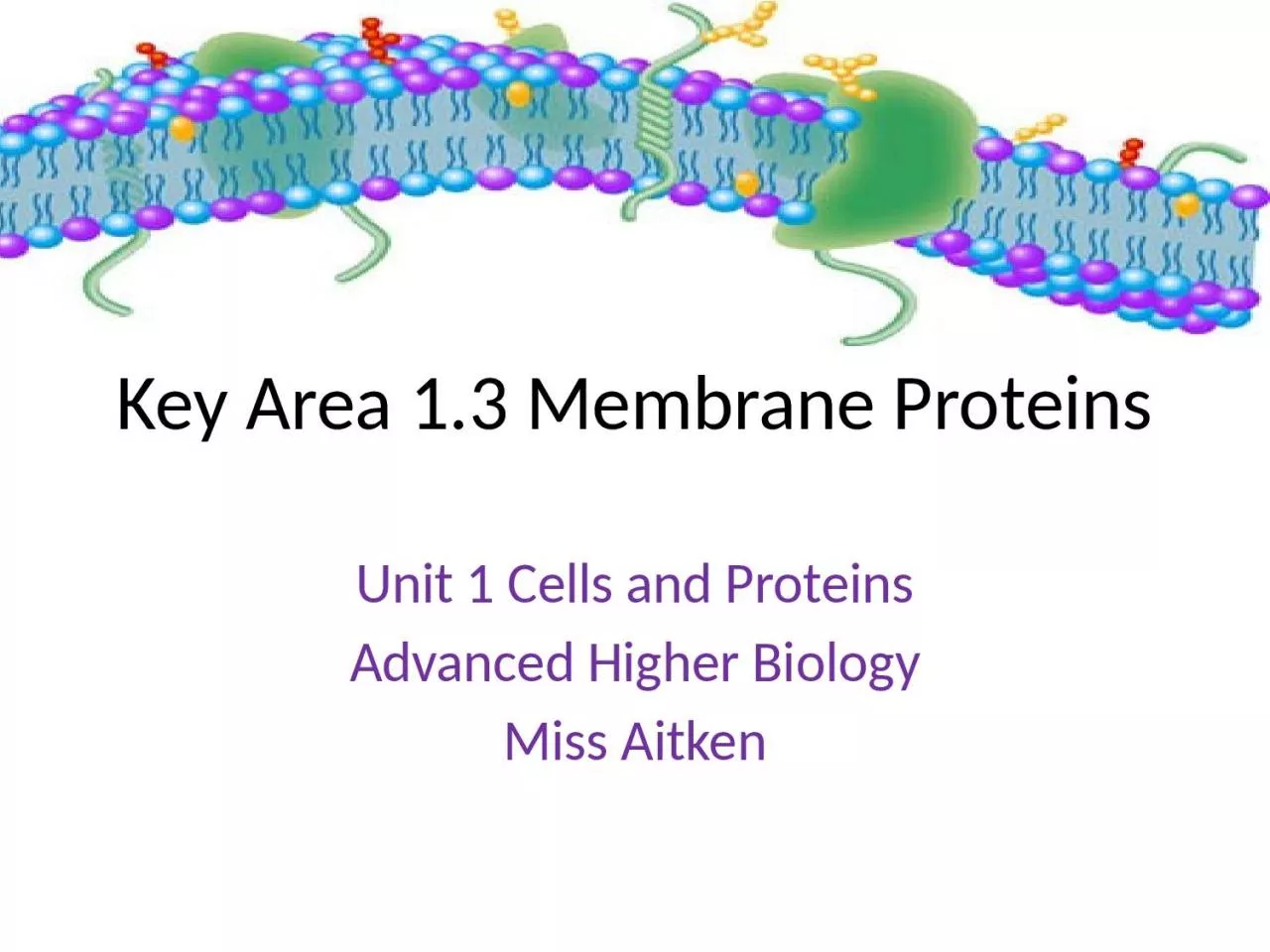
Unit 1 Cells and Proteins Advanced Higher Biology Miss Aitken Recap on the Cell Membrane In N5 we learned that the membrane of cells is made up of a bilayer of phospholipids with proteins embedded in the layers ID: 1042750

sodium membrane potassium ions membrane sodium ions potassium proteins cell pump molecules channel protein integral transporter ion pumpstep gradient

Share:

Link:

Embed:

Download Presentation from below link

Download Presentation The PPT/PDF document "Key Area 1.3 Membrane Proteins" is the property of its rightful owner. Permission is granted to download and print the materials on this web site for personal, non-commercial use only, and to display it on your personal computer provided you do not modify the materials and that you retain all copyright notices contained in the materials. By downloading content from our website, you accept the terms of this agreement.


Presentation Transcript

1. Key Area 1.3 Membrane ProteinsUnit 1 Cells and ProteinsAdvanced Higher BiologyMiss Aitken

2. Recap on the Cell Membrane In N5, we learned that the membrane of cells is made up of a bi-layer of phospholipids with proteins embedded in the layers.A phospholipid molecule has a hydrophilic head and a hydrophobic tail.

3.

4. Membrane ProteinsThere are two types of proteins found in the cell membrane:Integral proteinsPeripheral proteins

5. Integral ProteinsPenetrate the hydrophobic interior and form strong hydrophobic interactions with regions of hydrophobic R groups. Some only extend partly through the membrane and some are transmembrane proteins (span the width of the membrane).

6. Peripheral ProteinsNot embedded in the membrane – instead they form ionic and hydrogen bonds to the surface of the membrane. Have hydrophillic R groups on their surfaceMany peripheral proteins interact with the surfaces of integral proteins.

7. Membrane proteinsReworded slightly to help you understand and remember:“The main difference between integral and peripheral proteins is that integral proteins are embedded in the whole bilayer whereas peripheral proteins are located on the inner or outer surface of the phospholipid bilayer.”

8.

9. What can pass through the cell membrane directly?Oxygen (by diffusion)Carbon dioxide (by diffusion)Hydrophobic signalling moleculesThyroxineSteroidsOnly non-polar molecules can pass through the cell membrane directly.

10. What cannot pass through the cell membrane directly?Charged ionsH+ and Na+Hydrophilic amino acidsPolar moleculesWaterGlucoseThese molecules require an alternative route

11. Facilitated DiffusionDEFINITION: The passive transport of substances across the membrane through specific transmembrane proteins.

12. How do molecules cross the cell membrane?To perform specialised functions, different types of cell have different types of proteins in their cell membrane to help move molecules across.

13. Channel ProteinsA type of integral proteinThese proteins function by having a hydrophillic channel that certain molecules or ions can use as a tunnel through the membrane. Most channels are highly selective – they only let one type of molecule through.For example, the channel protein aquaporin facilitates the passage of water molecules.

14. Channel ProteinsSome channel proteins can be opened or closed to allow/disallow diffusion of a molecule/ion. These are called gated channels – there are 2 types.Ligand-gated: A signal molecule binds to the channel protein and changes its shape/opens or closes gate.Voltage-gated: A change in ion concentration across the cell membrane causes a change in the channel protein and it opens or closes.

15. Transporter ProteinsAnother type of integral proteinSometimes called carrier proteinsSpecific to one substanceThese proteins hold onto their passengers and change their shape in a way that shuttles them across the membrane. Transporters alternate between two conformations so that the binding site is exposed to one side of the membrane, then the other.

16. Movement of Ions and Molecules Across MembranesIons/Polar molecules must go through a protein channel in order to pass through the cell membraneThere are specific transmembrane proteins which act as sodium channels etc.Some transmembrane proteins control the concentration gradient across a membrane by controlling numbers of ions on either side.

17. Transporter ProteinsAlso involved in facilitated diffusionAlso specific to one type of ion/moleculeMain difference: Transporter proteins bind to ions or molecules and this causes a conformational change in the protein. Protein actually passes ions or molecules across the membrane rather than providing a route through.Example: GLUT4 glucose transporter

18. The Sodium Potassium PumpThe sodium potassium pump is a vital transmembrane transporter protein.It does active transport – moving sodium ions out of the cell whilst moving potassium ions into the cell. It uses ATP to do this as it is moving ions against their concentration gradient.When thinking about the sodium potassium pump, it is vital to remember that Na+ ions and K+ ions are both POSITIVE.

19. The Sodium Potassium PumpThe sodium potassium pump moves 3 sodium ions out for every 2 potassium ions it moves into the cell.The sodium potassium pump has two stable conformational states.There are six steps in the movement of the sodium potassium pump.

20. The Sodium Potassium PumpStep 1: The transporter protein has it’s ion binding sites exposed to the cytoplasm (inside of cell).This conformation has an affinity for sodium ions and this means 3 sodium ions will bind.

21. The Sodium Potassium PumpStep 2: When the ions attach to the pump, the pump is then able to hydrolyse ATP.Breaking down ATP to ADP + Pi means that the phosphate can be used to phosphorylate the pump and this causes a conformational change.

22. The Sodium Potassium PumpStep 3: This new shape has it’s ion binding sites exposed to the outside of the cell. The affinity for sodium ions is now low, so the pump releases them.

23. The Sodium Potassium PumpStep 4: At the same time, the affinity for potassium ions is high so 2 potassium ions bind to the binding sites.This triggers dephosphorylation, the removal of the phosphate group.

24. The Sodium Potassium PumpStep 5: Dephosphorylation causes the protein to change it’s shape to the first conformation, thus exposing the binding sites to the cytoplasm again.

25. The Sodium Potassium PumpStep 6: This conformation has a low affinity for potassium ions so the pump lets them go into the cytoplasm.The pump then reverts back to stage 1 as it has a high affinity for sodium ions, and so on...

26.

27.

28. The Sodium Potassium PumpWhy is this so important? It’s found in most animal cells.Reasons:Maintains osmotic balance in cellsAllows sodium ion gradient in glucose symport Generates a gradient in kidney tubulesAllows resting potential in neurons

29. Coupled TransportThe movement of one molecule ‘downhill’ along it’s concentration gradient powers the movement of another ‘uphill’ against it’s concentration gradient.Example: glucose symport process

30. SpecialisationDifferent tissues in a multicellular organism have different types of channel and transporter proteins in their membrane to move different materials across their cell membranes.e.g: glucose symporters are only found in the cells lining the small intestine and kidney nephron.