/
Fetal Distress Condition Fetal Distress Condition

Fetal Distress Condition - PDF document

trinity
trinity . @trinity
Follow
343 views
Uploaded On 2021-08-27

Fetal Distress Condition - PPT Presentation

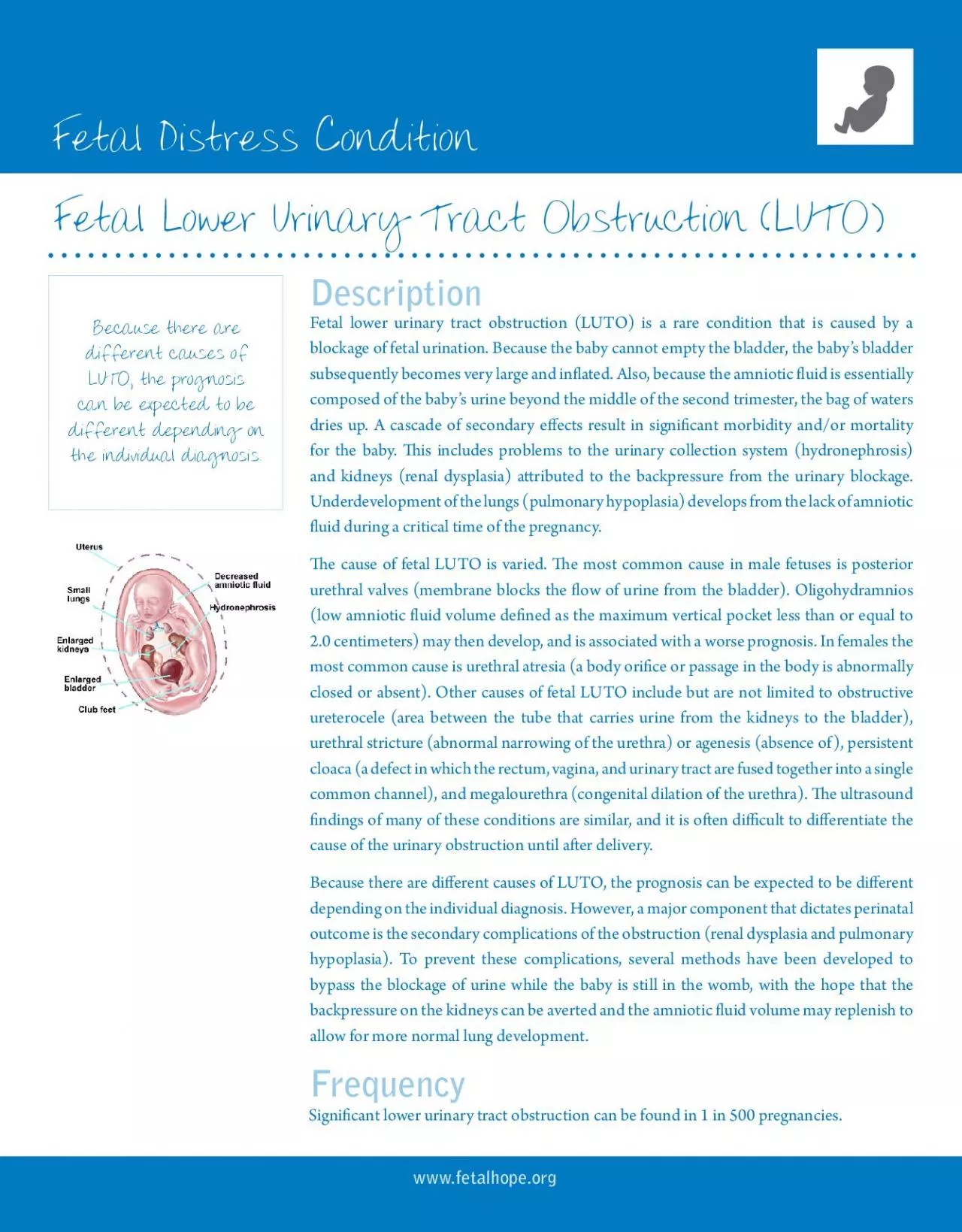
wwwfetalhopeorgFetal Lower Urinary Tract Obstruction LUTODescriptionFetal lower urinary tract obstruction LUTO is a rare condition that is caused by a subsequently becomes very large and in31ated Also ID: 873050

bladder fetal urinary baby fetal bladder baby urinary 146 luto ultrasound amniotic shunt urine obstruction urethral kidney uid performed

Share:

Link:

Embed:

Download Presentation from below link

Download Pdf The PPT/PDF document "Fetal Distress Condition" is the property of its rightful owner. Permission is granted to download and print the materials on this web site for personal, non-commercial use only, and to display it on your personal computer provided you do not modify the materials and that you retain all copyright notices contained in the materials. By downloading content from our website, you accept the terms of this agreement.


Presentation Transcript

1 Fetal Distress Condition www.fetalhope.o
Fetal Distress Condition www.fetalhope.org Fetal Lower Urinary Tract Obstruction (LUTO) Description Fetal lower urinary tract obstruction (LUTO) is a rare condition that is caused by a subsequently becomes very large and inated. Also, because the amniotic uid is essentially composed of the baby’s urine beyond the middle of the second trimester, the bag of waters dries up. A cascade of secondary eects result in signicant morbidity and/or mortality for the baby. is includes problems to the urinary collection system (hydronephrosis) Underdevelopment of the lungs (pulmonary hypoplasia) develops from the lack of amniotic uid during a critical time of the pregnancy. e cause of fetal LUTO is varied. e most common cause in male fetuses is posterior urethral valves (membrane blocks the ow of urine from the bladder). Oligohydramnios (low amniotic uid volume dened as the maximum vertical pocket less than or equal to most common cause is urethral atresia (a body orice or passage in the body is abnormally closed or absent). Other causes of fetal LUTO include but are not limited to obstructive ureterocele (area between the tube that carries urine from the kidneys to the bladder), urethral stricture (abnormal narrowing of the urethra) or agenesis (absence of), persistent common channel), and megalourethra (congenital dilation of the urethra). e ultrasound ndings of many of these conditions are similar, and it is oen dicult to dierentiate the cause of the urinary obstruction until aer delivery. Because there are dierent causes of LUTO, the prognosis can be expected to be dierent depending on the individual diagnosis. However, a major component that dictates perinatal outcome is the secondary complications of the obstruction (renal dysplasia and pulmonary hypoplasia). To prevent these complications, several methods have been developed to bypass the blockage of urine while the baby is still in the womb, with the hope that the backpressure on the kidneys can be averted and the amniotic uid volume may replenish to allow for more normal lung development. different causes of LUTO, the prognosis can be expected to be different depending on the individual diagnosis. Frequency Signicant lower urinary tract obstruction can be found in 1 in 500 pregnancies. Fetal Distress Condition www.fetalhope.org e diagnosis of LUTO is made by prenatal targeted ultrasound. Typically, the baby’s bladder is very distended (swollen). e presence of a “key-hol

2 e” sign is suggestive of posterior
e” sign is suggestive of posterior urethral valves, particularly in a male fetus. ere may be variable degrees of dilation of the upper urinary collection system. e ultrasound ndings of the baby’s kidneys should be carefully assessed for evidence of damage. Assessment of amniotic uid volume as well as the presence of other potential structural abnormalities is sought. Once the diagnosis of LUTO is established, the prognosis for survival is then assessed. e baby’s outcomes have been correlated to the kidney function as assessed prior to treatment. ere are two methods to determine the prognosis before surgery. ese methods are called fetal vesicocentesis, which samples the baby’s urine, and cordocentesis, which samples the baby’s blood. Genetic studies are also performed. 1. Fetal Vesicocentesis: Fetal kidney function may be obtained by performing sampling of the baby’s urine by placing a thin needle into the baby’s bladder and draining the urine. If the results of the rst drainage are below the threshold values then fetal therapy may be oered. If the rst vesicocentesis shows values above the threshold, a repeat vesicocentesis will be performed in 48 hours. 2. Cordocentesis: An alternative to vesicocentesis is to perform a cordocentesis. Under ultrasound guidance, a needle is placed in the umbilical cord. Fetal blood is drawn and sent for a serum Beta-2-microglobulin level. If the serum Beta- 2-microglobulin level is less than 5.6 then fetal therapy may be oered. Diagnosis & Prognostic Criteria e ability to evaluate kidney function is somewhat imprecise. is probably reects the dierent diagnoses responsible for the sonographic ndings, which do not have a similar prognosis despite comparable fetal urinary ndings or vice versa. Despite this limitation, the follow treatment options are available: 1. Expectant Management: is approach entails frequent ultrasound assessment to assess progression during the pregnancy. Aer delivery, pediatric specialists will evaluate the baby and subsequently oer treatments at that time. e risk of this approach is that further kidney and lung damage may occur during the pregnancy. In the seing of oligohydramnios (low amniotic uid volume), the expected perinatal mortality rate is 77%. Pregnancies where the obstruction only aects one kidney have a very good prognosis. ese pregnancies can be monitored regularly by ultra

3 sound and will likely deliver at full te
sound and will likely deliver at full term. 2. Vesicoamniotic Shunt: Fetal urinary diversion procedures have been performed since the 1980’s. Essentially, a shunt is placed between the baby’s bladder and the amniotic uid, thus relieving the blockage. is approach is meant to prevent further kidney and lung damage. e nal treatment of the obstruction is performed aer the birth of the child. In cases that have been determined to be in the favorable prognostic category (see above), the approximate perinatal survival using this treatment approach is 66%, of which half will have signicant kidney damage and may need dialysis or kidney transplant. Risks of shunt placement include dislodgement/blockage/malfunction (25%) thus requiring multiple shunt placements during the pregnancy, urinary ascites (leakage of urine from the bladder into the baby’s abdomen), and fetal death (4%). Management Options & Outcomes Fetal Distress Condition www.fetalhope.org 3. Fetal Cystoscopy and Treatment of LUTO: is approach allows for direct visualization of the baby’s bladder, proximal urethra, and ureteral orices. is approach, which was developed by Dr. Ruben Quintero, has the theoretic advantage of providing a more precise diagnosis. e ability to establish the correct diagnosis prenatally may improve the counseling capacity. In addition, ablation of posterior urethral valves or other in utero endoscopic treatments of fetal lower urinary tract obstruction may be performed. Whether these theoretical advantages translates into improved perinatal outcomes remains to be proven, which is why this and other centers are conducting studies in this regard. Risks from placing a needle or trocar into the fetal bladder include: infection, bleeding, trauma to the baby such as iatrogenic gastroschisis (hole in the abdominal wall lack the protrusion of the abdominal contents into the amniotic cavity), thermal damage to surrounding structures, or fetal death (4%). e details of the possible diagnostic and treatment approaches are detailed below. e following criteria generally must be met to oer fetal therapy for LUTO: 1. Gestational age: 16 weeks 0 days to 30 weeks 0 days. 2. Sonographic evidence of LUTO. 3. Fetal renal values below threshold shown in items 5 and 6 listed below. 4. Twin gestation may be included. Candidacy for Fetal Treatment e procedure(s) will be performed under local anesthesia. Fetal anesthesi

4 a will be delivered to the fetus with a
a will be delivered to the fetus with a ne needle under ultrasound guidance prior to entering the fetal bladder. Techniques 1. Vesicoamniotic catheter placement: A vesicoamniotic shunt will be placed in all cases over a wire guide. is can be done under ultrasound guidance. Endoscopic documentation of adequate shunt placement may be performed as well. Vesicoamniotic shunting may be necessary several times during the pregnancy due to dislodgement of the shunt or continued urinary outlet obstruction. 2. Diagnostic Fetal Cystoscopy: Aer a 2-3 millimeter skin incision is made, and under ultrasound guidance, an operative fetoscope will be inserted through the mother’s abdomen and uterus into the fetal bladder. e bladder wall, mucosa, trigone, and urethra will be the targets of the endoscopic assessment. 3. Operative Cystoscopy: If posterior urethral valves are identied, the valves will be ablated using laser energy through a contact YAG laser ber. Similarly, an obstructive ureterocele may be incised within the fetal bladder with a contact laser ber. ere may be rare circumstances that fetal urethral patency (unblocked condition) may be assessed with a so wire guide. If patency is documented, a thin transurethral catheter may be placed over a wire guide. Finally, spontaneous or iatrogenic urinary ascites (diversion of urine from the bladder into the baby’s abdomen) may occur. In these cases, access to the fetal bladder may best be achieved by fetal hydrolaparoscopy. An incision is made over the bladder dome with YAG laser energy, until the inside of the bladder can be seen. If the urethra can be Details of Procedure Fetal Distress Condition www.fetalhope.org accessed and the posterior urethral valves can be ablated, this is done at this point. Otherwise, surgery is completed by placing a peritoneoamniotic shunt. Before birth, it is recommended that ultrasound assessment occur every week for the rst four weeks, then every three to four weeks thereaer. Ultrasound parameters of particular importance include the amount of amniotic uid volume, measurement of the fetal bladder, assessment of the fetal kidneys and urinary collection system, presence of urinary ascites, and location of the vesicoamniotic shunt. Aer birth, the child will be evaluated by pediatric specialist and may require further tests and treatments. Postoperative Care Additional Resources For more information on LUTO, please visit www.fetalhope.or g