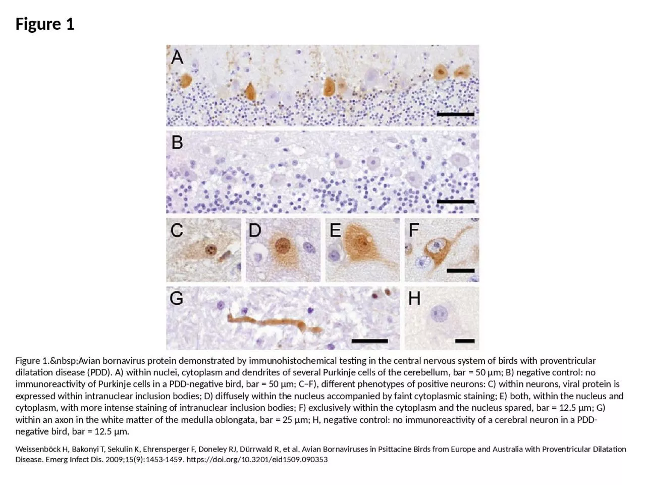
/
Figure 1 Figure 1. Avian bornavirus protein demonstrated by immunohistochemical Figure 1 Figure 1. Avian bornavirus protein demonstrated by immunohistochemical

Figure 1 Figure 1. Avian bornavirus protein demonstrated by immunohistochemical - PowerPoint Presentation

trinity
trinity . @trinity
Follow
27 views
Uploaded On 2024-02-09

Figure 1 Figure 1. Avian bornavirus protein demonstrated by immunohistochemical - PPT Presentation

Weissenböck H Bakonyi T Sekulin K Ehrensperger F Doneley RJ Dürrwald R et al Avian Bornaviruses in Psittacine Birds from Europe and Australia with Proventricular Dilatation Disease Emerg Infect Dis 200915914531459 httpsdoiorg103201eid1509090353 ID: 1044898

bar µm nucleus negative µm bar negative nucleus cytoplasm pdd disease bird immunoreactivity control intranuclear cells purkinje inclusion bodies

Share:

Link:

Embed:

Download Presentation from below link

Download Presentation The PPT/PDF document "Figure 1 Figure 1. Avian bornav..." is the property of its rightful owner. Permission is granted to download and print the materials on this web site for personal, non-commercial use only, and to display it on your personal computer provided you do not modify the materials and that you retain all copyright notices contained in the materials. By downloading content from our website, you accept the terms of this agreement.


Presentation Transcript

1. Figure 1Figure 1. Avian bornavirus protein demonstrated by immunohistochemical testing in the central nervous system of birds with proventricular dilatation disease (PDD). A) within nuclei, cytoplasm and dendrites of several Purkinje cells of the cerebellum, bar = 50 µm; B) negative control: no immunoreactivity of Purkinje cells in a PDD-negative bird, bar = 50 µm; C–F), different phenotypes of positive neurons: C) within neurons, viral protein is expressed within intranuclear inclusion bodies; D) diffusely within the nucleus accompanied by faint cytoplasmic staining; E) both, within the nucleus and cytoplasm, with more intense staining of intranuclear inclusion bodies; F) exclusively within the cytoplasm and the nucleus spared, bar = 12.5 µm; G) within an axon in the white matter of the medulla oblongata, bar = 25 µm; H, negative control: no immunoreactivity of a cerebral neuron in a PDD-negative bird, bar = 12.5 µm.Weissenböck H, Bakonyi T, Sekulin K, Ehrensperger F, Doneley RJ, Dürrwald R, et al. Avian Bornaviruses in Psittacine Birds from Europe and Australia with Proventricular Dilatation Disease. Emerg Infect Dis. 2009;15(9):1453-1459. https://doi.org/10.3201/eid1509.090353