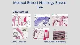
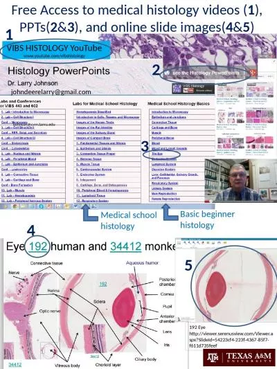
Eye and Ear Histology Orientation Images Eyelids Netter pl 76 UCSF 173 human eyelid Levator palpebrae sm S Tarsal muscle Orbicularis oris m Superior tarsus Tarsal gland Apocrine gland Lacrimal gland ID: 765302
Download Presentation The PPT/PDF document "Eye and Ear Histology" is the property of its rightful owner. Permission is granted to download and print the materials on this web site for personal, non-commercial use only, and to display it on your personal computer provided you do not modify the materials and that you retain all copyright notices contained in the materials. By downloading content from our website, you accept the terms of this agreement.
Eye and Ear Histology Orientation Images
Eyelids: Netter pl. 76
UCSF #173: human eyelid Levator palpebrae s.m. S. Tarsal muscle Orbicularis oris m. Superior tarsus Tarsal gland Apocrine gland Lacrimal gland Palpebral conjunctiva
Three Tunics of the Eye
Cornea Iris Anterior chamber Posterior chamber Ciliary body and processes Vitreous body Hyaloid canal Optic nerve Central retinal artery & vein Retina Choroid Sclera # Eye-2
Cornea Descemet’s membrane Corneal endothelium Corneal epithelium Bowman’s membrane Stroma Corneal stroma
Anterior chamber Cornea Posterior chamber Trabecular meshwork Canal of Schlemm Iris
A: Trabecular meshwork B: Canal of Schlemm A B B A
Lens Lens capsule Lens epithelium Lens fibers Zonule (of Zinn)
Lens fiber formation at the equator region equator Zonule
Ciliary body and ciliary processes Ciliary processes Ciliary body Pigmented and non-pigmented ciliary epithelial cells
Sphincter (constrictor) pupillae Dilator pupillae (smooth muscle) (myoepithelial cells) Eye-2
Sclera Choroid Bruch’s membrane Choriocapillary layer Retina Inner limiting membrane Optic nerve fibers Ganglion cell layer Inner plexiform layer Inner nuclear layer Outer plexiform layer Outer nuclear layer Outer limiting membrane Rods and cones Retinal pigment epithelium
EYE-1 Fovea centralis EYE-2 Ora serrata
Eye-2: Optic papilla (disc) Central retinal artery & vein Optic nerve
Ear Outer, middle and inner ear
Semicircular ducts Endolymphatic sac and duct Cochlear duct Saccule Utricle
Ear-2 Netter pl. 90 Cochlear duct (basal turn) Ampulla(e) Utricle Saccule Vestibular ganglion and nerve Cochlear nerve CN VIII: vestibulocochlear nerve ampulla Utricle Saccule
# Ear-4 Cochlear nerve Spiral (cochlear) ganglion Cochlear nerve
Ear-2 Vestibulocochlear Nerve (CN VIII) Cochlear Vestibule Cochlear Nerve Vestibular nerve Vestibular ganglion Vestibular nerve Vestibulocochlear Nerve (CNS VIII) Vestibular ganglion
EAR-2 Macula Crista ampullaris Otolithic membrane with otoconia (otoliths) Cupula Ear-4
Ear-3 and other ear slides Saccule ? Saccule Cochlear duct Stapes
Cochlear Nerve and spiral ganglia Helicotrema Cochlear Nerve Base Spiral ganglion Ear - 4
Organ of Corti in Scala Media Scala vestibuli Scala tympani Scala media ? ? ?
Stria vascularis Scala media (cochlear duct) Basilar membrane Vestibular membrane Tectorial m. Scala vestibuli Scala tympani
Organ of Corti Outer hair cells Inner hair cell Inner and outer pillar cells O. phalangeal cells I. phalangeal cell Inner tunnel
Perilymph and endolymph filled spaces in cochlea Blue: perilymph Yellow: endolymph
Helicotrema Osseous spiral lamina Cochlear duct Low pitch sound (longer basilar membrane) High pitch sound (shorter basilar membrane) ? ? EAR-3